Stroke & neurovascular medicine, from anatomy to the bedside
A comprehensive, illustrated, neurologist-level companion across the full breadth of cerebrovascular disease — an original diagram for every major topic, interactive score calculators, live search, and a board-style self-test. One self-contained file, online or off.
A diagram per topic
Open any topic for a relevant original figure — arterial territory, brainstem syndrome, score, or mechanism.
Search & browse
Live search highlights matches; 26 numbered chapters organise everything in the sidebar.
Bedside calculators
NIHSS, CHA₂DS₂-VASc, ICH Score, ABCD², Hunt-Hess and more compute as you click.
Board-style self-test
Worked, guideline-referenced questions — filter by chapter, practice or exam mode.
Neurovascular Atlas
51 original teaching diagrams spanning the anatomy, imaging and mechanisms behind stroke. Every one is drawn as scalable vector art (crisp at any zoom, dark-mode aware) and each also appears alongside its relevant chapter topic.
Interactive Score Calculators
Common bedside and prognostic scores — they compute live as you select each item. Educational decision-support only; confirm every input clinically and apply the appropriate guideline.
Self-Test
Board-style single-best-answer questions with worked, guideline-referenced explanations. Filter by chapter, choose a mode, and start.
Acute Ischemic Stroke — Overview & Definitions
Definition & epidemiology
Acute ischemic stroke (AIS) is an episode of neurological dysfunction caused by focal cerebral, spinal, or retinal infarction. The modern definition is tissue-based rather than time-based: infarction is confirmed by imaging (typically restricted diffusion on MRI) or neuropathology, or inferred from a persistent clinical deficit lasting >24 hours with other causes excluded. A transient ischemic attack (TIA) is by contrast a transient focal deficit without acute infarction on imaging — duration is no longer the discriminator.
Stroke is the second-leading cause of death worldwide and a leading cause of acquired adult disability. Ischemic stroke accounts for roughly 62–87% of all strokes (the remainder being intracerebral and subarachnoid hemorrhage), the proportion varying by population. Non-modifiable risk factors include age, sex, race/ethnicity and heredity; the dominant modifiable factors are hypertension (the single largest attributable risk), atrial fibrillation, diabetes, dyslipidemia, smoking, physical inactivity and obesity.
Pathophysiology: ischemic core vs penumbra
Arterial occlusion produces a spatial gradient of cerebral blood flow (CBF). The ischemic core is tissue with CBF so low that membrane-pump (Na⁺/K⁺-ATPase) failure and cytotoxic edema occur within minutes — irreversibly infarcted. Surrounding it is the ischemic penumbra: hypoperfused tissue that is electrically silent (functionally inactive, hence the clinical deficit) but structurally intact and potentially salvageable if flow is restored before it progresses to infarction. Between the penumbra and normal brain lies benign oligemia, which does not infarct even without reperfusion. The penumbra is the therapeutic target of all reperfusion therapy; its size and survival are governed chiefly by collateral circulation and time.
Time is brain. In supratentorial large-vessel occlusion, an estimated 1.9 million neurons, 14 billion synapses and 12 km of myelinated fibers are lost per minute of untreated ischemia; each hour without reperfusion ages the brain by the equivalent of roughly 3.6 years (Saver, 2006). This quantifies the imperative for rapid recanalization and underlies every door-to-treatment metric.
Etiologic classification (TOAST)
| TOAST subtype | Typical mechanism / features |
|---|---|
| Large-artery atherosclerosis | ≥50% stenosis/occlusion of extra- or intracranial artery; artery-to-artery embolism or hemodynamic; cortical/cerebellar/brainstem or subcortical infarcts >1.5 cm |
| Cardioembolism | High-risk source (AF, mechanical valve, recent MI, LV thrombus, endocarditis); often multiple territories, cortical, hemorrhagic transformation |
| Small-vessel occlusion (lacunar) | Lipohyalinosis of penetrating arteries; classic lacunar syndrome, infarct <1.5–2 cm in deep/brainstem territory |
| Other determined etiology | Dissection, vasculitis, hypercoagulable state, sickle cell, moyamoya, RCVS, genetic (CADASIL, Fabry) |
| Undetermined (cryptogenic) | Two competing causes, negative workup, or incomplete evaluation; overlaps with the ESUS construct (embolic stroke of undetermined source) |
Diagnostic workup
Immediate goals are to exclude hemorrhage, identify treatable large-vessel occlusion (LVO), and estimate core vs penumbra. Non-contrast CT (NCCT) excludes hemorrhage and is scored with ASPECTS; CT angiography localizes occlusion and assesses collaterals; CT perfusion or MRI (DWI/PWI) estimates core and salvageable tissue for extended-window selection. Point-of-care glucose is mandatory before thrombolysis (hypo- and hyperglycemia are mimics/modifiers).
Acute management (framework)
Two pillars: reperfusion — intravenous thrombolysis (tenecteplase or alteplase) and/or endovascular thrombectomy for LVO — layered on supportive neuroprotective physiology (blood pressure, glucose, temperature, oxygenation, aspiration prevention) delivered in an organized stroke unit. These are detailed in the dedicated topics that follow.
Clinical pearls
- Duration no longer defines TIA vs stroke — DWI positivity does. Up to a third of clinically defined "TIAs" show infarction.
- A normal early NCCT does not exclude stroke; it excludes hemorrhage and gross established infarct. DWI is the most sensitive test for acute ischemia.
- The penumbra is a physiological, not anatomical, entity — its extent depends on collaterals, so two patients with identical occlusions can have very different salvageable tissue and time windows.
Key references: AHA/ASA Guideline for the Early Management of Patients With Acute Ischemic Stroke, 2019 & 2019 update, and 2026 update; Sacco et al. AHA/ASA updated definition of stroke/TIA, Stroke 2013; Saver, "Time is Brain — Quantified," Stroke 2006; Adams et al., TOAST classification, Stroke 1993.
Stroke Recognition & Symptoms
Clinical features & recognition tools
Stroke presents as the sudden onset of a focal neurological deficit referable to a vascular territory. Public and prehospital screening uses FAST (Face droop, Arm drift, Speech disturbance, Time to call emergency services). BE-FAST adds Balance (acute gait/truncal ataxia) and Eyes (sudden diplopia, visual loss, or gaze deviation) to capture posterior-circulation events that plain FAST misses — FAST alone fails to detect roughly 1 in 7 strokes, predominantly posterior. Sudden severe "worst-ever" headache, decreased consciousness, or vomiting should prompt consideration of hemorrhage or basilar/cerebellar stroke.
Vascular anatomy & clinical syndromes
Localizing the syndrome guides territory, LVO likelihood and mimic probability.
| Territory | Hallmark features |
|---|---|
| MCA — superior division | Contralateral face/arm > leg weakness & sensory loss; eyes deviate toward lesion; Broca (expressive) aphasia if dominant hemisphere |
| MCA — inferior division | Contralateral homonymous hemianopia/quadrantanopia; Wernicke aphasia (dominant); hemineglect, anosognosia, constructional apraxia (non-dominant); little/no weakness |
| MCA — deep (lenticulostriate) | Contralateral pure motor hemiparesis (face, arm, leg equally) from internal capsule involvement |
| ACA | Contralateral leg > arm weakness/sensory loss; abulia, akinetic mutism, transcortical motor aphasia, urinary incontinence, grasp reflex |
| PCA | Contralateral homonymous hemianopia with macular sparing; alexia without agraphia (dominant splenium); thalamic (Déjerine–Roussy) sensory loss/pain; visual agnosia, prosopagnosia |
| Vertebrobasilar / brainstem | Crossed findings (ipsilateral cranial nerve + contralateral body), diplopia, vertigo, dysarthria, dysphagia, ataxia, decreased consciousness; "5 D's" plus crossed signs |
| Lateral medullary (Wallenberg, PICA/vertebral) | Ipsilateral facial sensory loss, Horner, ataxia, dysphagia/hoarseness (nucleus ambiguus); contralateral body pain/temperature loss; vertigo, nystagmus — no limb weakness |
| Basilar (top-of-the-basilar / pontine) | Fluctuating consciousness, vertical gaze palsy, pupillary abnormalities; locked-in syndrome with ventral pontine infarction (quadriplegia, preserved vertical gaze/blink) |
Cortical vs subcortical vs posterior. Cortical signs (aphasia, neglect, gaze deviation, hemianopia) indicate large-artery/cardioembolic cortical involvement and predict LVO. Classic lacunar (subcortical) syndromes lack cortical signs: pure motor hemiparesis (posterior limb internal capsule/basis pontis), pure sensory stroke (thalamic VPL), ataxic hemiparesis, dysarthria–clumsy hand, and sensorimotor stroke. Posterior-circulation strokes are the most frequently missed and under-scored.
NIHSS overview
The National Institutes of Health Stroke Scale (NIHSS) is a 15-item, 0–42 quantification of deficit severity used for triage, thrombolysis/thrombectomy decisions, and serial monitoring. Domains: level of consciousness (1a–c), best gaze, visual fields, facial palsy, motor arm (L/R), motor leg (L/R), limb ataxia, sensory, best language, dysarthria, and extinction/inattention (neglect).
| NIHSS total | Severity band |
|---|---|
| 0 | No stroke symptoms |
| 1–4 | Minor |
| 5–15 | Moderate |
| 16–20 | Moderate–severe |
| 21–42 | Severe |
Caveats: the NIHSS is weighted toward left (dominant) hemisphere function (language items carry ~7 points), so a dominant-hemisphere stroke scores higher than a non-dominant stroke of equal volume. It is relatively insensitive to posterior-circulation and right-hemisphere strokes — a disabling brainstem or cerebellar infarct may score only 1–3. NIHSS ≥6 is a common (not absolute) threshold suggesting LVO warranting vessel imaging.
Differential diagnosis (stroke mimics)
- Seizure / Todd paralysis — postictal focal weakness; a witnessed convulsion or gradual "spreading" onset favors seizure.
- Hypoglycemia / hyperglycemia — can reproduce focal deficits; always check glucose first.
- Migraine with aura — spreading, marching positive symptoms (scintillations, then numbness) over minutes; younger patients.
- Functional (conversion) deficit — non-anatomical, give-way weakness, Hoover sign positive.
- Structural — tumor, subdural hematoma (subacute, fluctuating).
- Toxic-metabolic / infective — hepatic or septic encephalopathy, hyponatremia, Wernicke, HSV encephalitis.
- Peripheral vertigo — vestibular neuritis vs posterior stroke; the bedside HINTS exam (Head-Impulse, Nystagmus, Test-of-Skew) outperforms early MRI in acute vestibular syndrome — a normal head-impulse, direction-changing nystagmus, or skew deviation points to a central (stroke) cause.
Clinical pearls
- Isolated acute vertigo with a normal head-impulse test is a red flag for cerebellar/brainstem stroke, not labyrinthitis.
- Gaze deviation localizes: in hemispheric stroke the eyes look toward the lesion (away from the hemiparesis); in a pontine gaze palsy they look away from the lesion.
- A low NIHSS never excludes LVO — always image the vessels in posterior-circulation presentations.
Key references: AHA/ASA AIS Guideline 2019/2026; Aroor et al. BE-FAST, Stroke 2017; NIH Stroke Scale (Brott et al., 1989); Kattah et al., HINTS, Stroke 2009.
Early Management of Suspected Stroke
Prehospital care
Emergency dispatch should treat suspected stroke as a time-critical emergency. Field priorities: confirm a prehospital screen (FAST/BE-FAST or Cincinnati/Los Angeles scales), establish last known well (LKW) time, check glucose, obtain a witness/contact and medication history (especially anticoagulants), and pre-notify the receiving center. Prehospital notification measurably shortens door-to-imaging and door-to-needle times. Mobile stroke units (ambulances with onboard CT and telemedicine) enable thrombolysis in the field and, per randomized evidence (BEST-MSU, B_PROUD), improve functional outcomes; the 2026 AHA/ASA guideline gives them a Class 1 recommendation where feasible.
Prehospital LVO triage scales
Beyond detecting stroke, EMS increasingly stratifies severity to route probable LVO patients directly to thrombectomy-capable centers. No scale is perfect; each trades sensitivity against specificity.
| Scale | Components | LVO-positive threshold |
|---|---|---|
| RACE | Facial palsy, arm & leg motor, gaze, aphasia/agnosia | ≥5 |
| LAMS | Facial droop, arm drift, grip strength | ≥4 |
| C-STAT / CPSSS | Gaze, LOC commands/questions, arm | ≥2 |
| FAST-ED | Face, arm, speech, eye deviation, denial/neglect | ≥4 |
| VAN | Arm weakness + Visual/Aphasia/Neglect | Weakness plus any cortical sign |
In-hospital "code stroke"
A parallel-process pathway compresses evaluation: simultaneous triage, focused history/exam with NIHSS, point-of-care glucose, IV access and labs, and immediate transfer to CT. Advanced imaging (CTA ± CT perfusion) is obtained without delaying IV thrombolysis when the patient is otherwise eligible. Thrombolysis eligibility (time window, BP, contraindications) is determined in parallel so the bolus can be given in the scanner suite.
Time-target (door-to-treatment) metrics
| Interval | Benchmark target |
|---|---|
| Door-to-physician | ≤10 min |
| Door-to-CT (imaging initiated) | ≤20–25 min |
| Door-to-needle (IV thrombolysis) | ≤60 min in ≥50% (stretch goal ≤45 min in ≥50%; ≤30 min achievable) |
| Door-to-groin/puncture (EVT, direct arrivals) | ≤90 min |
| Door-in–door-out (transfer for EVT) | ≤60 min (aspirational) |
These benchmarks derive from the AHA Target: Stroke quality initiative; each 15-minute reduction in door-to-needle time yields measurable gains in independent survival and reductions in symptomatic hemorrhage and mortality.
Differential diagnosis at triage
Roughly a fifth of "code stroke" activations are mimics (see Stroke Recognition topic). The two exclusions that must be made before thrombolysis are hemorrhage (NCCT) and hypoglycemia (glucose). Reassuringly, treating a mimic with IV thrombolysis carries a low symptomatic-hemorrhage rate, so uncertainty should not cause the treatable stroke patient to miss the window.
Clinical pearls
- "Last known well," not symptom-discovery time, starts the clock — critical for wake-up and aphasic/unwitnessed patients.
- Do not wait for coagulation results to treat unless the patient is on anticoagulants or has a bleeding-risk history; institutional protocols allow empiric thrombolysis when suspicion of coagulopathy is low.
- Routing decisions balance a short delay to reach an EVT center against the benefit of earlier IV thrombolysis at a closer primary center ("drip-and-ship" vs "mothership").
Key references: AHA/ASA AIS Guideline 2019/2026; AHA Target: Stroke Phase III; BEST-MSU (Grotta et al., NEJM 2021) and B_PROUD (Ebinger et al., JAMA 2021); Pérez de la Ossa et al., RACE scale, Stroke 2014.
General Management of the Acute Stroke Patient
Airway, breathing & oxygenation
Protect the airway in patients with depressed consciousness (large hemispheric, brainstem, or basilar strokes) or bulbar dysfunction with aspiration risk. Supplemental oxygen is indicated only to maintain SpO₂ >94%; routine supplemental oxygen in non-hypoxic patients confers no benefit and is not recommended.
Glucose
Both extremes worsen outcome. Treat hypoglycemia (<60 mg/dL) promptly. Target a serum glucose of roughly 140–180 mg/dL in the first 24 hours. Intensive insulin protocols targeting near-normoglycemia (e.g., 80–130 mg/dL) do not improve outcomes and increase severe hypoglycemia (SHINE trial); the 2026 guideline recommends against them.
Temperature
Fever worsens ischemic injury and independently predicts poor outcome. Identify and treat the source; give antipyretics (e.g., acetaminophen) for temperature >38 °C. Therapeutic hypothermia is not recommended outside of trials — it has not improved outcomes and adds complications (pneumonia, shivering).
Dysphagia / swallow screening
Perform a validated bedside swallow screen before any oral intake (including oral medications) — dysphagia affects up to half of acute stroke patients and drives aspiration pneumonia. Keep patients NPO until they pass; refer failures for formal speech-language/instrumental (video-fluoroscopy or FEES) evaluation.
Venous thromboembolism (VTE) prophylaxis
| Measure | Guidance |
|---|---|
| Intermittent pneumatic compression (IPC) | Recommended for immobile patients starting on admission (CLOTS-3 reduced DVT and improved survival) |
| Pharmacologic prophylaxis (LMWH/UFH) | Consider for immobile patients once hemorrhage excluded; individualize timing after thrombolysis/large infarct; benefit for DVT/PE balanced against bleeding |
| Graduated compression stockings | Not recommended — ineffective and cause skin breakdown (CLOTS-1/2) |
Early mobilization & supportive care
Avoid prolonged bed rest, but very early, high-dose out-of-bed mobilization within 24 hours was harmful in AVERT — favor frequent, shorter, individualized mobilization beginning after the first day once stable. Additional bundle elements: aspiration precautions, bowel/bladder care (avoid indwelling catheters — infection and delirium risk), pressure-area care, early nutrition (nasogastric feeding within days if dysphagic; PEG reserved for prolonged dysphagia), glycemic and BP protocols, and depression screening.
Stroke unit care
Admission to an organized, geographically discrete stroke unit with a coordinated multidisciplinary team is one of the most robust interventions in all of stroke medicine: it reduces death and dependency across all severities and ages, independent of thrombolysis, chiefly by preventing and rapidly treating complications. Continuous cardiac monitoring for ≥24 hours detects atrial fibrillation and arrhythmia.
Differential considerations in deterioration
Early neurological worsening should trigger evaluation for cerebral edema, hemorrhagic transformation, seizure, recurrent/extending ischemia, or systemic causes (hypoxia, hypotension, hyper/hypoglycemia, hyponatremia, infection) — several are reversible (see Acute Stroke Complications and Stroke in Evolution topics).
Clinical pearls
- The swallow screen is the single highest-yield nursing intervention for preventing aspiration pneumonia — enforce NPO until passed.
- Do not chase "tight" glucose control; 140–180 mg/dL is the sweet spot.
- Stroke-unit care benefits patients regardless of whether they received reperfusion therapy — it is the default disposition for every stroke.
Key references: AHA/ASA AIS Guideline 2019/2026; SHINE trial (Johnston et al., JAMA 2019); CLOTS-1/2/3 (Lancet 2009–2013); AVERT trial (Lancet 2015); Stroke Unit Trialists' Collaboration (Cochrane).
Blood Pressure Management in Acute Ischemic Stroke
Principles
Most acute ischemic strokes present with reactive hypertension that spontaneously declines. In the ischemic penumbra, cerebral autoregulation is impaired and perfusion becomes pressure-passive — aggressive BP lowering can extend infarction, whereas extreme hypertension risks hemorrhagic transformation and edema. Management therefore differs sharply by reperfusion status.
Targets by scenario
| Scenario | Blood-pressure target |
|---|---|
| Not receiving reperfusion therapy | Permissive hypertension: treat only if >220/120 mm Hg, or if there is a concurrent indication (acute coronary event, heart failure, aortic dissection, hemorrhagic transformation, preeclampsia). Reasonable initial goal ~15% reduction in the first 24 h. |
| Before IV thrombolysis | Must be <185/110 mm Hg before the bolus; lower cautiously with labetalol or nicardipine if above. |
| During & first 24 h after thrombolysis | Maintain <180/105 mm Hg; monitor BP frequently (e.g., q15 min ×2 h, then q30 min ×6 h, then hourly). |
| Before / during thrombectomy | Avoid hypotension; commonly keep SBP roughly 140–180 mm Hg peri-procedurally to preserve collateral flow until reperfusion. |
| After successful thrombectomy (anterior circulation) | Do not intensively lower — avoid reducing SBP <140 mm Hg in the first 72 h (2026 guideline, Class III/harm). Individualize a ceiling (often <180 mm Hg); higher risk of hemorrhage favors the lower end of the range. |
Randomized trials of intensive post-thrombectomy BP lowering — ENCHANTED2/MT (SBP <120 vs <140–180) and OPTIMAL-BP (SBP <140 vs 140–180) — both showed worse functional outcomes with aggressive control after successful reperfusion, and BEST-II found no benefit to lower targets. Hence the current stance against driving BP down after recanalization.
Preferred agents
| Agent | Dose / administration | Notes |
|---|---|---|
| Labetalol | 10–20 mg IV push over 1–2 min; may repeat once; or infusion | Avoid in bradycardia, high-grade block, decompensated heart failure, bronchospasm |
| Nicardipine | Infusion 5 mg/h, titrate by 2.5 mg/h every 5–15 min (max ~15 mg/h) | Smooth, titratable dihydropyridine; first-line infusion in many centers |
| Clevidipine | Infusion 1–2 mg/h, double every 90 s toward effect (max ~21 mg/h) | Ultra-short-acting; rapid titration; lipid emulsion (egg/soy allergy caution) |
| Hydralazine / enalaprilat | Second-line boluses | Less predictable; enalaprilat useful when tachycardia limits other agents |
Nitroprusside is reserved for refractory hypertension (>220/120) given cyanide toxicity and potential to raise intracranial pressure. Titratable infusions (nicardipine/clevidipine) are generally preferred over repeated boluses when tight control is needed peri-thrombolysis.
Clinical pearls
- Before lytic, the number is 185/110; after lytic, 180/105; without reperfusion, do not treat until 220/120. Memorize these three thresholds.
- After successful thrombectomy the instinct to "protect the brain" by lowering BP is counterproductive — the reperfused but vulnerable tissue tolerates hypotension poorly.
- Always exclude a full bladder, pain, and raised ICP as reversible causes of hypertension before pharmacologic treatment.
Key references: AHA/ASA AIS Guideline 2019/2026; ENCHANTED2/MT (Yang et al., Lancet 2022); OPTIMAL-BP (Nam et al., JAMA 2023); BEST-II (Mistry et al., JAMA 2023).
Acute Stroke Complications
Cerebral edema & raised intracranial pressure
Cytotoxic then vasogenic edema peaks at 2–5 days. Large hemispheric ("malignant" MCA) and space-occupying cerebellar infarcts cause herniation. Warning signs: declining consciousness, new pupillary asymmetry, worsening NIHSS, Cushing response. Cerebellar infarcts additionally threaten fourth-ventricle compression (obstructive hydrocephalus) and direct brainstem compression. Management: head elevation ~30°, normocapnia, osmotherapy (hypertonic saline or mannitol) as a bridge, avoidance of hypotonic fluids and fever; surgical decompression is the definitive life-saving therapy — hemicraniectomy for malignant MCA infarction and suboccipital craniectomy ± EVD for cerebellar infarction (see dedicated hemicraniectomy topic). Corticosteroids are not effective for cytotoxic ischemic edema.
Hemorrhagic transformation (HT)
HT ranges from asymptomatic petechial staining to a space-occupying parenchymal hematoma. Risk rises with infarct size, cardioembolic mechanism, reperfusion therapy, hyperglycemia, and hypertension. The radiographic ECASS classification is clinically useful:
| ECASS grade | Description |
|---|---|
| HI1 | Small petechiae along infarct margin |
| HI2 | Confluent petechiae within infarct, no mass effect |
| PH1 | Hematoma ≤30% of infarct with mild mass effect |
| PH2 | Hematoma >30% of infarct with significant mass effect / remote hemorrhage |
Symptomatic ICH (sICH) — hematoma plus neurological deterioration (definitions vary: NINDS any decline; ECASS III ≥4 NIHSS points with PH2) — complicates roughly 2–7% of thrombolysis cases. Management of post-lytic sICH: stop the infusion, urgent NCCT, reverse fibrinolysis with cryoprecipitate (to replace fibrinogen) ± tranexamic acid or aminocaproic acid, correct coagulopathy, control BP, and neurosurgical consultation.
Seizures
Early (within 7 days) seizures occur in a minority, more with cortical and hemorrhagic infarcts. Treat clinical or electrographic seizures with standard antiseizure medication; prophylactic antiseizure drugs are not recommended. Consider nonconvulsive status epilepticus (EEG) when consciousness is unexplainedly depressed or fluctuating.
Infection & aspiration
Aspiration pneumonia and urinary tract infection are the commonest medical complications and directly worsen outcome — prevention hinges on swallow screening, oral care, aspiration precautions and avoiding indwelling catheters. Prophylactic antibiotics do not improve outcomes (STROKE-INF, PASS) and are not recommended; treat established infection promptly.
Cardiac complications
Stroke — especially insular cortex involvement — triggers autonomic dysregulation producing arrhythmias (new AF, QT prolongation), troponin elevation, neurogenic (Takotsubo) cardiomyopathy, and ECG changes mimicking ischemia. Continuous telemetry ≥24 h is standard. Distinguish demand ischemia/neurogenic injury from primary acute coronary syndrome, as management (particularly antithrombotics) differs.
Other
- VTE — DVT/PE from immobility (see prophylaxis in General Management).
- Hyponatremia — SIADH or cerebral salt wasting; worsens edema.
- Falls, pressure injury, depression, central/shoulder pain, spasticity — subacute complications requiring rehabilitation-phase attention.
Clinical pearls
- A deteriorating cerebellar stroke is a neurosurgical emergency — decompression/EVD is highly effective and time-sensitive; do not attribute drowsiness to "sedation."
- Reverse post-thrombolysis hemorrhage with cryoprecipitate (fibrinogen replacement), not simply platelets or vitamin K.
- Troponin elevation after stroke is common and usually neurogenic — avoid reflex antithrombotic escalation that could precipitate hemorrhagic transformation.
Key references: AHA/ASA AIS Guideline 2019/2026; ECASS/NINDS HT classifications; STROKE-INF (Kalra et al., Lancet 2015) & PASS (Westendorp et al., Lancet 2015); Wijdicks et al. AHA cerebellar/space-occupying infarct statement, Stroke 2014.
Ischemic Penumbra & Cerebral Blood Flow Thresholds
Concept
The penumbra (Astrup, Symon & Siesjö, 1981) is tissue in which CBF has fallen below the threshold for electrical function but remains above the threshold for membrane/metabolic failure — it is electrically silent yet structurally viable, and its fate (recovery vs infarction) depends on depth and duration of hypoperfusion and on collateral support. It is the operational target of every reperfusion therapy; without reperfusion, penumbra progressively recruits into core over hours.
CBF thresholds (approximate, flow in mL/100 g/min)
| Zone | CBF (mL/100 g/min) | Physiology |
|---|---|---|
| Normal gray matter | ~50–55 | Full function |
| Benign oligemia | ~22–50 | Protein-synthesis inhibition; will not infarct even without reperfusion |
| Penumbra | ~10–22 | Electrical failure (loss of evoked/spontaneous activity ~16–18); neurons silent but ion pumps preserved — salvageable |
| Ischemic core | <~8–10 | Membrane-pump/energy failure, anoxic depolarization, cytotoxic edema — irreversible |
These thresholds are time-dependent, not fixed: tissue tolerates moderate flow reduction far longer than severe reduction. The relationship between flow depth and time to infarction is the physiological basis for individualized ("tissue-clock") rather than purely time-based selection.
Imaging surrogates & operational definitions
| Parameter | Operational definition |
|---|---|
| Core (CT perfusion) | Relative CBF <30% of contralateral (rCBF<30%) |
| Critically hypoperfused / penumbra (CTP) | Tmax >6 s |
| Core (MRI) | Diffusion restriction (low ADC) on DWI |
| Penumbra (MRI) | Diffusion–perfusion mismatch (PWI lesion > DWI lesion) |
| "Target mismatch" (DEFUSE/DAWN) | Mismatch ratio ≥1.8, mismatch volume ≥15 mL, core <70 mL (plus low Tmax>10 s volume) |
Automated software (e.g., RAPID-type post-processing) generates these volumes and drove patient selection in the extended-window thrombectomy trials DAWN (6–24 h, clinical–core mismatch) and DEFUSE-3 (6–16 h, perfusion–core mismatch), and the thrombolysis trials EXTEND and WAKE-UP.
Collateral circulation
Leptomeningeal (pial) collaterals from adjacent ACA/PCA territories, plus the circle of Willis, sustain the penumbra. Good collaterals slow core growth, enlarge the salvageable region, widen the treatment window and predict better reperfusion outcomes; poor collaterals produce rapid "malignant" core growth ("fast progressors"). Collateral status is assessed on multiphase CTA or from the CTP hypoperfusion-intensity ratio.
Differential / mimics of imaging mismatch
Perfusion "mismatch" is not specific to stroke — seizure (peri-ictal hyperperfusion or post-ictal hypoperfusion), migraine aura, and venous pathology can produce perfusion abnormalities that do not respect an arterial territory. Interpret CTP alongside vessel imaging and the clinical syndrome.
Clinical pearls
- The mismatch concept lets physiology, not just the clock, select patients — a "slow progressor" with excellent collaterals may retain salvageable penumbra well beyond 24 h.
- A large core with a small penumbra ("matched defect") predicts little benefit and higher hemorrhage risk; a small core with large penumbra is the ideal reperfusion candidate.
- Perfusion thresholds are operational conveniences — they approximate, but do not equal, the true pathophysiological flow thresholds and can over- or under-estimate core depending on timing and software.
Key references: Astrup, Siesjö & Symon, "Thresholds in cerebral ischemia — the ischemic penumbra," Stroke 1981; DAWN (Nogueira et al., NEJM 2018); DEFUSE-3 (Albers et al., NEJM 2018); AHA/ASA AIS Guideline 2019/2026.
Reperfusion Injury, No-Reflow Phenomenon & Blood–Brain Barrier Disruption
Overview
Restoring flow is necessary but not by itself sufficient — the return of oxygenated blood to ischemic tissue can itself injure the brain (ischemia–reperfusion injury), microvascular flow may fail to recover despite arterial recanalization (no-reflow), and the blood–brain barrier (BBB) may break down, producing edema and hemorrhagic transformation. These processes constrain the net benefit of recanalization and are active targets of adjunctive neuroprotection research.
Ischemia–reperfusion injury: mechanisms
- Excitotoxicity — energy failure causes glutamate accumulation; NMDA/AMPA over-activation drives Ca²⁺ influx.
- Calcium overload — activates proteases, lipases, endonucleases and nitric oxide synthase.
- Oxidative & nitrosative stress — reperfusion delivers O₂ to dysfunctional mitochondria, generating a burst of reactive oxygen/nitrogen species that overwhelm antioxidant defenses and peroxidize lipids.
- Mitochondrial permeability transition — pore opening triggers apoptotic and necrotic death.
- Inflammation — microglial activation, cytokine release (TNF-α, IL-1β, IL-6), adhesion-molecule up-regulation and leukocyte infiltration extend injury.
No-reflow phenomenon
Despite successful proximal recanalization (e.g., TICI 2b–3 angiographically), tissue-level microcirculatory perfusion can remain impaired — angiographic reperfusion without tissue reperfusion. Proposed mechanisms: pericyte constriction of capillaries persisting after reflow, capillary plugging by leukocytes and platelet–fibrin aggregates, endothelial and astrocyte end-foot swelling narrowing the lumen, and microthrombosis. No-reflow contributes to the "futile recanalization" seen when a technically successful thrombectomy fails to yield clinical improvement.
Blood–brain barrier disruption
Ischemia degrades the neurovascular unit: matrix metalloproteinases (notably MMP-9) and other proteases cleave tight-junction proteins (claudin-5, occludin, ZO-1) and basal lamina, increasing permeability. BBB opening is often biphasic — an early transient opening followed by a later, more severe phase associated with vasogenic edema and hemorrhagic transformation. The consequences are:
| Consequence | Correlate |
|---|---|
| Vasogenic edema | Adds to cytotoxic edema; raises mass effect / ICP |
| Hemorrhagic transformation | ECASS HI1–PH2 spectrum; risk ↑ with reperfusion, large core, hyperglycemia, HTN |
| Contrast staining / extravasation post-EVT | Hyperdensity on immediate post-procedure CT; dual-energy CT distinguishes contrast staining (benign, clears) from true hemorrhage |
Clinical & therapeutic relevance
These mechanisms explain why faster reperfusion yields larger benefit (less accumulated injury before reflow), why hyperglycemia and marked hypertension worsen HT, and why post-reperfusion BP should not be driven too low (impaired autoregulation + no-reflow render tissue perfusion pressure-dependent). Numerous cytoprotectants targeting these pathways (free-radical scavengers, glutamate antagonists, anti-inflammatories) have failed in trials; interest persists in agents given as adjuncts to reperfusion, but no cytoprotectant has definitive proven benefit.
Differential diagnosis (post-EVT hyperdensity)
A frequent bedside problem is the hyperdense lesion on immediate post-thrombectomy CT: contrast staining (blood–brain barrier leak of iodinated contrast — usually resolves within 24 h, Hounsfield units typically lower, respects gyral pattern) versus true hemorrhage. Delayed CT (contrast clears; hemorrhage persists/grows) or dual-energy CT resolves the distinction and prevents unnecessary reversal of antithrombotics.
Clinical pearls
- "Futile recanalization" — an open vessel with no clinical benefit — often reflects no-reflow, already-completed core, or distal embolization, not procedural failure.
- Contrast staining after EVT is common and benign; do not reflexively diagnose hemorrhage — obtain delayed/dual-energy imaging.
- Every minute saved to reperfusion reduces cumulative reperfusion injury and hemorrhage risk — speed matters even once the case is "in the lab."
Key references: Moskowitz, Lo & Iadecola, "The science of stroke: mechanisms in search of treatments," Neuron 2010; del Zoppo & Mabuchi, microvascular no-reflow reviews; Jha et al./Yang & Rosenberg, MMP and BBB disruption reviews; AHA/ASA AIS Guideline 2019/2026.
Malignant Cerebral (MCA) Infarction & Decompressive Hemicraniectomy
Definition & epidemiology
"Malignant MCA infarction" denotes a large hemispheric (typically complete MCA ± ACA/PCA) territory infarct that develops space-occupying cytotoxic edema, causing herniation and death in up to ~80% of medically managed patients. Edema peaks at days 2–5. It complicates a minority of MCA strokes but accounts for a disproportionate share of early stroke mortality, often in younger patients with poor collaterals and proximal (ICA-terminus/M1) occlusion.
Clinical & imaging predictors
- High baseline NIHSS (often >15 non-dominant, >20 dominant) with early decreased consciousness, nausea/vomiting.
- Early hypodensity involving >50% of the MCA territory on NCCT; hyperdense MCA sign; ICA-terminus ("T") occlusion; poor collaterals.
- Large early DWI infarct volume (e.g., >~82 mL within 6 h, >~145 mL by 14 h) predicts malignant course.
- The EDEMA score (basal cistern effacement, midline shift, glucose, prior stroke, reperfusion status) and related models help stratify risk of malignant edema, though thresholds are approximate and should not replace serial clinical/imaging assessment.
Medical management (temporizing)
Head elevation ~30°, normocapnia, osmotherapy (hypertonic saline or mannitol), fever and glucose control, avoidance of hypotonic fluids and hypoxia. These buy time but do not prevent the natural history — decompressive surgery is the only intervention shown to reduce mortality. Corticosteroids are ineffective and not recommended.
Decompressive hemicraniectomy: the evidence
| Trial(s) | Population | Key result |
|---|---|---|
| DECIMAL, DESTINY, HAMLET (pooled, Vahedi 2007) | Age 18–60; surgery within 48 h of onset | Absolute mortality reduction ~50% (≈71%→22%); more survivors with mRS ≤4 and ≤3; NNT ≈2 to prevent death, ≈4 for mRS ≤3 |
| DESTINY II (Jüttler 2014) | Age >60 (mean ~70); within 48 h | Survival markedly improved, but most survivors left with moderately severe–severe disability (mRS 4–5); very few achieved mRS ≤2 |
Procedure: a large fronto-temporo-parietal bone flap (diameter ≥12 cm, and ≥14 cm preferred) with duraplasty; an inadequately sized craniectomy fails to relieve pressure and risks venous infarction at the bony edge. Timing is ideally early — within 24–48 h and before clinical herniation; waiting for fixed pupillary dilation forfeits much of the benefit.
Patient selection & shared decision-making
| Factor | Consideration |
|---|---|
| Age | Strongest benefit ≤60; benefit >60 is survival with greater residual disability — individualize |
| Timing | Within 48 h, before herniation, gives best functional results |
| Hemisphere | Dominant-hemisphere infarction (aphasia) does not preclude surgery, but expected quality of life must be discussed frankly |
| Goals of care | Surgery converts likely death into survival often with significant disability — informed proxy/patient value judgment is essential |
Cerebellar (posterior fossa) counterpart
Space-occupying cerebellar infarction is managed analogously but by suboccipital decompressive craniectomy (± external ventricular drain for obstructive hydrocephalus, ± infarct resection). Because the posterior fossa is small, deterioration is rapid; decompression is highly effective and should not be delayed.
Clinical pearls
- Hemicraniectomy is a life-saving, not function-restoring, operation — frame counseling around survival with disability, especially over age 60.
- Operate before the pupil blows: pre-herniation surgery drives the outcome advantage.
- An EVD alone does not relieve brainstem compression from a cerebellar infarct — suboccipital craniectomy is the definitive treatment.
Key references: Vahedi et al. pooled analysis of DECIMAL/DESTINY/HAMLET, Lancet Neurol 2007; Jüttler et al. DESTINY II, NEJM 2014; AHA/ASA scientific statement on cerebral & cerebellar space-occupying infarction (Wijdicks 2014); AHA/ASA AIS Guideline 2019/2026.
Wake-Up Stroke
Definition & epidemiology
A wake-up stroke is one in which the patient is neurologically normal on going to sleep and has a deficit on awakening, so the exact onset is unknown and "last known well" defaults to bedtime. Wake-up strokes constitute roughly a fifth of ischemic strokes. Historically excluded from reperfusion because the conventional time window could not be established, many are now treatable using imaging-based ("tissue-clock") selection that estimates lesion age or salvageable tissue rather than relying on the wall clock.
DWI–FLAIR mismatch
An acute infarct is visible on diffusion-weighted imaging (DWI) almost immediately but takes several hours to become hyperintense on FLAIR. A lesion that is bright on DWI but not yet visible on FLAIR (DWI–FLAIR mismatch) therefore implies the infarct is relatively young — approximately within 4.5 hours — identifying patients likely to benefit from thrombolysis despite unknown onset.
| Sequence | Finding in early (<4.5 h) infarct |
|---|---|
| DWI | Hyperintense (restricted diffusion) — positive |
| FLAIR | Not yet hyperintense — negative |
| Interpretation | DWI+/FLAIR− "mismatch" ⇒ lesion likely young ⇒ thrombolysis candidate |
Evidence
- WAKE-UP (Thomalla, NEJM 2018): unknown-onset stroke with DWI–FLAIR mismatch randomized to alteplase 0.9 mg/kg vs placebo — significantly more patients achieved favorable outcome (mRS 0–1: ~53% vs ~42%), with a non-significant increase in symptomatic hemorrhage. The trial stopped early for funding.
- EXTEND (Ma, NEJM 2019): ischemic stroke 4.5–9 h from onset or wake-up (using time from midpoint of sleep) with automated perfusion mismatch — alteplase improved excellent outcome.
- Individual-patient meta-analysis of wake-up/late thrombolysis trials confirmed net benefit with imaging selection.
- For large-vessel occlusion wake-up strokes, extended-window thrombectomy (DAWN 6–24 h, DEFUSE-3 6–16 h) applies using clinical–core or perfusion–core mismatch.
Current guideline position
Imaging-selected thrombolysis for wake-up / unknown-onset stroke is endorsed: DWI–FLAIR mismatch (MRI) or perfusion mismatch (CTP) identifies candidates. The 2026 AHA/ASA guideline supports IV thrombolysis (tenecteplase or alteplase) as reasonable in the 4.5–9 h window and in wake-up stroke when advanced imaging demonstrates salvageable tissue, and extends both thrombolysis and thrombectomy consideration to selected patients up to 24 h with favorable imaging.
Differential diagnosis
Because onset is unwitnessed, mimics loom larger: nocturnal seizure with Todd paralysis, metabolic disturbance discovered on waking, and functional deficits. A DWI-positive lesion in a vascular territory anchors the diagnosis; a normal DWI in a persisting deficit should prompt reconsideration.
Clinical pearls
- Do not reflexively exclude the wake-up patient — obtain MRI (or CTP) and let the tissue clock decide.
- DWI+/FLAIR− is the practical MRI signature of a treatable young infarct; FLAIR positivity suggests the lesion is likely >4.5 h old.
- For wake-up LVO, pursue thrombectomy evaluation in parallel — the same imaging that selects for lysis also screens for endovascular therapy.
Key references: WAKE-UP (Thomalla et al., NEJM 2018); EXTEND (Ma et al., NEJM 2019); Thomalla et al. individual-patient meta-analysis, Lancet 2020; DAWN & DEFUSE-3 (NEJM 2018); AHA/ASA AIS Guideline 2019/2026.
Stroke in Evolution & Early Recurrence; Poststroke Recrudescence
Overview & why the distinction matters
Neurological worsening after an initial stroke presentation has three importantly different explanations that are frequently conflated: the original infarct is progressing (stroke in evolution), a new infarct has occurred (early recurrence), or an old deficit has transiently re-emerged without new infarction (poststroke recrudescence). Their workups and treatments diverge sharply — one needs urgent reperfusion/secondary-prevention escalation, another needs only correction of a systemic trigger — so distinguishing them at the bedside is a high-yield clinical skill.
Stroke in evolution (progressing stroke / early neurological deterioration)
Defined as objective worsening of the presenting deficit within the first hours to days (commonly ≥2–4-point NIHSS increase). It reflects growth of the same ischemic process. Causes and their remedies:
| Mechanism | Clue / action |
|---|---|
| Thrombus propagation / re-occlusion | Repeat vessel imaging; consider reperfusion if window/eligibility permit |
| Hemodynamic failure (BP drop, watershed) | Avoid over-treatment of BP; ensure euvolemia; flat positioning may help select cases |
| Cerebral edema / mass effect | Serial imaging; osmotherapy; decompression for malignant infarct |
| Hemorrhagic transformation | Urgent NCCT; reverse coagulopathy; BP control |
| Systemic (hypoxia, fever, hypo/hyperglycemia, hyponatremia, infection, seizure) | Correct the physiological derangement |
A recognizable subtype is the capsular warning syndrome — stuttering, recurrent pure-motor lacunar symptoms from a single perforator, with high risk of completed capsular/pontine infarction.
Early recurrence
A genuinely new ischemic event, typically in a different territory, driven by an active embolic or unstable atherosclerotic source. Highest early-recurrence risk accompanies symptomatic large-artery (carotid) stenosis, cardioembolism (especially atrial fibrillation), and intracranial atherosclerosis. Recurrence is confirmed by a new lesion on DWI. Prevention/management centers on rapid etiologic workup and mechanism-specific therapy:
- Short-term dual antiplatelet therapy for minor stroke or high-risk TIA — aspirin + clopidogrel for ~21 days (CHANCE/POINT; started within 24 h, for NIHSS ≤3 or high-risk TIA), or ticagrelor + aspirin (THALES, NIHSS ≤5) — then single-agent, reduces 90-day recurrence.
- Anticoagulation for cardioembolic sources (timing individualized by infarct size/hemorrhage risk).
- Expedited carotid revascularization for symptomatic high-grade stenosis.
- Risk-factor control (statin, BP, glucose).
Poststroke recrudescence
Recrudescence is the transient re-emergence of deficits from a prior, already-established stroke, provoked by a physiological stressor rather than any new ischemia. It is a stroke mimic. Characteristic features:
| Feature | Recrudescence |
|---|---|
| Deficit pattern | Reproduces the patient's old stroke deficit (maps to the prior infarct), not a new syndrome |
| Trigger | Infection/sepsis, hypotension, hypo- or hyperglycemia, hyponatremia, sedatives/benzodiazepines, fever, sleep deprivation, fatigue |
| Imaging | No new DWI lesion; only the old encephalomalacia/infarct |
| Course | Resolves as the trigger is corrected |
Recognizing recrudescence avoids unnecessary thrombolysis and instead directs treatment at the underlying cause (e.g., antibiotics, fluids, glucose/sodium correction). Because it can be indistinguishable from recurrence at the bedside, DWI is the arbiter: a new bright lesion means recurrence; its absence in a patient with a known prior stroke and an identified stressor supports recrudescence.
Distinguishing the three
| Deficit | New DWI lesion? | Primary treatment | |
|---|---|---|---|
| Evolution | Worsening of the presenting deficit | Growth of index lesion | Reperfusion/edema/HT management; correct hemodynamics |
| Recurrence | New deficit, often new territory | Yes — new lesion | Mechanism-specific prevention (DAPT, anticoagulation, revascularization) |
| Recrudescence | Re-emergence of an old deficit | No new lesion | Treat the systemic trigger |
Clinical pearls
- In a patient with a known prior stroke who worsens during an infection or with hypotension, think recrudescence before activating thrombolysis — but confirm with DWI.
- Capsular warning syndrome is fragile: stuttering lacunar symptoms often complete into a fixed deficit — monitor closely and optimize perfusion.
- Early recurrence risk is front-loaded (highest in the first days to weeks) — the value of a fast etiologic workup and early secondary prevention is greatest right after the index event.
Key references: AHA/ASA AIS Guideline 2019/2026; CHANCE (Wang et al., NEJM 2013), POINT (Johnston et al., NEJM 2018) & THALES (Johnston et al., NEJM 2020); Topcuoglu et al., recrudescence of deficits as a stroke mimic; Seners et al., early neurological deterioration reviews.
Recanalization Therapy — Overview (IVT, EVT, Bridging & Workflow)
Acute reperfusion of the ischemic penumbra is the single most powerful modifiable determinant of outcome in acute ischemic stroke (AIS). Two complementary pillars exist: intravenous thrombolysis (IVT) with a fibrinolytic (alteplase or tenecteplase) and endovascular thrombectomy (EVT) for large-vessel occlusion (LVO). The governing principle is that untreated proximal occlusion destroys an estimated ~1.9 million neurons per minute; every 15-minute reduction in onset-to-reperfusion time yields a measurable gain in disability-free survival.
The two pillars and how they combine
- IVT dissolves fibrin systemically and is the default for any eligible patient within the time window regardless of vessel, but recanalization of large proximal clots (ICA terminus, proximal M1, basilar) with lytic alone is poor (~10–30%).
- EVT physically retrieves clot and is the definitive therapy for anterior-circulation LVO (intracranial ICA, M1, proximal M2) and basilar occlusion; recanalization (mTICI 2b–3) exceeds 85% in contemporary practice.
- Bridging therapy = IVT immediately followed by transfer for EVT. IVT is not withheld to wait for EVT; the lytic is started as soon as eligibility is confirmed while the angiography suite is mobilized.
Bridging vs direct (EVT-alone) — the ongoing debate
Six randomized trials tested whether skipping IVT in LVO patients presenting directly to a thrombectomy-capable center is non-inferior to bridging:
| Trial (year) | Region | n | Result |
|---|---|---|---|
| DIRECT-MT (2020) | China | 656 | Non-inferiority met (margin-dependent) |
| DEVT (2021) | China | 234 | Non-inferiority met (stopped early) |
| SKIP (2021) | Japan | 204 | Non-inferiority NOT met |
| MR CLEAN-NO IV (2022) | Europe | 539 | Non-inferiority/superiority NOT shown |
| SWIFT-DIRECT (2022) | Europe/Canada | 408 | Non-inferiority NOT met |
| DIRECT-SAFE (2022) | Intl | 295 | Non-inferiority NOT met |
Meta-analytic synthesis (including the IRIS collaboration) shows bridging retains a small advantage in functional independence and pre-EVT reperfusion without a definitive safety penalty. Current guidance: give IVT to all eligible patients even when EVT is planned; do not skip the lytic. Direct EVT is reserved for patients with an IVT contraindication.
Workflow metrics and pre-hospital triage
- Door-to-needle (DTN): target <60 min, best-practice <30 min (Target: Stroke Phase III).
- Door-to-groin puncture: target <90 min for direct presenters; door-in–door-out <60 min for transfers.
- Last known well (LKW) anchors all windows, not symptom-discovery time.
- Pre-hospital LVO screens (RACE, LAMS, VAN, C-STAT) route suspected LVO to thrombectomy centers. Mobile stroke units (CT-equipped ambulances; BEST-MSU, B_PROUD) shorten onset-to-thrombolysis and improve outcomes.
Key references: Powers WJ et al. 2019 AHA/ASA Guidelines for the Early Management of AIS (Stroke 2019;50:e344) and the 2026 AHA/ASA update; Turc G et al. ESO-ESMINT thrombectomy guidelines 2019/2023; Fischer U et al. SWIFT-DIRECT (Lancet 2022); Majoie CB et al. IRIS individual-patient meta-analysis (Lancet 2023).
Intravenous Thrombolysis with Alteplase
Recombinant tissue plasminogen activator (alteplase) was the first proven reperfusion therapy (NINDS, 1995) and remains a standard agent, now increasingly displaced by tenecteplase. It is a fibrin-selective plasminogen activator with a short plasma half-life (~4–5 min), mandating a continuous infusion.
Indications
- Disabling AIS with treatment feasible ≤4.5 h from LKW (Class I ≤3 h; Class I for 3–4.5 h).
- Age ≥18 (benefit also established ≥80 y within 3 h; IST-3 supported broader age treatment).
- Measurable, disabling deficit — including otherwise-eligible patients with milder but disabling symptoms; NIHSS has no upper cutoff that excludes treatment per se.
Dosing & administration
| Parameter | Specification |
|---|---|
| Total dose | 0.9 mg/kg (actual body weight), maximum 90 mg |
| Bolus | 10% of total dose as IV push over 1 min |
| Infusion | Remaining 90% over 60 min |
| Weight cap | Dose calculated on true weight but capped at 90 mg (i.e. ≥100 kg receives 90 mg, not more) |
Worked examples: 70 kg → 63 mg total (6.3 mg bolus, 56.7 mg over 60 min). 85 kg → 76.5 mg (7.65 mg bolus, 68.85 mg infusion). 110 kg → capped 90 mg (9 mg bolus, 81 mg infusion). Overdose from estimated weight is a leading cause of avoidable thrombolysis-related hemorrhage — verify weight.
Practical protocol
- BP <185/110 mmHg before bolus (labetalol 10–20 mg IV, nicardipine 5 mg/h titrated, or clevidipine); maintain <180/105 for 24 h with q15min → q30min → q60min neuro/BP checks.
- Only laboratory result required before treatment in most patients is glucose; do not delay for INR/platelets unless anticoagulant use or bleeding diathesis is suspected.
- No antiplatelet/anticoagulant for 24 h; obtain follow-up CT/MRI at 24 h before starting antithrombotics.
- Avoid arterial punctures, central lines, NG tubes, and bladder catheters during and shortly after infusion where feasible.
Evidence
| Trial | Window | Key result |
|---|---|---|
| NINDS (1995) | ≤3 h | Absolute ~13% increase in mRS 0–1 at 3 mo; sICH 6.4% vs 0.6%; no mortality difference |
| ECASS III (2008) | 3–4.5 h | mRS 0–1 52.4% vs 45.2% (OR 1.34); sICH 2.4% vs 0.2% |
| IST-3 (2012) | ≤6 h | No shift in primary at 6 mo but benefit on ordinal analysis; supported treatment >80 y |
| Emberson pooled (2014) | ≤4.5 h | Earlier treatment → greater benefit; net benefit preserved despite sICH |
Complications
Symptomatic ICH (~2–6% depending on definition), orolingual angioedema (~1–5%, higher with ACE inhibitors), systemic bleeding, and rare reperfusion injury. (See dedicated hemorrhagic-transformation topic.)
Clinical pearls
- The 3–4.5 h window historically excluded age >80, NIHSS >25, oral anticoagulant use (any INR), and diabetes-plus-prior-stroke; AHA/ASA now considers many of these patients treatable — individualize rather than reflexively exclude.
- “Rapidly improving” deficits should be treated if the residual deficit is disabling; do not withhold for improvement that still leaves a functionally significant deficit (PRISMS informed but did not resolve the truly-minor question).
- Alteplase does not preclude subsequent EVT — it is bridging.
Key references: NINDS rt-PA Study Group (NEJM 1995); Hacke W et al. ECASS III (NEJM 2008); IST-3 Collaborative Group (Lancet 2012); Emberson J et al. (Lancet 2014); Powers WJ et al. 2019 AHA/ASA Guideline and 2026 update.
Tenecteplase in Acute Stroke
Tenecteplase (TNK) is a genetically engineered variant of alteplase with three amino-acid substitutions conferring greater fibrin specificity, resistance to PAI-1, and a longer half-life (~20–24 min) — permitting a single IV bolus. This pharmacology is ideal for stroke workflow, particularly drip-and-ship transfers for EVT. In a landmark change, the FDA approved TNKase (tenecteplase) for AIS on 3 March 2025 — the first new stroke thrombolytic in roughly 30 years.
Dosing & administration
| Parameter | Specification |
|---|---|
| Dose | 0.25 mg/kg, maximum 25 mg |
| Administration | Single IV bolus over ~5 seconds (no infusion) |
| Window | ≤4.5 h from LKW (standard); extended-window data emerging |
| Avoid | 0.40 mg/kg — higher dose gives no added benefit and more hemorrhage |
Note: The cardiac TNK dose/reconstitution differs; stroke dosing is 0.25 mg/kg (max 25 mg). Reconstitution and rounding errors are a recognized safety issue — use weight-based charts.
Evidence
| Trial (year) | Comparison / population | Key result |
|---|---|---|
| EXTEND-IA TNK (2018) | 0.25 mg/kg vs alteplase, LVO pre-EVT | Pre-EVT reperfusion 22% vs 10%; better mRS — established TNK for bridging |
| EXTEND-IA TNK part 2 (2020) | 0.40 vs 0.25 mg/kg | No added reperfusion; 0.25 is the dose |
| NOR-TEST (2017) | 0.40 mg/kg vs alteplase | No difference (mostly minor strokes/mimics) |
| NOR-TEST 2A (2022) | 0.40 mg/kg, moderate–severe | Worse outcomes/more ICH — confirms 0.40 too high |
| AcT (2022, Canada) | 0.25 mg/kg vs alteplase, n=1600 | Non-inferior; mRS 0–1 36.9% vs 34.8%; similar sICH |
| TRACE-2 (2023, China) | 0.25 mg/kg vs alteplase, n=1430 | Non-inferior for mRS 0–1 |
| ATTEST-2 (2024, UK) | 0.25 mg/kg vs alteplase, n=1858 | Non-inferior; largest head-to-head |
Current status & LVO/bridging
The 2026 AHA/ASA guideline places tenecteplase 0.25 mg/kg and alteplase 0.9 mg/kg on equal footing within 4.5 h, noting TNK’s practical advantages (single bolus, simpler workflow). Many comprehensive stroke centers have switched to TNK as the default, especially for LVO/bridging where faster administration and higher early recanalization matter. For the extended and unknown-onset window, TNK data diverge by EVT status (see extended-window topic: TRACE-III positive without EVT; TIMELESS neutral in an EVT-predominant population).
Clinical pearls
- The single-bolus format eliminates infusion-pump interruptions during inter-hospital transfer — a real workflow advantage for bridging.
- Non-inferiority (not proven superiority) underlies the head-to-head data; TNK is favored on pragmatics and reperfusion physiology, not a large efficacy gap.
- Do not confuse the vial and dose with cardiac TNK protocols.
Key references: Campbell BCV et al. EXTEND-IA TNK (NEJM 2018) and part 2 (JAMA 2020); Menon BK et al. AcT (Lancet 2022); Wang Y et al. TRACE-2 (Lancet 2023); Muir KW et al. ATTEST-2 (Lancet Neurol 2024); FDA TNKase AIS approval (Mar 2025); 2026 AHA/ASA Guideline update.
Contraindications to Intravenous Thrombolysis
Thrombolysis eligibility balances hemorrhage risk against reperfusion benefit. The 2019 AHA/ASA guideline reclassified many historical “absolute” exclusions as relative or removed them. The list below reflects contemporary practice; when a criterion is relative, weigh individually and document.
Absolute contraindications
| Category | Contraindication |
|---|---|
| Imaging | Intracranial hemorrhage on CT/MRI; extensive established hypodensity |
| BP | Sustained BP >185/110 mmHg not lowerable/maintainable |
| Coagulation | Platelets <100,000/µL; INR >1.7 or PT >15 s; aPTT >40 s; therapeutic LMWH within 24 h; DOAC within 48 h (unless sensitive assay normal) |
| Glucose | Glucose <50 mg/dL with deficit fully explained by hypoglycemia (correct and reassess) |
| Recent CNS event | Ischemic stroke or severe head trauma within 3 months; intracranial/intraspinal surgery within 3 months; prior ICH (any) |
| Structural | Intra-axial intracranial neoplasm; known/suspected aortic arch dissection; active internal bleeding; infective endocarditis |
| GI | GI malignancy or GI bleed within 21 days |
Relative contraindications / special situations
| Situation | Guidance |
|---|---|
| Minor, non-disabling deficit | Reasonable to withhold if truly non-disabling (informed by PRISMS); treat if disabling |
| Rapidly improving symptoms | Treat if residual deficit is disabling |
| Seizure at onset | Treat if deficit is from ischemia, not post-ictal (Todd) phenomenon |
| Recent major surgery (≤14 d) | Individualize; risk of surgical-site bleeding vs stroke benefit |
| Recent MI (within 3 mo) | Reasonable if NSTEMI or STEMI not involving right/inferior myocardium; caution re: pericardial risk |
| Recent GU/GI tract or arterial puncture at noncompressible site | Individualize; menstruation is NOT a contraindication (counsel) |
| Pregnancy / early postpartum | May be considered when benefit outweighs uterine bleeding risk; limited data |
| Unruptured aneurysm (<10 mm) / small unruptured vascular malformation | Reasonable to treat; giant aneurysm/high-risk lesion — caution |
| Cerebral microbleeds | High burden (>10) increases sICH risk modestly; not an absolute bar |
| Dementia / pre-existing disability | Not an exclusion; may still benefit |
| Stroke mimic (migraine, conversion) | Risk of treating a mimic is low (~sICH <1%); do not over-investigate to the point of window loss |
| Extensive early ischemic change / low ASPECTS | Large established infarct raises hemorrhage risk; individualize |
Clinical pearls
- Do not delay the bolus for coagulation panels in patients without anticoagulant exposure or bleeding history — only glucose is mandatory.
- DOAC exposure: treat if last dose >48 h with normal renal function, or if a calibrated drug-specific assay (anti-Xa for apixaban/rivaroxaban, dilute thrombin time/ecarin for dabigatran) is below the detection threshold.
- “INR >1.7” applies to warfarin; a mildly elevated INR from other causes still contraindicates.
Key references: Powers WJ et al. 2019 AHA/ASA Guideline (Stroke 2019;50:e344); Fugate JE, Rabinstein AA. Contraindications to IV thrombolysis (Neurohospitalist 2015); Re-examining Acute Eligibility for Thrombolysis (RAET) analyses.
Extended & Unknown-Onset Window Thrombolysis
Roughly 20–25% of strokes are “wake-up” or unwitnessed with unknown onset, historically excluded from lysis. Advanced imaging — DWI–FLAIR mismatch (tissue clock: infarct visible on DWI but not yet on FLAIR implies onset <~4.5 h) or perfusion core/penumbra mismatch — identifies patients likely to benefit beyond the conventional window.
Imaging selection paradigms
- DWI–FLAIR mismatch: acute DWI lesion without corresponding parenchymal FLAIR hyperintensity → treat (WAKE-UP paradigm).
- Perfusion mismatch (CTP/MR): small core with salvageable penumbra → treat (EXTEND paradigm). Typical thresholds mirror EVT trials: core (rCBF <30%) modest, mismatch ratio >1.2–1.8, mismatch volume >10–15 mL.
Evidence
| Trial (year) | Selection / window | Key result |
|---|---|---|
| WAKE-UP (2018) | Unknown onset, DWI–FLAIR mismatch; alteplase | mRS 0–1 53.3% vs 41.8% (OR 1.61); sICH 2.0% vs 0.4%; stopped early for funding |
| EXTEND (2019) | 4.5–9 h or wake-up, perfusion mismatch; alteplase | mRS 0–1 35.4% vs 29.5% (adj RR 1.44); sICH 6.2% vs 0.9% |
| THAWS (2020) | Wake-up, DWI–FLAIR; alteplase (Japan, 0.6 mg/kg) | Neutral individually (underpowered) |
| ECASS-4 EXTEND (2019) | 4.5–9 h perfusion; alteplase | Neutral individually (small) |
| Thomalla pooled (2020) | Individual-patient meta-analysis | Imaging-selected extended/unknown-onset lysis improves outcome; favorable net benefit |
| TWIST (2023) | Wake-up, no advanced imaging; tenecteplase | No benefit without penumbral selection — imaging selection matters |
Tenecteplase in the late window
| Trial (year) | Population | Result |
|---|---|---|
| TRACE-III (2024) | LVO, 4.5–24 h, perfusion mismatch, not undergoing EVT; TNK 0.25 | Positive — less disability vs standard care (more mRS 0–1); modest increase in sICH |
| TIMELESS (2024) | LVO, 4.5–24 h, perfusion-selected; most also received EVT; TNK 0.25 | Neutral — no improvement in 90-day disability (EVT likely dominated benefit); safe |
Current guidance (2026 update)
The 2026 AHA/ASA update endorses imaging-guided IVT in the extended window: reasonable to give IVT 4.5–9 h after onset or from the midpoint of sleep in wake-up stroke when advanced imaging shows salvageable tissue; and for LVO patients who cannot access EVT, IVT may be considered up to 4.5–24 h with favorable perfusion imaging (informed by TRACE-III). When EVT is available and indicated, EVT remains the priority intervention in the late window.
Clinical pearls
- “Time is brain” becomes “tissue is brain” in the late window — the scan, not the clock, drives eligibility.
- TWIST (no imaging) vs TRACE-III (imaging) crystallizes the lesson: penumbral selection is non-negotiable beyond 4.5 h.
- Do not use late-window IVT as a substitute for EVT in an EVT-eligible LVO patient with access to thrombectomy.
Key references: Thomalla G et al. WAKE-UP (NEJM 2018); Ma H et al. EXTEND (NEJM 2019); Thomalla G et al. pooled analysis (Lancet 2020); Xiong Y et al. TRACE-III (NEJM 2024); Albers GW et al. TIMELESS (NEJM 2024); 2026 AHA/ASA update.
Intra-Arterial Thrombolysis
Catheter-directed intra-arterial (IA) fibrinolysis delivers lytic at high local concentration to the clot face. Once a stand-alone strategy, it has been largely superseded by stent-retriever/aspiration thrombectomy but retains defined niche and adjunctive roles.
Historical basis
- PROACT II (1999): IA recombinant pro-urokinase within 6 h for M1/M2 MCA occlusion improved recanalization (66% vs 18%) and mRS 0–2 (40% vs 25%) despite higher early sICH — proof of concept for endovascular reperfusion.
- The device era (2015 onward) demonstrated mechanical retrieval is faster and achieves higher, more durable recanalization than IA lytic alone.
Current niche & adjunctive use
- Distal / medium-vessel occlusions (distal M2/M3, A2/A3, P2) not safely reachable by a retriever — low-dose IA alteplase/TNK or tirofiban may be used as rescue.
- Rescue after incomplete EVT — residual distal branch occlusion or no-reflow despite proximal recanalization.
- Adjunctive IA lytic after successful thrombectomy to improve microvascular (capillary-level) reperfusion — tested by CHOICE.
Evidence for adjunctive IA lytic
| Trial (year) | Design | Result |
|---|---|---|
| CHOICE (2022) | IA alteplase (0.225 mg/kg) vs placebo after mTICI 2b–3 EVT; n=113 | mRS 0–1 59.0% vs 40.4% (adj RD 18.4%); no excess sICH; stopped early (small, hypothesis-generating) |
CHOICE is promising but underpowered; larger trials (e.g. of adjunctive IA lytic and IA tirofiban) are ongoing. This is not yet standard of care.
Clinical pearls
- IA lytic dosing is empirical and much lower than IV; there is no universally validated regimen — use institutional protocols.
- Consider IA rescue for a symptomatic branch the retriever cannot engage, weighing hemorrhage risk against the deficit.
Key references: Furlan A et al. PROACT II (JAMA 1999); Renu A et al. CHOICE (JAMA 2022); ESO-ESMINT guideline commentary on medium-vessel occlusion.
Mechanical Thrombectomy
Endovascular thrombectomy (EVT) with modern devices is the most effective treatment in medicine for anterior-circulation LVO by number-needed-to-treat — roughly NNT 2.6 for reduced disability in the early-window pooled data. Success is defined angiographically as mTICI 2b–3, with a target of 2c/3.
Indications (early window, ≤6 h)
- Age ≥18; pre-stroke mRS 0–1; causative occlusion of ICA or M1; ASPECTS ≥6; NIHSS ≥6; groin puncture ≤6 h from LKW (Class I).
- Reasonable (Class IIa/IIb) for proximal M2, selected A1/A2, and carefully chosen patients outside the strict pivotal criteria (lower ASPECTS, mRS >1, NIHSS <6).
Extended window (6–24 h) — imaging-selected
| Trial | Window | Selection |
|---|---|---|
| DAWN (2018) | 6–24 h | Clinical–core mismatch: age-adjusted NIHSS vs DWI/CTP core (e.g. ≥80 y: NIHSS ≥10 & core <21 mL; <80 y: NIHSS ≥10 & core <31 mL, or NIHSS ≥20 & core <51 mL) |
| DEFUSE-3 (2018) | 6–16 h | Perfusion mismatch: core <70 mL, mismatch ratio ≥1.8, mismatch volume ≥15 mL |
Evidence (early window, HERMES)
The HERMES individual-patient meta-analysis pooled the five 2015 stent-retriever trials (MR CLEAN, ESCAPE, EXTEND-IA, SWIFT PRIME, REVASCAT; n=1287): mRS shift common OR 2.49; mRS 0–2 46.0% vs 26.5%; benefit across age, sex, NIHSS, and with/without IVT. DAWN and DEFUSE-3 extended benefit to 24 h with dramatic effect sizes (see large-core/summary topics).
Devices & technique
| Approach | Notes / key trials |
|---|---|
| Stent retriever (SR) | Self-expanding stent deployed across clot, retrieved with aspiration; basis of pivotal trials |
| Direct aspiration (ADAPT) | Large-bore catheter aspiration first-pass; ASTER and COMPASS showed non-inferiority to SR for reperfusion |
| Combined (SR + aspiration) | Widely used; may improve first-pass effect in selected clots |
ASTER (2017) and COMPASS (2019) established aspiration-first as a reasonable primary technique; choice is operator/anatomy dependent. First-pass effect (mTICI 2c/3 on a single pass) correlates with best outcomes.
ASPECTS in patient selection
The Alberta Stroke Program Early CT Score (0–10; deduct 1 per involved MCA region) quantifies early ischemic change. Pivotal early-window trials generally required ASPECTS ≥6. Lower scores historically excluded patients — a boundary now overturned by large-core trials (see next topic).
Complications
Vessel perforation/dissection, distal embolization to new territory, symptomatic ICH, groin/access-site hematoma, contrast nephropathy, and reperfusion hemorrhage. Procedural vascular complications were notably higher in some large-core trials.
Clinical pearls
- Reperfusion time is the dominant modifiable variable — every pass and every minute counts; aim for mTICI 2c/3.
- Late-window eligibility hinges on penumbral imaging (CTP/MR perfusion or clinical-core mismatch), not the clock alone.
- Do not withhold IVT before EVT in an IVT-eligible patient to “save time” — bridging remains standard.
Key references: Goyal M et al. HERMES (Lancet 2016); Nogueira RG et al. DAWN (NEJM 2018); Albers GW et al. DEFUSE-3 (NEJM 2018); Lapergue B et al. ASTER (JAMA 2017); Turk AS et al. COMPASS (Lancet 2019); 2019 AHA/ASA Guideline; ESO-ESMINT 2019/2023.
Large-Core & Basilar Thrombectomy
Two frontiers extended EVT beyond the original pivotal criteria: patients with an already-large infarct core (low ASPECTS) and patients with basilar-artery occlusion (BAO), both previously excluded or of uncertain benefit.
Large-core anterior-circulation trials
| Trial (year) | n | Core criteria | Window | Key result |
|---|---|---|---|---|
| RESCUE-Japan LIMIT (2022) | 203 | ASPECTS 3–5 | ≤6 h (or 6–24 h, no FLAIR change) | mRS 0–3 31.0% vs 12.7% (RR 2.43); more sICH |
| SELECT2 (2023) | 352 | ASPECTS 3–5 or core ≥50 mL | ≤24 h | mRS shift cOR 1.51 (1.20–1.89); mRS 0–2 20% vs 7%; sICH 0.6% vs 1.1% |
| ANGEL-ASPECT (2023) | 456 | ASPECTS 3–5, or core 70–100 mL | ≤24 h | mRS shift cOR 1.37 (1.11–1.69); mRS 0–2 30% vs 11.6%; sICH 6.1% vs 2.7% |
| TENSION (2023) | 253 | ASPECTS 3–5 | ≤12 h | mRS shift adj cOR 2.58 (1.60–4.15); lower mortality; stopped early for efficacy |
| LASTE (2024) | 324 | ASPECTS 0–5 (<80 y); 4–5 (≥80 y) | ≤6.5 h | mRS shift cOR 1.63 (1.29–2.06); mortality 36.1% vs 55.5%; sICH 9.6% vs 5.7% |
| TESLA (2024) | 300 | ASPECTS 2–5 | ≤24 h | Utility-weighted mRS did not cross the prespecified Bayesian threshold; secondary endpoints favored EVT (interpreted as supportive) |
Synthesis: across trials, EVT for large cores improves functional outcomes and (in LASTE and TENSION) reduces mortality despite a higher absolute rate of symptomatic hemorrhage. Meta-analyses (after TESLA, TENSION, LASTE) confirm benefit even down to ASPECTS 0–2, though absolute good-outcome rates are low and shared decision-making is essential. The 2023 AHA/ASA Science Advisory endorses EVT for selected large-core patients (roughly ASPECTS 3–5, or core up to ~100 mL).
Basilar-artery occlusion
Early BAO RCTs (BEST 2020, BASICS 2021) were individually neutral, undermined by crossover and slow enrollment. Two subsequent Chinese trials were decisively positive:
| Trial (year) | n | Window | Key result |
|---|---|---|---|
| ATTENTION (2022) | 340 | ≤12 h (NIHSS ≥10) | mRS 0–3 46% vs 23% (RR 2.06); mortality 37% vs 55%; sICH 5.3% vs 0.5% |
| BAOCHE (2022) | 217 | 6–24 h | mRS 0–3 46% vs 24% (adj RR 1.81); sICH ~6% vs 1%; lower mortality |
EVT is now indicated for BAO with a salvageable brainstem/posterior circulation, extending to 24 h in selected patients. Posterior-circulation ASPECTS (pc-ASPECTS) and the pons-midbrain index help gauge established infarct; extensive brainstem infarction portends futility.
Clinical pearls
- A large core is no longer an automatic exclusion — but counsel families that many survivors remain dependent; the benefit is a shift toward less-severe disability and survival, not routine independence.
- For BAO, treat aggressively within 24 h if the brainstem is not already infarcted; outcomes without recanalization are often fatal or locked-in.
- Malignant edema is a major early risk after large-core reperfusion — anticipate decompressive surgery.
Key references: Yoshimura S et al. RESCUE-Japan LIMIT (NEJM 2022); Sarraj A et al. SELECT2 (NEJM 2023); Huo X et al. ANGEL-ASPECT (NEJM 2023); Bendszus M et al. TENSION (Lancet 2023); Costalat V et al. LASTE (NEJM 2024); Yoo AJ et al. TESLA (JAMA 2024); Tao C et al. ATTENTION (NEJM 2022); Jovin TG et al. BAOCHE (NEJM 2022); AHA/ASA Large-Core Science Advisory (Stroke 2023).
Mechanical Recanalization Trials — Summary Table
Consolidated reference of the landmark EVT randomized controlled trials that define contemporary practice, grouped by era and indication. Effect sizes are for the primary functional endpoint unless noted.
Early-window pivotal (2015) & pooled
| Trial | Year | n | Key outcome |
|---|---|---|---|
| MR CLEAN | 2015 | 500 | mRS 0–2 32.6% vs 19.1%; first positive SR trial |
| ESCAPE | 2015 | 316 | mRS 0–2 53% vs 29.3%; mortality reduced |
| EXTEND-IA | 2015 | 70 | Perfusion-selected; reperfusion 100% vs 37%; better mRS |
| SWIFT PRIME | 2015 | 196 | mRS 0–2 60% vs 35% |
| REVASCAT | 2015 | 206 | mRS 0–2 43.7% vs 28.2%; ≤8 h |
| HERMES (pooled) | 2016 | 1287 | mRS shift cOR 2.49; mRS 0–2 46.0% vs 26.5%; NNT ~2.6 |
Extended window (imaging-selected)
| Trial | Year | n | Key outcome |
|---|---|---|---|
| DAWN | 2018 | 206 | 6–24 h; mRS 0–2 49% vs 13%; cOR ~5.7 |
| DEFUSE-3 | 2018 | 182 | 6–16 h; mRS 0–2 45% vs 17% |
Large core
| Trial | Year | n | Key outcome |
|---|---|---|---|
| RESCUE-Japan LIMIT | 2022 | 203 | ASPECTS 3–5; mRS 0–3 31.0% vs 12.7% |
| SELECT2 | 2023 | 352 | mRS shift cOR 1.51; benefit to core ≥50 mL |
| ANGEL-ASPECT | 2023 | 456 | mRS shift cOR 1.37; mRS 0–2 30% vs 11.6% |
| TENSION | 2023 | 253 | mRS shift cOR 2.58; mortality reduced |
| LASTE | 2024 | 324 | ASPECTS 0–5; cOR 1.63; mortality 36% vs 56% |
| TESLA | 2024 | 300 | Bayesian primary not met; secondaries favored EVT |
Basilar / posterior circulation
| Trial | Year | n | Key outcome |
|---|---|---|---|
| BEST | 2020 | 131 | Neutral (ITT); crossover-confounded |
| BASICS | 2021 | 300 | Neutral overall; trend in severe strokes |
| ATTENTION | 2022 | 340 | ≤12 h; mRS 0–3 46% vs 23% |
| BAOCHE | 2022 | 217 | 6–24 h; mRS 0–3 46% vs 24% |
Bridging vs direct EVT
| Trial | Year | n | Result |
|---|---|---|---|
| DIRECT-MT | 2020 | 656 | Non-inferiority met (China) |
| DEVT | 2021 | 234 | Non-inferiority met (China) |
| SKIP | 2021 | 204 | Non-inferiority NOT met |
| MR CLEAN-NO IV | 2022 | 539 | Neither non-inferiority nor superiority |
| SWIFT-DIRECT | 2022 | 408 | Non-inferiority NOT met |
| DIRECT-SAFE | 2022 | 295 | Non-inferiority NOT met |
Key references: Individual trial primary publications (NEJM/Lancet/JAMA 2015–2024) as cited in the respective topics; Goyal M et al. HERMES (Lancet 2016); Majoie CB et al. IRIS bridging meta-analysis (Lancet 2023).
Recanalization in Anticoagulated Patients
Prior anticoagulation complicates thrombolysis (bleeding risk) far more than thrombectomy (mechanical, no systemic lytic). The general principle: EVT is preferred and generally safe in anticoagulated LVO patients, whereas IVT is constrained by coagulation status.
Warfarin (vitamin-K antagonist)
- IVT permitted if INR ≤1.7 (and PT ≤15 s); INR >1.7 is a contraindication to lysis.
- EVT is appropriate regardless of INR when LVO is present.
Direct oral anticoagulants (DOACs)
| Scenario | IVT guidance |
|---|---|
| Last dose >48 h ago, normal renal function | IVT generally considered acceptable |
| Last dose ≤48 h / uncertain | Avoid IVT unless a drug-specific assay confirms low levels |
| Dabigatran | Normal dilute thrombin time (dTT)/ecarin clotting time, or reversal with idarucizumab then lyse (case-series supported) |
| Factor-Xa inhibitors (apixaban, rivaroxaban, edoxaban) | Calibrated anti-Xa below threshold (often <~0.5 IU/mL) supports IVT; andexanet alfa reversal before IVT is NOT recommended (procoagulant, may worsen thrombosis) |
Reversal-then-lyse strategy
- Dabigatran + idarucizumab → IVT is the best-established reversal-then-thrombolysis pathway (registry/case-series evidence; generally safe).
- For Xa inhibitors, reversal-then-lyse is not advised because andexanet’s procoagulant profile and its interference with heparin (relevant if EVT follows) create hazard; proceed to EVT instead when LVO is present.
Practical algorithm
- Confirm agent, last dose timing, renal function, and available assays immediately.
- LVO + recent DOAC: proceed to EVT (no lytic exposure needed) rather than delaying for reversal.
- Non-LVO stroke on a DOAC within 48 h: usually forego IVT unless assays confirm safety; treat with best medical care and reassess.
- Antiplatelet therapy (even dual) is not a contraindication to IVT, though it modestly raises sICH risk.
Clinical pearls
- The single most important move in an anticoagulated LVO patient is to route to thrombectomy, not to chase coagulation reversal.
- Do not administer andexanet to enable IVT — it can promote clot propagation and complicate periprocedural heparinization.
- Document assay values and timing; “on a DOAC” alone is insufficient to decide.
Key references: 2019 AHA/ASA Guideline (Stroke 2019;50:e344) § anticoagulation; Kermer P et al. idarucizumab-before-thrombolysis registry (Eur J Neurol / Int J Stroke); Shahjouei S et al. DOAC-thrombolysis meta-analyses; ESO thrombolysis guidance.
Hemorrhagic Transformation & Complications after Reperfusion
Hemorrhagic transformation (HT) is the principal hazard of reperfusion. It spans clinically silent petechiae to space-occupying parenchymal hematoma. Standardized radiographic and clinical definitions are essential for prognosis and trial comparison.
ECASS radiographic classification
| Type | Description |
|---|---|
| HI1 | Small petechiae along infarct margin |
| HI2 | Confluent petechiae within infarct, no mass effect |
| PH1 | Hematoma ≤30% of infarct with mild mass effect |
| PH2 | Hematoma >30% of infarct with significant mass effect, or any remote clot |
PH2 carries the strongest association with neurological deterioration and death.
Definitions of symptomatic ICH (sICH)
| Definition | Threshold |
|---|---|
| NINDS | Any ICH + any neurological worsening (most inclusive) |
| ECASS II | ICH + ≥4-point NIHSS increase |
| ECASS III | ICH judged the predominant cause of ≥4-point NIHSS worsening |
| SITS-MOST | PH2 (local or remote) + ≥4-point NIHSS increase or death (most stringent) |
Reported sICH rates differ severalfold depending on which definition is used — always specify.
Risk-prediction scores
| Score | Components |
|---|---|
| HAT (Hemorrhage After Thrombolysis) | Baseline NIHSS, serum glucose/history of diabetes, CT hypodensity extent (0–5; higher → higher sICH) |
| SEDAN | Blood Sugar, Early infarct signs, hyperdense cerebral artery, Age, NIHSS |
| SPAN-100 / others | Age + NIHSS composites |
Management of post-thrombolysis sICH
- Stop the alteplase infusion immediately (if still running).
- Emergent non-contrast CT; send fibrinogen, PT/aPTT, platelets, type & cross.
- Cryoprecipitate 10 units IV (repeat to keep fibrinogen ≥150 mg/dL) — the primary reversal for lytic-associated hemorrhage.
- Tranexamic acid 1000 mg IV over 10 min (or ε-aminocaproic acid) as antifibrinolytic adjunct.
- Platelet transfusion if thrombocytopenic or on antiplatelets (individualized; PATCH cautions against routine platelets in spontaneous ICH but the post-lytic context differs).
- BP control, reverse any concurrent anticoagulant, neurosurgical consult for evacuation/EVD, ICU care.
Other reperfusion complications
- Orolingual angioedema (~1–5%; ACE-inhibitor & insular/frontal cortex association): often contralateral to the ischemic hemisphere. Stop lytic if severe; give antihistamine (H1+H2), corticosteroid, and secure the airway; icatibant/C1-esterase inhibitor for refractory bradykinin-mediated cases; watch for biphasic reactions.
- Malignant cerebral edema after large-territory reperfusion — consider early decompressive hemicraniectomy (DESTINY/DECIMAL/HAMLET; ≤48 h, age-dependent benefit).
- Reperfusion injury / hyperperfusion, contrast-induced encephalopathy or nephropathy (post-EVT), access-site hematoma, and distal embolization.
Clinical pearls
- Acute neuro-worsening during/after lysis is ICH until proven otherwise — scan now, do not wait.
- Cryoprecipitate (fibrinogen replacement) is the cornerstone antidote for lytic hemorrhage; TXA is adjunctive.
- Silent HI1/HI2 does not by itself mandate a change in management, but delays antithrombotic initiation.
Key references: Hacke W et al. ECASS classification; Wahlgren N et al. SITS-MOST (Lancet 2007); Lou M et al. HAT score (Neurology 2008); Strbian D et al. SEDAN (Ann Neurol 2012); 2019 AHA/ASA Guideline; Frontera JA et al. reversal of antithrombotic-associated ICH guideline (Neurocrit Care 2016).
Adjunctive & Other Acute Therapies
Beyond IVT and EVT, several adjuncts and alternative reperfusion/neuroprotection strategies have been tested. Most are investigational or niche; awareness of the evidence prevents both under- and over-use.
Glycoprotein IIb/IIIa inhibitors & periprocedural antithrombotics
- Tirofiban, eptifibatide, abciximab are not routine in AIS. Intra-arterial or IV GP IIb/IIIa may be used as rescue for procedural re-occlusion, in-stent thrombosis (e.g. after emergent carotid stenting/tandem lesions), or refractory reperfusion.
- RESCUE BT (2022, China): IV tirofiban before EVT did not improve the primary functional outcome overall; possible signal in specific etiologies (e.g. large-artery atherosclerosis). Bleeding risk requires caution.
- Emergent carotid stenting during EVT for tandem occlusion often requires antiplatelet loading — balance against reperfusion-hemorrhage risk.
Intra-arterial adjuncts
- CHOICE (2022): IA alteplase after successful thrombectomy (mTICI 2b–3) improved mRS 0–1 without excess sICH in a small trial — hypothesis-generating, targeting residual microvascular no-reflow. Larger trials pending; not standard of care.
- IA tirofiban after EVT is under study for the same indication.
Sonothrombolysis
- CLOTBUST (2004) phase 2 suggested transcranial-Doppler augmentation of lytic recanalization; the pivotal CLOTBUST-ER (2019) was neutral. Microbubble/ultrasound approaches carried ICH concerns in some studies. Not recommended outside research.
Neuroprotection
| Agent / strategy | Evidence status |
|---|---|
| Nerinetide (NA-1, PSD-95 inhibitor) | ESCAPE-NA1 (2020): neutral overall; benefit in the no-alteplase subgroup (plasmin degrades the peptide). ESCAPE-NEXT tested the no-lytic hypothesis |
| Uric acid | URICO-ICTUS: neutral primary; possible benefit in women/subgroups |
| Therapeutic hypothermia | Not shown to improve outcome in AIS; logistically difficult; not recommended |
| Magnesium, citicoline, NXY-059, others | Consistently neutral in large trials |
| Remote ischemic conditioning | Under investigation (e.g. RESIST); not yet standard |
No pharmacologic neuroprotectant is approved for AIS. Reperfusion remains the only proven “neuroprotection.”
General supportive measures that matter
- Normoglycemia (avoid >180 mg/dL and hypoglycemia), normothermia, permissive hypertension in non-reperfused tissue, euvolemia, early dysphagia screening, and DVT prophylaxis.
Clinical pearls
- Do not deploy sonothrombolysis or unproven neuroprotectants in place of established reperfusion.
- GP IIb/IIIa agents are a rescue tool for procedural thrombosis, not a routine pre-treatment.
- The strongest “adjunct” remains fast, complete recanalization plus meticulous physiologic support.
Key references: Hill MD et al. ESCAPE-NA1 (Lancet 2020); Chamorro A et al. URICO-ICTUS (Lancet Neurol 2014); Alexandrov AV et al. CLOTBUST-ER (Lancet Neurol 2019); Qiu Z et al. RESCUE BT (JAMA 2022); Renu A et al. CHOICE (JAMA 2022).
Reperfusion Grading — TICI / mTICI and Angiographic Outcome
Angiographic reperfusion is graded to standardize procedural success and predict outcome. The Thrombolysis in Cerebral Infarction (TICI) scale and its modifications are the lingua franca of thrombectomy reporting.
Original TICI scale
| Grade | Definition |
|---|---|
| 0 | No perfusion / no antegrade flow beyond occlusion |
| 1 | Penetration past occlusion but minimal distal filling |
| 2a | Partial filling of <50% of the downstream territory |
| 2b | Partial filling of ≥50% of the territory |
| 3 | Complete perfusion of the distal territory |
Modified TICI (mTICI) and expanded (eTICI)
| Grade | mTICI | eTICI (finer strata) |
|---|---|---|
| 2a | <50% territory | 50–66% |
| 2b | ≥50% territory | 67–89% |
| 2c | Near-complete, 90–99% (minor distal emboli/slow flow) | 90–99% |
| 3 | Complete (100%) | 100% |
Note the 2b threshold difference: original TICI 2b was ≥50% wording but mTICI operationalizes it as ≥50% substantial reperfusion; eTICI subdivides 2b into finer bands (67–89%) and formalizes 2c, improving outcome prediction and inter-rater reliability.
Definitions that drive practice
- Successful reperfusion = mTICI 2b–3. This is the benchmark endpoint of thrombectomy trials.
- Excellent reperfusion = mTICI 2c–3, the contemporary goal — outcomes with 2c/3 clearly exceed 2b.
- First-pass effect (FPE): mTICI 2c/3 achieved with a single device pass and no rescue — the strongest procedural predictor of good functional outcome; “modified FPE” = 2b–3 first pass.
Why it matters
- Functional independence rises stepwise with reperfusion grade; each increment from 2a → 2b → 2c → 3 improves mRS distribution.
- Persisting distal occlusion after proximal recanalization (2b) is a rationale for adjunctive rescue (IA lytic/tirofiban, additional passes) — weighed against hemorrhage and procedural risk.
- Grading should be reported for the target territory at the end of the procedure using the final control run.
Clinical pearls
- Aim for 2c/3, not merely 2b — “good enough” recanalization leaves outcome on the table, but balance additional passes against escalating vessel-injury and hemorrhage risk.
- Document the number of passes and FPE status — they are quality metrics and prognostic markers.
- TICI describes angiographic reperfusion, not tissue-level (microvascular) reperfusion, which no-reflow phenomena can dissociate from — the target of adjunctive strategies like CHOICE.
Key references: Higashida RT, Furlan AJ. TICI recommendations (Stroke 2003); Zaidat OO et al. mTICI/consensus reperfusion grading (Stroke 2013); Liebeskind DS et al. eTICI (J Neurointerv Surg 2019); Zaidat OO et al. first-pass effect (Stroke 2018).
Cardioembolic Stroke
Definition & epidemiology
Cardioembolic stroke is arterial occlusion by thromboembolic material originating in the heart or, by extension, the proximal aorta. It accounts for roughly 20–30% of ischemic strokes and its share rises steeply with age because of the age-dependence of atrial fibrillation (AF). Cardioembolic strokes are, on average, the most severe ischemic subtype (highest admission NIHSS, highest early mortality, highest rate of hemorrhagic transformation) and carry the highest recurrence risk when the underlying source is left untreated, which is why prompt source identification is decisive.
Pathophysiology & vascular anatomy
An embolus travels distally until it impacts at a vessel of matching caliber, typically at a bifurcation. The middle cerebral artery (MCA) territory receives the largest share of emboli owing to flow dynamics, with the superior and inferior divisions and the M2 branches commonly involved; the posterior cerebral artery (PCA) and the basilar apex (“top of the basilar”) are also frequent targets. Hallmarks that suggest an embolic mechanism include involvement of multiple vascular territories (e.g., simultaneous MCA and PCA, or bilateral/anterior-and-posterior circulation infarcts), a wedge-shaped cortical or cortico-subcortical topography, early recanalization producing the “spectacular shrinking deficit,” and hemorrhagic transformation from reperfusion of ischemic tissue.
Sources are stratified by embolic risk. This distinction matters because high-risk sources justify anticoagulation whereas minor/uncertain sources often do not.
| Risk tier | Representative sources |
|---|---|
| High-risk (major) | Non-valvular AF and atrial flutter; mechanical prosthetic valve; rheumatic mitral stenosis; left atrial/LAA thrombus; recent MI (<4 weeks) with mural thrombus; left ventricular thrombus; dilated cardiomyopathy / EF <30–35%; infective and non-bacterial thrombotic (marantic) endocarditis; atrial myxoma and papillary fibroelastoma; sick sinus syndrome |
| Minor / uncertain | Patent foramen ovale (PFO) ± atrial septal aneurysm; mitral annular calcification; mitral valve prolapse; calcific aortic stenosis; left ventricular apical akinesis/aneurysm; atrial cardiopathy (LA enlargement, elevated NT-proBNP, P-wave terminal force in V1); spontaneous echo contrast; complex aortic arch atheroma (≥4 mm, mobile) |
Clinical features
Onset is typically abrupt with a maximal deficit at outset (in contrast to the stuttering course of small-vessel or branch atheromatous disease). Suggestive features include depressed level of consciousness, prominent cortical signs (aphasia, neglect, hemianopia, gaze deviation), seizure at onset, and a deficit that improves rapidly as the clot fragments. None of these is pathognomonic; source imaging remains essential.
Diagnostic workup
- Rhythm detection (the central task): admission 12-lead ECG and continuous inpatient telemetry detect only a minority of paroxysmal AF. Escalate to ambulatory monitoring — 24–48 h Holter, then 14–30 day event/patch monitors, and an insertable cardiac monitor (ICM) when suspicion remains high. EMBRACE (30-day event recorder) and CRYSTAL-AF (ICM: AF detection ~8.9% at 6 months rising to ~30% at 3 years) established that longer monitoring markedly increases yield. STROKE-AF and the LOOP study extended this to non-cryptogenic and community populations, though the clinical benefit of treating short, device-detected subclinical AF is being refined by NOAH-AFNET 6 and ARTESIA.
- Cardiac structural imaging: transthoracic echocardiography for ventricular function, wall-motion abnormality and valves; transesophageal echocardiography (TEE) is superior for LAA thrombus, aortic arch atheroma, PFO with bubble study, vegetations and prosthetic valves. Cardiac CT/MRI can characterize thrombus and cardiomyopathy.
- Vascular and parenchymal imaging to demonstrate the embolic pattern and exclude large-artery stenosis; blood cultures and valve imaging if endocarditis is suspected.
Management
Secondary prevention centers on the source. For non-valvular AF, direct oral anticoagulants (DOACs: apixaban, rivaroxaban, edoxaban, dabigatran) are preferred over warfarin for most patients (lower intracranial hemorrhage, no INR monitoring). Warfarin remains indicated for mechanical valves and moderate-to-severe mitral stenosis, where DOACs are contraindicated (RE-ALIGN). Timing of anticoagulation after an acute AF-related infarct has been clarified by ELAN (early initiation, ~day 3–4 for larger strokes, was safe and reasonable), OPTIMAS (early initiation ≤4 days non-inferior to delayed), and the CATALYST individual-patient-data meta-analysis (favoring early DOAC), superseding the older empirical “1–3–6–12 day” rule that keyed timing to infarct size. Left atrial appendage occlusion (e.g., WATCHMAN; PROTECT-AF, PREVAIL) is an option when long-term anticoagulation is contraindicated. Antibiotics (not anticoagulation) treat infective endocarditis; treat the underlying malignancy and use LMWH/anticoagulation for marantic endocarditis.
Prognosis
Cardioembolic infarcts are larger and more disabling on average, but anticoagulation of a high-risk source produces one of the largest relative risk reductions in all of stroke prevention (roughly two-thirds reduction for AF), making accurate diagnosis high-yield.
Clinical pearls
- Multiterritory or bilateral acute infarcts on DWI strongly imply a proximal (cardiac or aortic) embolic source until proven otherwise.
- A single admission ECG effectively never excludes paroxysmal AF; commit to prolonged monitoring in embolic-appearing strokes.
- Hemorrhagic transformation is common and reflects reperfusion—anticipate it when timing anticoagulation.
- Do not anticoagulate infective endocarditis for stroke prevention—it increases intracranial hemorrhage risk.
Key references: Kleindorfer et al., 2021 AHA/ASA Guideline for the Prevention of Stroke in Patients With Stroke/TIA (Stroke). Sanna et al., CRYSTAL-AF (NEJM 2014). Gladstone et al., EMBRACE (NEJM 2014). Fischer et al., ELAN (NEJM 2023). Werring et al., OPTIMAS (Lancet 2024). CATALYST collaboration (Lancet 2025).
Lacunar Stroke & Classic Lacunar Syndromes
Definition & epidemiology
Lacunar (small-vessel) infarcts are small (<15–20 mm) subcortical lesions caused by occlusion of a single deep penetrating arteriole. They constitute roughly 20–25% of ischemic strokes. C. Miller Fisher defined both the pathology and the classic clinical syndromes; the term “lacune” (little lake) describes the small cavity left after the infarct resolves.
Pathophysiology & vascular anatomy
The culprit vessels are small (40–400 µm) end-arteries with little collateral: the lenticulostriate branches of the M1 MCA (supplying putamen, globus pallidus, internal capsule, caudate, corona radiata), the thalamoperforators/thalamogeniculate branches of the PCA and posterior communicating artery (thalamus), the recurrent artery of Heubner (anterior limb of the internal capsule/caudate), and the paramedian pontine perforators of the basilar (basis pontis). Two microangiopathic mechanisms dominate: lipohyalinosis (fibrinoid degeneration of the vessel wall, classically producing the smallest lacunes) and microatheroma at the origin of a single perforator. The principal risk factors are chronic hypertension and diabetes; the mechanism is intrinsic small-vessel disease rather than embolism, so an extensive embolic search is usually low-yield when the clinical and imaging picture is classic. A key clinical rule: lacunar syndromes lack cortical signs (no aphasia, neglect, hemianopia, or depressed consciousness).
Clinical features — the five classic syndromes
| Syndrome | Deficit | Typical lesion site(s) |
|---|---|---|
| Pure motor hemiparesis (most common) | Face + arm + leg weakness, no sensory/cortical/visual signs | Posterior limb of internal capsule; corona radiata; basis pontis; cerebral peduncle |
| Pure sensory stroke | Hemibody numbness/paresthesias without weakness | Ventral posterolateral (VPL) nucleus of thalamus |
| Ataxic hemiparesis | Ipsilateral pyramidal weakness (leg > arm) with disproportionate cerebellar-type ataxia | Posterior limb internal capsule; corona radiata; basis pontis (junction upper/middle third) |
| Dysarthria–clumsy hand | Dysarthria, facial weakness, tongue deviation, clumsiness of one hand | Basis pontis; genu/anterior limb of internal capsule |
| Sensorimotor stroke | Combined hemiparesis and hemisensory loss, no cortical signs | Thalamocapsular (VPL thalamus + adjacent posterior limb internal capsule) |
Differential diagnosis
Branch atheromatous disease (BAD) produces a larger, often fluctuating/progressive deficit from occlusion at the origin of a perforator; small striatocapsular or cortical embolic infarcts can mimic a lacunar syndrome but usually show a cardioembolic source or additional cortical signs; small deep hemorrhage can present identically and must be excluded with imaging.
Diagnostic workup
DWI-MRI confirms the small deep infarct and its perforator territory (up to a fifth of clinically lacunar strokes are DWI-negative early—re-image if the syndrome is classic). Vascular imaging and cardiac evaluation are warranted to exclude a large-artery or embolic mimic, but yield is lower than in cortical strokes. Look for coexisting markers of cerebral small-vessel disease (confluent white-matter hyperintensities, microbleeds, enlarged perivascular spaces, old lacunes) that reinforce the microangiopathic diagnosis.
Management
Antiplatelet therapy, aggressive risk-factor control (particularly blood pressure), and high-intensity statin. The SPS3 trial informs two points specific to lacunar disease: long-term dual antiplatelet therapy (aspirin + clopidogrel) increased bleeding and mortality without preventing recurrence and should be avoided, and a systolic target of <130 mmHg was reasonable and trended toward fewer hemorrhages. Short-course DAPT still applies to the acute minor-stroke window (see TIA topic).
Prognosis
Early functional recovery is often good, but lacunar disease is not benign: it drives stepwise cognitive decline and vascular dementia, gait disorder, and carries a substantial recurrent-stroke rate reflecting ongoing small-vessel pathology.
Clinical pearls
- A lacunar syndrome plus any cortical sign should prompt reclassification—you are probably dealing with an embolus or BAD.
- Ataxic hemiparesis and dysarthria–clumsy hand overlap and both localize commonly to the pons or internal capsule.
- Avoid long-term dual antiplatelet therapy in established lacunar disease (SPS3).
Key references: Fisher CM, lacunar syndromes (Neurology, classic series). SPS3 Investigators, dual antiplatelet and blood-pressure targets (NEJM 2012; Lancet 2013). Wardlaw et al., STRIVE neuroimaging standards for small-vessel disease (Lancet Neurology 2013).
Vertebrobasilar / Posterior Circulation Stroke
Definition & epidemiology
Posterior circulation strokes involve the vertebrobasilar system and account for roughly 20–25% of ischemic strokes. They are disproportionately misdiagnosed at first contact because symptoms (dizziness, imbalance, nausea) overlap with benign vestibular disorders and because early CT and even early DWI are less sensitive in the posterior fossa.
Pathophysiology & vascular anatomy
The vertebral arteries (V1–V4) give rise to the posterior inferior cerebellar arteries (PICA) and unite to form the basilar artery, which supplies the pons via paramedian and short/long circumferential perforators, gives off the anterior inferior cerebellar arteries (AICA) and superior cerebellar arteries (SCA), and terminates in the paired PCAs supplying the midbrain, thalamus (via thalamoperforators/thalamogeniculate branches and the artery of Percheron), medial temporal and occipital lobes. Mechanisms include large-artery atherosclerosis (vertebral origin, intracranial vertebral and basilar), artery-to-artery and cardiac embolism (basilar apex a favored target), small-vessel occlusion of pontine perforators, and vertebral artery dissection (a leading cause in the young).
Clinical features — brainstem & cerebellar syndromes
The signature of a brainstem stroke is “crossed” findings (ipsilateral cranial nerve deficit with contralateral long-tract deficit) and the classic “5 D's”—dizziness/vertigo, diplopia, dysarthria, dysphagia, dystaxia—often with nausea, hiccups, and Horner syndrome.
| Syndrome | Vessel / level | Key features |
|---|---|---|
| Lateral medullary (Wallenberg) | Vertebral / PICA; lateral medulla | Ipsilateral facial pain-temperature loss, Horner, ataxia, dysphagia/hoarseness (nucleus ambiguus), vertigo, nystagmus; contralateral body pain-temperature loss; face and body dissociation |
| Medial medullary (Dejerine) | Vertebral/anterior spinal; medial medulla | Contralateral arm/leg weakness, contralateral loss of proprioception (medial lemniscus), ipsilateral tongue weakness (CN XII) |
| Lateral pontine (AICA) | AICA; lateral pons | Wallenberg-like plus ipsilateral facial weakness (CN VII) and deafness/vertigo (labyrinthine/CN VIII infarction) |
| Medial pontine (Foville/Millard–Gubler) | Basilar paramedian perforators | Contralateral hemiparesis with ipsilateral CN VI/VII palsy; horizontal gaze palsy toward lesion (Foville) |
| Midbrain (Weber / Benedikt / Claude) | PCA/basilar perforators; midbrain | Ipsilateral CN III palsy with: contralateral hemiparesis (Weber, peduncle), red-nucleus tremor/chorea (Benedikt), or cerebellar ataxia (Claude) |
| Basilar apex / “top of the basilar” | Rostral basilar embolism | Somnolence, vertical gaze palsy, convergence & pupillary abnormalities, memory loss, bilateral visual/occipital deficits, peduncular hallucinosis |
| Locked-in syndrome | Ventral pons (basilar occlusion) | Quadriplegia, anarthria, preserved consciousness and vertical eye/lid movement |
| PCA territory | PCA; occipital ± thalamus | Contralateral homonymous hemianopia (macular sparing), alexia without agraphia (dominant), visual agnosia; thalamic/memory involvement |
| Artery of Percheron | Single perforator to bilateral paramedian thalami ± midbrain | Sudden coma/hypersomnia, vertical gaze palsy, memory impairment—a notorious mimic of metabolic encephalopathy |
| Cerebellar (SCA/PICA) | SCA, PICA | Ataxia, vertigo, vomiting; danger of malignant posterior-fossa edema with brainstem compression/hydrocephalus |
Diagnostic pitfalls & the HINTS examination
Isolated vertigo can be central; features raising alarm include inability to stand/walk unaided, direction-changing or vertical/torsional nystagmus, and any associated brainstem sign. In the acute vestibular syndrome (continuous vertigo with nystagmus, nausea and head-motion intolerance), the bedside HINTS battery outperforms early MRI-DWI for distinguishing central from peripheral causes:
- Head Impulse: a normal (no corrective saccade) horizontal head-impulse test is paradoxically worrying—it points central; an abnormal test (corrective saccade) suggests a peripheral vestibulopathy.
- Nystagmus: direction-changing or vertical/torsional nystagmus is central.
- Test of Skew: any vertical skew deviation on cover-uncover is central.
The mnemonic INFARCT (Impulse Normal, Fast-phase Alternating, Refixation on Cover Test) flags a central lesion. HINTS is validated only for continuous acute vestibular syndrome with nystagmus, applied by trained examiners—not for episodic dizziness. Remember early DWI misses ~15–20% of posterior-fossa infarcts in the first 24–48 h; a negative scan does not exclude the diagnosis, and repeat imaging is warranted when suspicion is high.
Diagnostic workup
MRI-DWI, and CTA/MRA of the vertebrobasilar system to detect stenosis, dissection, or basilar occlusion. Basilar occlusion is a neurological emergency—low threshold for vessel imaging in any patient with fluctuating brainstem signs, decreased consciousness, or a “herald” pattern.
Management
IV thrombolysis by standard criteria. For basilar artery occlusion, endovascular thrombectomy is supported by BAOCHE and ATTENTION (benefit within extended windows), following the earlier neutral BASICS trial; the wide time window reflects the devastating natural history of untreated basilar occlusion. Suboccipital decompression is life-saving in malignant cerebellar infarction with brainstem compression or obstructive hydrocephalus.
Prognosis
Highly variable—from full recovery in a small cerebellar infarct to fatal or locked-in outcomes in basilar occlusion. Early recognition and reperfusion are the main modifiable determinants.
Clinical pearls
- “Crossed” deficits localize to the brainstem; the crossing cranial nerve identifies the level.
- A patient who cannot stand or walk despite seemingly “benign” vertigo has a central lesion until proven otherwise.
- A normal head-impulse test in acute vestibular syndrome is a red flag, not reassurance.
- Sudden bilateral thalamic dysfunction with vertical gaze palsy = think artery of Percheron.
Key references: Kattah et al., HINTS (Stroke 2009). Caplan LR, Posterior Circulation Ischemia. Tao et al., ATTENTION (NEJM 2022); Jovin et al., BAOCHE (NEJM 2022); Langezaal et al., BASICS (NEJM 2021).
Cryptogenic Stroke & Embolic Stroke of Undetermined Source (ESUS)
Definition & epidemiology
A cryptogenic stroke is an ischemic stroke with no identified cause after standard evaluation; depending on the depth of workup it represents 20–40% of ischemic strokes. ESUS (Hart et al., Cryptogenic Stroke/ESUS International Working Group, 2014) is a more specific construct: a non-lacunar infarct presumed embolic in the absence of a demonstrable high-risk cardioembolic source or significant proximal artery stenosis. The premise was that if these strokes are embolic, anticoagulation might outperform antiplatelets—a hypothesis that has since been tested and largely refuted.
Criteria
| ESUS requires ALL of the following |
|---|
| Ischemic stroke detected by CT/MRI that is non-lacunar (not a small deep infarct ≤1.5 cm in a perforator territory; ≤2.0 cm on MRI) |
| Absence of extracranial or intracranial atherosclerosis causing ≥50% luminal stenosis in an artery supplying the ischemic area |
| No major-risk cardioembolic source (e.g., AF, intracardiac thrombus, prosthetic valve, mitral stenosis, recent MI, EF <30%, endocarditis, atrial myxoma) |
| No other specific cause identified (e.g., arteritis, dissection, migraine/vasospasm, drug use) |
The minimum diagnostic workup to assign ESUS includes brain imaging, 12-lead ECG plus ≥24 h cardiac rhythm monitoring, transthoracic echocardiography, and imaging of the extracranial and intracranial arteries.
Diagnostic workup & candidate mechanisms
Because ESUS is a “source-not-yet-found” label, evaluation focuses on the plausible occult sources: covert paroxysmal AF (prolonged monitoring/ICM), atrial cardiopathy even without AF (LA enlargement, elevated NT-proBNP, P-wave terminal force in V1), PFO (especially in younger patients), non-stenotic but complex/ulcerated carotid or aortic arch plaque, occult malignancy with hypercoagulability (marantic endocarditis, elevated D-dimer, multiterritory infarcts), and left ventricular disease.
Secondary prevention evidence
Three landmark randomized trials failed to show benefit of empiric anticoagulation over antiplatelet therapy in unselected ESUS, so antiplatelet therapy remains standard:
- NAVIGATE-ESUS (rivaroxaban 15 mg vs aspirin): stopped early—no reduction in recurrent stroke and more bleeding.
- RESPECT-ESUS (dabigatran vs aspirin): no significant reduction in recurrent stroke.
- ARCADIA (apixaban vs aspirin in ESUS with biomarker-defined atrial cardiopathy): neutral—anticoagulation did not reduce recurrence even in this enriched subgroup, undercutting the atrial-cardiopathy-as-treatable-target hypothesis.
For ESUS attributed to a PFO in a younger patient, percutaneous PFO closure plus antiplatelet therapy reduces recurrence versus medical therapy alone (RESPECT long-term, CLOSE, REDUCE, DEFENSE-PFO); the RoPE score and anatomic features (large shunt, associated atrial septal aneurysm) help identify a probably causal PFO. Closure is generally offered to patients roughly 18–60 years old with a nonlacunar cryptogenic stroke and a high-risk PFO after multidisciplinary evaluation.
Differential diagnosis
Reclassification is common on follow-up: newly detected AF, an emergent malignancy, or a delayed diagnosis of dissection frequently converts an “ESUS” into a defined subtype—hence the value of prolonged monitoring and vigilance for systemic clues.
Management & prognosis
Antiplatelet therapy, aggressive vascular risk-factor modification, high-intensity statin, and prolonged rhythm monitoring to capture treatable AF; PFO closure in selected younger patients. Annual recurrence is meaningful (~4–5%), underscoring that ESUS is a call to keep searching rather than a final etiology.
Clinical pearls
- ESUS is not an indication for empiric anticoagulation—three RCTs (NAVIGATE, RESPECT-ESUS, ARCADIA) were neutral.
- Continue looking: the most valuable “treatment” is often the extended monitor that finds AF.
- Multiterritory ESUS with elevated D-dimer should trigger a malignancy/hypercoagulability search.
Key references: Hart et al., ESUS definition (Lancet Neurology 2014). Hart et al., NAVIGATE-ESUS (NEJM 2018). Diener et al., RESPECT-ESUS (NEJM 2019). Kamel et al., ARCADIA (JAMA 2024). Kent et al., RoPE score; Saver et al., RESPECT long-term (NEJM 2017).
Stroke in Young Adults
Definition & epidemiology
Stroke in the young is typically defined as ischemic stroke before age 50 (some series use <45). It represents roughly 10–15% of ischemic strokes, and its incidence has been rising. The etiologic spectrum differs markedly from that of older patients—dissection, cardioembolism through a PFO, and uncommon vasculopathies and hematologic disorders replace atherosclerosis as leading causes—so the workup must be correspondingly broad, and a substantial proportion remain cryptogenic.
Pathophysiology & etiologic spectrum
| Category | Representative causes |
|---|---|
| Arterial dissection (a leading cause) | Spontaneous or traumatic cervical carotid/vertebral dissection; connective-tissue disease (Ehlers–Danlos type IV, Marfan, fibromuscular dysplasia) |
| Cardioembolism | PFO/atrial septal aneurysm (paradoxical embolism), rheumatic and prosthetic valves, endocarditis, cardiomyopathy, congenital heart disease, atrial myxoma, early-onset AF |
| Premature atherosclerosis / large-artery | Familial hyperlipidemia, early atherosclerosis from smoking/diabetes/hypertension, moyamoya |
| Hematologic / hypercoagulable | Antiphospholipid syndrome, protein C/S and antithrombin deficiency, factor V Leiden/prothrombin mutation, sickle cell disease, malignancy, oral contraceptives, pregnancy/puerperium, PNH, TTP |
| Non-atherosclerotic vasculopathy | Vasculitis (primary CNS, systemic—GCA, PAN, Behcet, SLE), reversible cerebral vasoconstriction syndrome (RCVS), infectious (VZV, HIV, syphilis, TB, COVID), radiation vasculopathy |
| Genetic / metabolic | CADASIL (NOTCH3), MELAS (mitochondrial), Fabry disease (alpha-galactosidase A), homocystinuria, sickle cell, COL4A1 |
| Substance-related & other | Cocaine, amphetamines, cannabis; migrainous infarction; cervical manipulation; pregnancy-associated (eclampsia, PRES, peripartum cardiomyopathy, cerebral venous thrombosis) |
Diagnostic workup
Layer the evaluation on top of the standard stroke workup:
- Vessel imaging with attention to the arterial wall: CTA/MRA head and neck for dissection, vasculopathy, moyamoya; high-resolution vessel-wall MRI to distinguish vasculitis/RCVS/dissection; catheter angiography when non-invasive imaging is inconclusive.
- Cardiac: TTE and TEE with agitated-saline (bubble) study for PFO/shunt; prolonged rhythm monitoring.
- Thrombophilia and immunologic panel: antiphospholipid antibodies (lupus anticoagulant, anticardiolipin, anti-beta2-glycoprotein I—repeat at 12 weeks), inherited thrombophilia when venous/paradoxical mechanism suspected, ANA/ENA, ESR/CRP, homocysteine.
- Targeted testing by phenotype: genetic testing (NOTCH3, mitochondrial mutations), alpha-galactosidase A/leukocyte enzyme for Fabry, lactate, CSF for vasculitis/infection, toxicology, pregnancy testing.
Management & prognosis
Treat the specific cause: antithrombotics (antiplatelet or anticoagulation, comparably effective for extracranial dissection per CADISS/TREAT-CAD), immunosuppression for vasculitis, calcium-channel blockers and trigger removal for RCVS, transfusion/hydroxyurea for sickle cell, enzyme replacement for Fabry, PFO closure in selected patients. Prognosis is generally better than in older cohorts, but long-term risks of recurrence, epilepsy, cognitive and psychosocial morbidity are substantial and warrant sustained follow-up.
Clinical pearls
- Neck pain or headache preceding a young person's stroke suggests dissection until vessel imaging proves otherwise.
- Thunderclap headache with multifocal segmental arterial narrowing that reverses = RCVS, not vasculitis—steroids may worsen it.
- Confirm antiphospholipid antibodies on a repeat sample ≥12 weeks apart before committing to lifelong anticoagulation.
- Consider Fabry, MELAS, and CADASIL when strokes recur without conventional risk factors or with suggestive systemic/imaging clues.
Key references: Ferro et al.; Ekker et al., trends in young stroke (Lancet Neurology). CADISS trial (Lancet Neurology 2015); TREAT-CAD (Lancet Neurology 2021). 2021 AHA/ASA Secondary Prevention Guideline (Stroke).
Capsular Warning Syndrome
Definition & epidemiology
Capsular warning syndrome (CWS) is a distinctive TIA pattern: recurrent, stereotyped, crescendo episodes of transient motor (or sensorimotor) deficit—typically involving face, arm and leg without cortical signs—referable to the internal capsule or corona radiata. It is uncommon (a small percentage of TIAs) but clinically important because it carries an exceptionally high short-term risk of progressing to a completed capsular lacunar infarct, on the order of 40–60% within a week, far exceeding the risk predicted by conventional TIA scores.
Pathophysiology & vascular anatomy
The prevailing mechanism is intermittent hemodynamic compromise of a single deep perforator—most often a lenticulostriate branch supplying the internal capsule, or a paramedian pontine perforator in the analogous “pontine warning syndrome.” Small-vessel disease at the perforator origin (microatheroma or lipohyalinosis) produces a critically narrowed end-artery with negligible collateral, so the territory oscillates between ischemia and reperfusion, generating repetitive stereotyped spells before infarction becomes fixed.
Clinical features
Repetitive, near-identical episodes (often three or more within 24–72 h) of pure motor or sensorimotor hemiparesis, characteristically without aphasia, neglect, or visual field loss. Between spells the patient may return to baseline. The stereotyped, escalating pattern is the diagnostic signature and should be treated as an emergency rather than reassuring transient events.
Differential diagnosis
Focal motor seizures (usually positive phenomena, march, post-ictal deficit—capture on EEG when uncertain), hemiplegic migraine, branch atheromatous disease with fluctuating course, and large-artery flow-limiting stenosis producing limb-shaking TIAs (which are usually posture/exertion related and point to carotid disease rather than a perforator).
Diagnostic workup
Urgent MRI-DWI (initially often negative, since infarction may not yet be established), MRA/CTA to exclude a proximal stenotic source, and cardiac evaluation. Consider EEG if seizure is plausible. Early DWI negativity does not exclude CWS and should not lower vigilance.
Management
There is no high-quality randomized evidence; management is pragmatic and aggressive. Approaches include short-course dual antiplatelet therapy (aspirin + clopidogrel), and—because the mechanism is thought to be hemodynamic—permissive hypertension / avoidance of aggressive blood-pressure lowering to maintain perforator perfusion. Case reports describe IV thrombolysis during an acute spell, but efficacy is unproven and must be weighed individually. Some centers use short-term intravenous antithrombotics (e.g., tirofiban or argatroban) in refractory crescendo cases. Admit for monitoring; deficits may fix rapidly.
Prognosis
High early risk of a completed capsular infarct with fixed deficit; once infarction occurs, subsequent recovery follows that of any lacunar stroke. Recognizing CWS is the key opportunity to intervene before the deficit becomes permanent.
Clinical pearls
- Stereotyped, repetitive pure-motor TIAs are a neurologic emergency—do not discharge on the basis of a normal early DWI.
- Resist reflexive blood-pressure lowering; the deficit is thought to be perfusion-dependent.
- The pontine equivalent (“pontine warning syndrome”) behaves similarly and carries the same high conversion risk.
Key references: Donnan et al., capsular warning syndrome (Neurology). Paul et al., outcomes and recurrence in CWS (Stroke). Camps-Renom et al., pontine warning syndrome series.
Branch Atheromatous Disease (BAD)
Definition & epidemiology
Branch atheromatous disease, a concept advanced by Caplan, refers to infarction from occlusion of a large-caliber deep penetrating artery at or near its origin, caused by microatheroma or a junctional plaque of the parent artery blocking the ostium of the branch. It occupies a mechanistic middle ground between classic lipohyalinotic lacunar disease and large-artery atherosclerosis, and it is increasingly recognized as a cause of “lacunar-appearing” strokes that behave badly.
Pathophysiology & vascular anatomy
Two territories are archetypal:
- Lenticulostriate territory: ostial atheroma at the origin of a lateral lenticulostriate branch from the M1 MCA produces a comma-shaped striatocapsular infarct that typically extends across several axial slices—larger than a lipohyalinotic lacune.
- Paramedian pontine territory: atheroma at the ostium of a paramedian perforator from the basilar produces a pontine infarct that characteristically abuts the ventral pontine surface (rather than sitting deep within the pons), a helpful imaging clue.
Because a single larger perforator is occluded at its mouth, the infarct is bigger and the penumbra more vulnerable to progression than in typical small-vessel disease.
Clinical features
Presents like a lacunar syndrome (often pure motor or sensorimotor, without cortical signs) but with two distinguishing behaviors: early neurological deterioration / stepwise or fluctuating progression in a substantial fraction of patients over the first hours to days, and a larger final infarct volume. This progressive course is the clinical hallmark that separates BAD from a stable lacune.
Differential diagnosis
| Feature | Lipohyalinotic lacune | Branch atheromatous disease |
|---|---|---|
| Vessel pathology | Small distal perforator (lipohyalinosis) | Ostial microatheroma of a large perforator / parent-artery plaque |
| Infarct size / shape | Small (≤15 mm), round | Larger; extends over multiple slices; pontine lesions reach the ventral surface |
| Clinical course | Usually stable | Frequent early progression / fluctuation |
| Prognosis | Generally good early recovery | Higher risk of deterioration and disability |
Diagnostic workup
MRI-DWI defines the size and surface relationship (ventral pontine contact); high-resolution vessel-wall MRI can sometimes demonstrate the parent-artery plaque at the perforator origin; CTA/MRA excludes a flow-limiting proximal stenosis or embolic mimic. Monitor closely for progression during the first 72 h.
Management
No dedicated large RCT defines therapy; management extrapolates from minor-stroke antithrombotic data with attention to the high rate of early progression. Options include short-course dual antiplatelet therapy and aggressive risk-factor control; some centers use intravenous antithrombotic agents (argatroban or the GP IIb/IIIa inhibitor tirofiban) for progressive deficits, though evidence is limited and evolving. Avoid excessive blood-pressure lowering acutely given probable perfusion-dependence.
Prognosis
Worse than classic lacunar stroke because of early deterioration and larger infarcts; identifying BAD prospectively allows closer monitoring and earlier escalation.
Clinical pearls
- A “lacunar” deficit that worsens stepwise over hours is BAD until proven otherwise.
- A pontine infarct touching the ventral surface points to a paramedian perforator ostial atheroma (BAD), not a deep lacune.
- Counsel patients and teams to expect possible progression and monitor accordingly.
Key references: Caplan LR, Intracranial branch atheromatous disease (Neurology 1989). Petrone et al., branch atheromatous disease review (Stroke). Nah et al., striatocapsular infarct topography and mechanism.
Transient Ischemic Attack (TIA)
Definition & epidemiology
TIA is now defined on a tissue basis (AHA/ASA 2009): a transient episode of neurologic dysfunction caused by focal brain, spinal cord, or retinal ischemia without acute infarction on imaging. This replaced the arbitrary time-based definition (deficit <24 h), because up to a third of clinically “transient” events show DWI infarction and are therefore strokes. TIA is a medical emergency: the 90-day stroke risk without treatment approaches 10–17%, with much of that risk concentrated in the first 48 hours—a warning that demands the same urgency as an acute stroke.
Pathophysiology & vascular anatomy
Mechanisms mirror those of ischemic stroke: artery-to-artery embolism from carotid or intracranial plaque, cardioembolism, small-vessel low-flow, and hemodynamic TIAs from critical stenosis (e.g., limb-shaking TIA of severe carotid disease; amaurosis fugax from retinal/ophthalmic artery embolism). The territory of symptoms guides the vascular search (carotid vs vertebrobasilar).
Risk stratification — the ABCD2 score
| Component | Points |
|---|---|
| Age ≥60 years | 1 |
| Blood pressure ≥140/90 mmHg | 1 |
| Clinical: unilateral weakness | 2 |
| Clinical: speech disturbance without weakness | 1 |
| Duration ≥60 min | 2 |
| Duration 10–59 min | 1 |
| Diabetes | 1 |
Total 0–7; higher scores (≥4) predict higher early stroke risk. ABCD2 is a useful triage aid but should not gate imaging or specialist referral—it misses cause-specific high-risk features (carotid stenosis, AF, DWI positivity), which independently mandate urgent evaluation regardless of score.
Differential diagnosis
Distinguish from TIA mimics: focal seizure with post-ictal deficit, migraine aura (positive, spreading, prolonged), transient global amnesia, hypoglycemia, syncope/pre-syncope, peripheral vestibular vertigo, functional episodes, and structural lesions producing intermittent symptoms. “Negative” symptoms of sudden onset without march favor ischemia; spreading positive phenomena favor migraine or seizure.
Diagnostic workup (rapid, ideally same-day)
- Urgent MRI with DWI (defines infarction and often the mechanism) or CT if MRI unavailable.
- Cervical and intracranial vessel imaging (CTA/MRA or carotid ultrasound) to identify symptomatic stenosis amenable to urgent revascularization.
- ECG and cardiac monitoring for AF; echocardiography as indicated; basic labs including glucose.
- Structured urgent pathways (the EXPRESS study and SOS-TIA) demonstrated that immediate assessment and treatment reduce subsequent stroke by roughly 80%, justifying rapid-access TIA clinics or admission.
Management
For high-risk TIA (ABCD2 ≥4) or minor ischemic stroke (NIHSS ≤3–5), start short-course dual antiplatelet therapy promptly:
- CHANCE and POINT established aspirin + clopidogrel started within 24 h reduces recurrent stroke; the benefit is front-loaded and bleeding accrues with time, so DAPT is limited to about 21 days (POINT/CHANCE pooled analysis) before de-escalation to monotherapy.
- INSPIRES extended the treatment window to within 72 h for minor stroke/high-risk TIA of presumed atherosclerotic origin, again favoring clopidogrel–aspirin over aspirin alone (with more, mostly minor, bleeding).
- CHANCE-2 showed that in carriers of CYP2C19 loss-of-function alleles (clopidogrel non-responders), ticagrelor + aspirin was superior to clopidogrel + aspirin—supporting genotype-guided selection where testing is available.
Add high-intensity statin, treat hypertension and diabetes, anticoagulate if AF is found, and pursue urgent carotid revascularization for symptomatic high-grade stenosis (ideally within 2 weeks).
Prognosis
Early treatment transforms outcome: the highest-risk window is the first 48 h, and structured urgent care with DAPT, statin, risk-factor control and revascularization dramatically lowers 90-day stroke risk.
Clinical pearls
- “Transient” symptoms with DWI lesion = stroke, not TIA—image everyone.
- A low ABCD2 does not license delay if there is carotid stenosis, AF, or a DWI lesion.
- Cap DAPT at ~21 days (up to 90 in selected high-risk atherosclerotic cases per INSPIRES) to avoid net harm from bleeding.
- Consider CYP2C19 status: ticagrelor–aspirin for loss-of-function carriers (CHANCE-2).
Key references: Easton et al., tissue-based TIA definition (Stroke 2009). Johnston et al., POINT (NEJM 2018); Wang et al., CHANCE (NEJM 2013); CHANCE-2 (NEJM 2021); INSPIRES (NEJM 2023). Rothwell et al., EXPRESS (Lancet 2007); Lavallee et al., SOS-TIA (Lancet Neurology 2007).
Transient Global Amnesia (TGA)
Definition & epidemiology
Transient global amnesia is a striking, self-limited amnestic syndrome: the abrupt onset of profound anterograde amnesia (inability to form new memories) with variable retrograde amnesia, in an otherwise alert patient with preserved personal identity and no other focal neurologic deficit, resolving within 24 hours. It typically affects adults 50–70 years old and often follows an identifiable precipitant (emotional stress, physical exertion, Valsalva, immersion in cold or hot water, sexual intercourse, or a medical procedure). The hallmark at the bedside is repetitive questioning (“Where am I? What are we doing?”) as each answer fails to consolidate.
Criteria (Hodges & Warlow)
| Diagnostic criteria for TGA |
|---|
| Attack witnessed and reported by a capable observer |
| Clear anterograde amnesia during the attack |
| No clouding of consciousness or loss of personal identity; cognition otherwise intact (no aphasia, apraxia) |
| No focal neurologic signs or epileptic features during or after the attack |
| No recent head injury or active epilepsy |
| Resolution within 24 hours |
Pathophysiology & imaging
The mechanism is debated; leading hypotheses include transient dysfunction of the hippocampal CA1 (Sommer sector, selectively vulnerable to metabolic stress), venous congestion of the mesial temporal lobe (association with internal jugular venous valve incompetence and Valsalva), and a spreading-depression–like phenomenon. Characteristically, MRI shows small punctate DWI hyperintensities in the hippocampus (CA1) that are frequently not visible in the first hours—sensitivity peaks around 24–72 hours, favored by thin-section high-b-value 3T imaging. These transient “dots” support the diagnosis but their absence does not exclude it, and they typically resolve without leaving a permanent lesion.
Differential diagnosis
| Condition | Distinguishing features from TGA |
|---|---|
| TIA / posterior circulation stroke | Amnesia is rarely isolated in ischemia; expect other brainstem/occipital signs; TGA lacks focal deficits and recurs far less |
| Transient epileptic amnesia (TEA) | Brief (<1 h), often on waking, recurrent and stereotyped, may have olfactory/automatism features; interictal EEG epileptiform; responds to antiseizure drugs |
| Complex partial / focal seizure | Automatisms, impaired awareness, post-ictal state, EEG changes |
| Transient global (dissociative/psychogenic) amnesia | Loss of personal identity, retrograde > anterograde, psychological context |
| Toxic/metabolic, drugs (e.g., benzodiazepines), Wernicke | Clouded consciousness, systemic features, relevant exposure history |
Diagnostic workup
The diagnosis is clinical. Obtain glucose and basic labs, and MRI-DWI when the presentation is atypical or to document hippocampal changes; consider EEG when recurrence, brevity, or stereotypy suggests transient epileptic amnesia. Extensive vascular or cardiac workup is generally unnecessary for a classic single episode, since TGA is not a marker of increased stroke risk.
Management & prognosis
No specific treatment is required; management is reassurance and observation until the episode resolves. TGA is benign: memory returns to normal (a permanent gap remains only for the event itself), and the recurrence rate is low (roughly 3–6% per year). Frequent, brief, recurrent spells should prompt reconsideration of transient epileptic amnesia.
Clinical pearls
- Preserved identity + isolated anterograde amnesia + repetitive questioning = TGA; loss of identity suggests a psychogenic cause.
- Early DWI may be normal—the hippocampal “dot” is most sensitive at 24–72 h and is confirmatory, not required.
- Short, recurrent, stereotyped amnestic spells (especially on waking) point to transient epileptic amnesia—get an EEG.
- TGA does not confer excess stroke risk and does not warrant an aggressive vascular workup.
Key references: Hodges & Warlow, diagnostic criteria (J Neurol Neurosurg Psychiatry 1990). Bartsch et al., CA1 hippocampal lesions in TGA (Brain). Szabo et al., DWI timing in TGA. Zeman et al., transient epileptic amnesia.
Stroke Mimics
Definition & epidemiology
Stroke mimics are non-vascular conditions producing acute focal neurologic deficits that resemble stroke; they represent roughly 20–30% of suspected strokes at presentation and up to a comparable fraction of “stroke code” activations. Distinguishing mimics matters for avoiding unnecessary treatment—yet, importantly, thrombolysis inadvertently given to mimics carries a low symptomatic hemorrhage risk, so uncertainty should not delay treatment of a plausible ischemic stroke within the window.
Clinical features & distinguishing clues
| Mimic | Distinguishing features |
|---|---|
| Seizure / Todd paralysis | Positive phenomena or witnessed convulsion, post-ictal negative deficit that gradually improves, tongue-biting/incontinence, prior epilepsy; deficit does not fit a single vascular territory |
| Migraine with aura (incl. hemiplegic migraine) | Gradual spreading/marching positive symptoms (scintillating scotoma, then paresthesia), evolution over minutes, headache, prior similar episodes, young patient |
| Hypoglycemia | Can produce focal deficits and aphasia; diaphoresis, tremor, low fingerstick glucose; resolves with dextrose—check glucose in every stroke code |
| Functional (conversion) disorder | Inconsistent/non-anatomic findings, Hoover sign, give-way weakness, midline splitting of sensation, normal reflexes, positive distraction testing |
| Multiple sclerosis / demyelination | Subacute onset over hours–days, younger patient, prior episodes, optic neuritis/INO, MRI white-matter lesions with characteristic morphology |
| Brain tumor / mass | Often subacute progression, seizures, headache; may present acutely with hemorrhage or peritumoral edema; MRI shows enhancing mass |
| Metabolic/toxic encephalopathy | Fluctuating global deficit, unmasking of an old stroke by infection/sepsis, hyponatremia, hepatic/renal failure, drug toxicity |
| Peripheral vestibulopathy | Isolated vertigo with unidirectional horizontal nystagmus, abnormal head-impulse test, ability to walk; reassuring HINTS pattern |
| Bell palsy (peripheral CN VII) | Involves the forehead (unlike central facial palsy), hyperacusis, taste change; no limb or cortical signs |
| Syncope / presyncope | Global transient loss of consciousness or lightheadedness without focal deficit—rarely a true stroke |
| PRES / hypertensive encephalopathy | Headache, seizures, visual symptoms, severe hypertension; posterior-predominant vasogenic edema on MRI |
| Transient global amnesia | Isolated anterograde amnesia with repetitive questioning; no other deficit |
| Wernicke encephalopathy, CNS infection, MELAS | Context-dependent: nutritional/alcohol history, fever/meningism, lactate and non-vascular MRI patterns |
Diagnostic approach
A focused history (onset tempo, positive vs negative symptoms, precipitants, prior episodes) and examination for non-anatomic or functional signs, plus an immediate glucose, resolve many cases. MRI-DWI is the most powerful discriminator when the diagnosis is uncertain. Prediction aids such as the FABS tool and TeleStroke mimic scores can support triage but do not replace clinical judgment or override the imperative to treat probable stroke within the window.
Management principle
When a plausible acute ischemic stroke is within the treatment window and imaging excludes hemorrhage, do not withhold thrombolysis merely because a mimic is possible—the risk of harm from treating a mimic is low, whereas the cost of missing a true stroke is high. Conversely, recognizing a clear mimic (documented hypoglycemia, obvious seizure with a positive march) avoids needless risk.
Clinical pearls
- Check a fingerstick glucose before every thrombolysis decision—hypoglycemia is an eminently reversible mimic.
- Forehead sparing distinguishes a central (stroke) facial palsy from Bell palsy.
- Spreading, marching, positive symptoms favor migraine or seizure over ischemia.
- Functional deficits show internal inconsistency (Hoover sign, give-way weakness, distractibility)—but remain a diagnosis of caution, not exclusion.
Key references: Libman et al., conditions mimicking stroke. Goyal et al./Zinkstok et al., safety of thrombolysis in mimics. Vilela P, stroke mimics review (Eur J Radiol). Powers et al., 2019 AHA/ASA acute stroke guideline.
Malignant MCA Infarction — Clinical Recognition
Definition & epidemiology
Malignant middle cerebral artery infarction is a large hemispheric infarct (typically ≥50% of the MCA territory, often with additional ACA/PCA involvement) that develops space-occupying cytotoxic edema, producing progressive mass effect, midline shift, and transtentorial (uncal/central) herniation. It complicates roughly 1–10% of supratentorial infarcts and, untreated, carries mortality approaching 70–80%, most deaths occurring between days 2 and 5 as edema peaks. Recognizing it early—ideally before herniation—is the entire clinical point, because decompressive surgery is time-sensitive.
Pathophysiology & vascular anatomy
The substrate is proximal (terminal ICA or M1) occlusion with poor collaterals, giving a complete MCA-territory infarct. Cytotoxic edema accumulates over 2–5 days; because the adult cranium is a fixed volume, tissue swelling raises intracranial pressure and shifts structures, compressing the diencephalon and brainstem and secondarily infarcting the PCA territory and the contralateral hemisphere. Younger patients (with less brain atrophy and therefore less compensatory reserve) are paradoxically at higher risk of fatal herniation.
Clinical features — recognizing deterioration
- Severe deficit at onset: high NIHSS (often >15 in the non-dominant and >20 in the dominant hemisphere), dense hemiplegia, forced eye and head gaze deviation toward the lesion, global aphasia or hemineglect.
- Declining level of consciousness over 24–96 h—the cardinal warning sign—often with early nausea/vomiting and headache.
- Evolving pupillary asymmetry (ipsilateral fixed, dilated pupil from CN III compression), Cushing response, and posturing signal impending or established herniation—late findings that should already have prompted intervention.
Diagnostic workup & radiographic predictors
| Predictor of malignant course | Threshold / note |
|---|---|
| Early CT hypodensity extent | >50% of MCA territory (or ASPECTS ≤7) within the first 6–12 h |
| DWI infarct volume (early) | ≥80–82 cm³ within 6 h (or ≥145 cm³ overall) predicts malignant edema |
| Additional territory involvement | Concurrent ACA and/or PCA infarction |
| Vessel occlusion | Terminal ICA / proximal M1 occlusion with poor collaterals; failed recanalization |
| Early midline shift / effacement | Septum pellucidum or pineal shift on serial CT within 48 h |
| Clinical/lab | High NIHSS, early nausea/vomiting, hyperglycemia, younger age (less reserve) |
Admit high-risk patients to a neuro-ICU/stroke unit for close neurologic monitoring and serial imaging; the goal is to detect the trajectory toward malignant edema before consciousness is lost.
Management
Medical measures (osmotherapy with hypertonic saline or mannitol, head-of-bed elevation, normocapnia, avoidance of hypotonic fluids and fever) temporize but do not prevent herniation in a truly malignant course. Definitive treatment is early decompressive hemicraniectomy (see the dedicated surgical topic for full detail). The pooled analysis of DECIMAL, DESTINY, and HAMLET showed that hemicraniectomy within 48 h in patients ≤60 years substantially reduces mortality and improves the odds of favorable outcome; DESTINY II extended a survival benefit to patients >60 years, though with more survivors left moderately-to-severely disabled—making shared decision-making about acceptable outcomes essential.
Prognosis
Without surgery, malignant MCA infarction is usually fatal. With timely hemicraniectomy, mortality falls dramatically, but many survivors—especially older patients—live with significant disability; outcome depends on age, hemispheric dominance, timing, and infarct extent.
Clinical pearls
- A large MCA infarct that is quiet on day 1 can herniate on day 3—anticipate the delayed edema peak and monitor accordingly.
- Falling consciousness, not the pupil, is the signal to act; a blown pupil is already late.
- Early DWI ≥~80 cm³ and >50% CT hypodensity are the most useful early red flags—consult neurosurgery proactively.
- Discuss goals of care early, particularly in patients >60, given the disability profile of survivors.
Key references: Vahedi et al., pooled DECIMAL/DESTINY/HAMLET analysis (Lancet Neurology 2007). Juttler et al., DESTINY II (NEJM 2014). Wijdicks et al., AHA/ASA scientific statement on management of cerebral and cerebellar infarction with swelling (Stroke 2014).
TOAST Classification
Definition & purpose
The Trial of Org 10172 in Acute Stroke Treatment (TOAST) classification, published in 1993, is the most widely used etiologic scheme for ischemic stroke. It assigns each stroke to one of five mechanistic categories based on clinical features and the results of ancillary testing (brain imaging, vascular imaging, cardiac evaluation, and laboratory workup). It was designed for clinical trials but has become the standard vocabulary for describing stroke etiology in practice and research.
Categories & criteria
| Category | Defining criteria |
|---|---|
| 1. Large-artery atherosclerosis (LAA) | Clinical/imaging evidence of ≥50% stenosis or occlusion of a major extracranial or intracranial artery supplying the infarct, presumed atherosclerotic; cortical/cerebellar/brainstem or subcortical infarct >1.5 cm; supporting signs (bruit, prior TIA in same territory). Cardioembolic source excluded. |
| 2. Cardioembolism | At least one high- or medium-risk cardiac source (e.g., AF, prosthetic valve, LV thrombus, recent MI, endocarditis). Infarct pattern usually cortical or multiterritory. Large-artery atherosclerosis excluded. High- vs medium-risk sources may be distinguished as “probable” vs “possible.” |
| 3. Small-vessel occlusion (lacunar) | Classic lacunar syndrome without cortical signs; normal imaging or a relevant subcortical/brainstem infarct ≤1.5 cm; absence of a large-artery or cardioembolic source. Risk factors of hypertension/diabetes support the category. |
| 4. Stroke of other determined etiology | Rare identified causes: dissection, vasculitis, non-atherosclerotic vasculopathy (e.g., moyamoya, FMD), hypercoagulable states, hematologic disorders, RCVS, genetic (CADASIL), etc., after exclusion of the common mechanisms. |
| 5. Stroke of undetermined etiology | Three scenarios: (a) two or more competing causes identified; (b) negative evaluation despite complete workup (cryptogenic); (c) incomplete evaluation. |
Use & limitations
- Strengths: simple, widely adopted, provides a common framework for trials and epidemiology; reasonable inter-rater reliability for the major categories.
- Limitations: the “undetermined” category is heterogeneous and large, lumping together truly cryptogenic strokes, incompletely worked-up strokes, and those with competing causes; it forces a single mechanism even when several coexist; the atherosclerosis threshold (≥50%) ignores non-stenotic but unstable plaque; and category assignment depends heavily on how thoroughly the patient was investigated, creating variability across centers. These shortcomings motivated the more granular successor systems (SSS-TOAST/CCS, ASCOD, CISS).
Key references: Adams et al., Classification of subtype of acute ischemic stroke: TOAST (Stroke 1993). Goldstein et al., inter-rater reliability of TOAST.
SSS-TOAST / Causative Classification System (CCS)
Definition & purpose
The Stop Stroke Study TOAST (SSS-TOAST), and its computerized implementation the Causative Classification System (CCS) developed by Ay and colleagues, refine TOAST to reduce the size of the “undetermined” group and improve reproducibility. Two innovations distinguish it: it grades each mechanism by the weight of evidence that it actually caused the stroke, and it is delivered as a web-based algorithm that standardizes the assignment across raters by converting inputs into a category automatically.
Categories & evidentiary weighting
CCS retains the five familiar etiologic groups but subdivides each by causal likelihood, and separately reports a “causative” (most probable single cause) versus a “phenotypic” (all abnormalities present) classification.
| Major category | Evidentiary subgroups |
|---|---|
| Large-artery atherosclerosis | Evident / Probable / Possible (e.g., ≥50% stenosis vs <50% with plaque and matching infarct topography, artery-to-artery embolism) |
| Cardioaortic embolism | Evident / Probable / Possible, distinguishing high-risk from minor-risk sources and the strength of the temporal/topographic link |
| Small-artery occlusion | Evident / Probable / Possible, based on lacunar syndrome, imaging lesion size/location, and absence of competing causes |
| Other uncommon causes | Evident / Probable / Possible (dissection, vasculitis, etc.) |
| Undetermined | Unknown–cryptogenic embolism; other cryptogenic; incomplete evaluation; unclassified (two or more competing causes) |
Use & limitations
- Strengths: higher inter-rater reliability than original TOAST; the causative-versus-phenotypic distinction preserves information about multiple coexisting mechanisms; the “evident/probable/possible” grading captures diagnostic certainty; the automated web tool reduces subjective drift and improves reproducibility for multicenter research.
- Limitations: more complex and time-consuming than TOAST; still depends on completeness of the diagnostic workup; requires the software/algorithm for consistent application; less familiar to non-researchers.
Key references: Ay et al., A computerized algorithm for etiologic classification of ischemic stroke: the Causative Classification System (Stroke 2007). Ay et al., An evidence-based causative classification system for acute ischemic stroke (Ann Neurol 2005).
Oxfordshire (Bamford) Clinical Classification — TACS / PACS / LACS / POCS
Definition & purpose
The Oxfordshire Community Stroke Project (OCSP) classification, described by Bamford and colleagues in 1991, is a purely clinical, bedside scheme that categorizes ischemic stroke by the pattern of maximal deficit—before imaging—into four syndromes. Its enduring value is that it predicts infarct territory, likely etiology, and, notably, prognosis using only the neurologic examination, making it useful for rapid triage and for outcome estimation in resource-limited or hyperacute settings.
Categories & criteria
| Syndrome | Clinical criteria | Territory | Prognosis (approx. 1-year) |
|---|---|---|---|
| TACS (Total Anterior Circulation Syndrome) | All three: (1) higher cortical dysfunction (aphasia, neglect); (2) homonymous hemianopia; (3) motor and/or sensory deficit of ≥2 of face/arm/leg | Large MCA (± ACA); often proximal MCA/ICA occlusion; frequently cardioembolic or large-artery | Worst: ~60% dead, ~35% dependent, very few independent |
| PACS (Partial Anterior Circulation Syndrome) | Two of three TACS components, OR isolated higher cortical dysfunction, OR a motor/sensory deficit more restricted than a lacune | Branch of MCA (division/branch cortical infarct) | Intermediate; higher early recurrence risk (often embolic) |
| LACS (Lacunar Syndrome) | Classic lacunar syndrome (pure motor, pure sensory, sensorimotor, ataxic hemiparesis, dysarthria–clumsy hand); no cortical signs, no visual field deficit | Deep perforator (small-vessel) | Best early survival/independence; but higher long-term recurrence & cognitive decline |
| POCS (Posterior Circulation Syndrome) | Any of: ipsilateral cranial nerve palsy with contralateral long-tract signs; bilateral motor/sensory deficit; conjugate gaze palsy; cerebellar dysfunction; isolated homonymous hemianopia or cortical blindness | Vertebrobasilar (brainstem, cerebellum, occipital, thalamus) | Variable; risk of basilar occlusion; overall intermediate mortality with meaningful recovery in survivors |
Use & limitations
- Strengths: requires no investigations—pure bedside classification; strongly prognostic (TACS predicts high mortality/dependency, LACS the best early outcome); useful for triage, communication, and epidemiology.
- Limitations: it classifies the clinical syndrome/territory, not the etiologic mechanism, so it does not direct secondary prevention by itself; clinical–radiological mismatch occurs (a “LACS” may prove cortical on MRI and vice versa); it can misclassify small or resolving deficits; and it does not incorporate modern imaging or vessel data.
Key references: Bamford et al., Classification and natural history of clinically identifiable subtypes of cerebral infarction (Lancet 1991). Pittock et al., validation of the OCSP classification.
ASCOD Phenotyping & CISS (Chinese Ischemic Stroke Subclassification)
Definition & purpose
Both ASCOD and CISS move beyond assigning a single “winner” mechanism. ASCOD (Amarenco et al.), an evolution of the earlier ASCO scheme, is a phenotyping system: rather than choosing one cause, it documents every potential mechanism present in a given patient and grades the causal likelihood of each. CISS (Gao et al.) is an etiology-and-mechanism system developed in China that gives special emphasis to intracranial atherosclerosis (highly prevalent in Asian populations) and to the specific mechanism by which a large-artery plaque causes infarction.
ASCOD — categories & grading
ASCOD records five phenotypic axes for every patient, each graded independently, so a patient can carry, for example, an A1 C3 O0 profile that transparently displays coexisting mechanisms.
| Axis | Phenotype |
|---|---|
| A | Atherosclerosis |
| S | Small-vessel disease |
| C | Cardiac pathology (cardioembolic sources) |
| O | Other cause |
| D | Dissection |
Each axis is graded by the strength of the causal relationship: 0 = disease absent; 1 = present and potentially causal; 2 = present, causal link uncertain; 3 = present but unlikely causal (incidental); 9 = incomplete workup. This preserves the full diagnostic picture and is well suited to patients with multiple competing risk factors.
CISS — categories & mechanism
CISS first classifies etiology, then—crucially—the underlying mechanism when large-artery atherosclerosis is responsible:
| CISS etiologic category | Notes / mechanism subtypes for atherosclerosis |
|---|---|
| Large-artery atherosclerosis (intracranial & extracranial, plus aortic arch) | Subclassified by mechanism: (1) parent-artery plaque occluding a penetrator; (2) artery-to-artery embolism; (3) hypoperfusion / impaired clearance; (4) mixed. Aortic arch atheroma (≥4 mm/complex) is grouped here. |
| Cardiogenic embolism | High-risk cardiac sources as in TOAST |
| Penetrating artery disease | Small deep infarct not attributable to parent-artery plaque covering the perforator ostium (distinguishing lipohyalinotic lacune from branch atheromatous disease) |
| Other etiology | Dissection, vasculitis, hypercoagulable, etc. |
| Undetermined | No cause, multiple causes, or incomplete evaluation |
Use & limitations
- Strengths: ASCOD's phenotyping avoids discarding information when several mechanisms coexist and is ideal for research and for patients with mixed disease; CISS explicitly separates the mechanism of large-artery stroke (penetrator occlusion vs artery-to-artery embolism vs hypoperfusion), which can directly inform management, and it captures intracranial atherosclerosis and branch atheromatous disease that TOAST tends to misclassify as “small-vessel.”
- Limitations: both are more complex and require detailed imaging (including vessel-wall/perforator assessment for CISS); they are less familiar and less used in Western trials; grading still depends on workup completeness; and phenotyping does not by itself designate the single actionable cause a clinician may need for a treatment decision.
Key references: Amarenco et al., The ASCOD phenotyping of ischemic stroke (Cerebrovasc Dis 2013); Amarenco et al., ASCO (Cerebrovasc Dis 2009). Gao et al., Chinese Ischemic Stroke Subclassification (CISS) (Front Neurol 2011).
Stroke Severity Classification (NIHSS Bands & Minor Stroke)
Definition & purpose
The National Institutes of Health Stroke Scale (NIHSS) is the standard quantitative measure of stroke severity, scored 0–42 across 11 items (level of consciousness, gaze, visual fields, facial and limb strength, ataxia, sensation, language, dysarthria, and extinction/inattention). It is used to communicate severity, guide acute treatment decisions and eligibility, track neurologic change, and predict outcome. Grouping scores into severity bands provides a shared shorthand, and defining “minor” stroke has direct therapeutic consequences (antiplatelet vs thrombolysis decisions, DAPT eligibility).
Categories — NIHSS severity bands
| NIHSS score | Severity band | Typical implication |
|---|---|---|
| 0 | No stroke symptoms | — |
| 1–4 | Minor stroke | Often good prognosis; DAPT candidate for high-risk minor stroke/TIA |
| 5–15 | Moderate stroke | Thrombolysis/thrombectomy considerations by territory |
| 16–20 | Moderate–to–severe stroke | Higher disability/mortality; ICU-level monitoring |
| 21–42 | Severe stroke | High risk of malignant edema, hemorrhagic transformation, poor outcome |
Criteria — defining “minor stroke”
There is no single universal cutoff; commonly used operational definitions include:
- NIHSS ≤3 or ≤5—the most frequent numeric thresholds in trials and practice.
- Non-disabling deficit—a definition based on function rather than a number: the deficit does not impair basic activities of daily living or would not itself preclude independence, regardless of the exact NIHSS. This distinction underlies thrombolysis decisions in mild stroke (e.g., the PRISMS trial addressed alteplase in non-disabling deficits).
- For DAPT eligibility (CHANCE/POINT/INSPIRES), “minor stroke” is typically NIHSS ≤3 (POINT/CHANCE) or ≤5 (INSPIRES), paired with high-risk TIA (ABCD2 ≥4).
Use & limitations
- Strengths: reproducible, quick, validated, strongly predictive of outcome and of large-vessel occlusion (higher scores raise LVO likelihood); a universal language for triage and trials.
- Limitations: the NIHSS is weighted toward the dominant (left) hemisphere—language items contribute heavily, so a disabling non-dominant (right) hemispheric stroke with neglect can score deceptively low; it under-captures posterior circulation and brainstem deficits (a life-threatening basilar stroke may have a modest score); it does not measure disability from a “low but disabling” deficit (isolated hemianopia, aphasia, or hand weakness affecting occupation); and inter-rater reliability, though good with certification, varies for ataxia and dysarthria items. Severity bands therefore inform, but do not replace, individualized clinical and functional assessment.
Key references: Brott et al., original NIHSS (Stroke 1989). Lyden P, Using the NIHSS (Stroke 2017). Khatri et al., PRISMS trial of alteplase in minor non-disabling stroke (JAMA 2018). Powers et al., 2019 AHA/ASA acute ischemic stroke guideline.
Secondary Prevention of Ischemic Stroke — Overview (Etiology-Driven Approach)
Rationale & evidence
Roughly one in four strokes occurs in a person who has already had a cerebrovascular event, and the highest recurrence hazard is concentrated in the first days to weeks. Untreated, the 90-day recurrence risk after minor stroke or high-risk TIA approaches 10–18%; structured secondary prevention lowers relative recurrence by more than 80% when antithrombotic, blood-pressure, lipid and lifestyle interventions are combined. The organising principle of modern secondary prevention is that the mechanism dictates the medicine: a rational regimen cannot be chosen until the stroke is phenotyped. Expedited work-up (vessel imaging of the arch-to-vertex circulation, cardiac rhythm monitoring, echocardiography, and targeted laboratory testing) should therefore begin during the index admission rather than after discharge.
Etiologic classification typically follows the TOAST framework — large-artery atherosclerosis, cardioembolism, small-vessel (lacunar) disease, other determined cause, and undetermined/cryptogenic — refined by the embolic stroke of undetermined source (ESUS) construct and the ASCOD phenotyping system, which grades every co-existing mechanism rather than forcing a single label. Up to 25–40% of ischemic strokes remain cryptogenic after standard evaluation, and a substantial fraction of these harbour occult paroxysmal atrial fibrillation detectable only with prolonged monitoring (CRYSTAL-AF, EMBRACE, STROKE-AF, PER DIEM).
Therapy — matching mechanism to intervention
| Mechanism | Antithrombotic backbone | Mechanism-specific measures |
|---|---|---|
| Large-artery atherosclerosis (extra/intracranial) | Antiplatelet (short-course DAPT then monotherapy); high-intensity statin mandatory | Carotid revascularisation if symptomatic 50–99% stenosis; intensive risk-factor control; consider intracranial angioplasty/stenting only after medical failure (SAMMPRIS lesson) |
| Cardioembolism (AF, mechanical valve, LV thrombus) | Oral anticoagulation (DOAC preferred for non-valvular AF; VKA for mechanical valves & rheumatic mitral stenosis) | Rhythm/rate control; LAA occlusion if anticoagulation contraindicated |
| Small-vessel (lacunar) | Single antiplatelet (long-term DAPT harmful — SPS3) | Aggressive BP control (target <130/80); statin; glycaemic control |
| ESUS / cryptogenic | Antiplatelet (empirical anticoagulation NOT superior — NAVIGATE-ESUS, RE-SPECT ESUS) | Prolonged rhythm monitoring; PFO closure in selected patients aged ≤60 with high-risk PFO (RoPE/PASCAL) |
| Other (dissection, vasculitis, hypercoagulable) | Cause-specific (antiplatelet or anticoagulant for dissection — CADISS/TREAT-CAD equivalence) | Immunosuppression, thrombophilia management, genetic counselling as indicated |
Targets — the universal bundle
Regardless of subtype, every patient should receive: blood pressure <130/80 mm Hg; LDL-C <70 mg/dL (<1.8 mmol/L) for atherosclerotic mechanisms, with <55 mg/dL (<1.4 mmol/L) an ESC option for very-high-risk patients; HbA1c individualised near <7%; complete tobacco cessation; Mediterranean-style diet; 10 or more MET-hours of physical activity weekly; and treatment of obstructive sleep apnoea and heavy alcohol use. The AHA/ASA 2021 guideline frames these as class I interventions applicable across mechanisms.
Clinical pearls
- Do not discharge a "cryptogenic" stroke without a monitoring plan — the diagnostic label often changes the drug (antiplatelet becomes anticoagulant) once AF is captured.
- Antiplatelet plus anticoagulant combinations should be time-limited and justified (e.g., recent PCI); routine dual pathway therapy after stroke adds bleeding without ischemic benefit for most.
- The steepest recurrence curve is early: statin, antiplatelet and BP therapy should start in hospital, not at the first outpatient visit.
- Adherence is the rate-limiting step; polypharmacy simplification and a single-visit initiation strategy (as in the EXPRESS/SOS-TIA models) sharply cut recurrence.
Key references: Kleindorfer et al. AHA/ASA 2021 Guideline for the Prevention of Stroke in Patients With Stroke and TIA (Stroke 2021); Adams et al. TOAST (Stroke 1993); Hart et al. ESUS construct (Lancet Neurol 2014); ESO Guidelines on secondary prevention.
Vascular Risk Factors — Overview (Modifiable/Non-Modifiable, Population-Attributable Risk, INTERSTROKE)
Rationale & evidence
The INTERSTROKE case-control study (nearly 27,000 participants across 32 countries) established that ten largely modifiable risk factors collectively account for about 90% of the population-attributable risk (PAR) of stroke — 89.7% for ischemic and 88.1% for intracerebral haemorrhage. Because PAR reflects both the strength of association and the prevalence of a factor in the population, common exposures such as hypertension dominate even when their individual odds ratios are moderate. The clinical corollary is that most strokes are preventable, and that population-level risk-factor control yields larger absolute gains than any single pharmacologic advance.
Targets — population-attributable risk (INTERSTROKE, all stroke)
| Risk factor | Approx. PAR (all stroke) | Comment |
|---|---|---|
| Hypertension (history or BP >140/90) | ~48% | Strongest single contributor; PAR even higher (~56%) for intracerebral haemorrhage |
| Physical inactivity | ~36% | Regular activity independently protective |
| Apolipoprotein B/A1 ratio (dyslipidaemia) | ~27% | ApoB ratio outperformed conventional lipids in the model |
| Poor diet / unhealthy dietary risk score | ~23% | High sodium, low fruit/vegetable, low fish |
| Abdominal obesity (waist-to-hip ratio) | ~19% | Central adiposity a better predictor than BMI |
| Smoking (current) | ~12% | Dose-dependent; strongest for large-artery disease |
| Cardiac causes (AF, prior MI, valvular) | ~9% | Higher PAR in high-income regions |
| Alcohol (high or heavy episodic intake) | ~6% | J-shaped relationship debated; heavy intake clearly harmful |
| Psychosocial stress / depression | ~6% | Under-recognised, potentially bidirectional |
| Diabetes mellitus | ~4% | Stronger for ischemic than haemorrhagic stroke |
Non-modifiable factors
- Age — the dominant non-modifiable factor; risk roughly doubles each decade after 55.
- Sex — higher lifetime incidence in men at most ages, but women have more events in absolute terms owing to longevity, plus sex-specific exposures (pregnancy/pre-eclampsia, hormonal therapy, migraine with aura).
- Race/ethnicity — higher incidence and earlier onset in Black, Hispanic and East Asian populations; intracranial atherosclerosis more prevalent in Asian, Black and Hispanic groups.
- Genetics/family history — polygenic burden plus monogenic syndromes (CADASIL, Fabry, sickle cell, familial hypercholesterolaemia); a positive family history roughly doubles risk.
- Prior stroke/TIA — the single strongest clinical predictor of a future event.
Clinical pearls
- PAR figures overlap (a patient may carry several factors), so summed percentages exceed the total attributable fraction; the practical message is cumulative, multiplicative risk.
- The relative contribution of factors shifts by region and stroke subtype — hypertension and diet dominate globally, whereas cardioembolic and lipid factors weigh more heavily in high-income settings.
- Targeting a single high-prevalence factor (blood pressure) across a population averts more strokes than perfecting management of a rare high-risk one.
Key references: O'Donnell et al. INTERSTROKE (Lancet 2010; and the expanded 2016 analysis, Lancet 2016); GBD Stroke Collaborators (Lancet Neurol 2021); Feigin et al. global risk-factor burden.
Arterial Hypertension & Stroke
Rationale & evidence
Hypertension is the most important modifiable risk factor for both ischemic and haemorrhagic stroke, with a continuous log-linear relationship between usual blood pressure and stroke risk down to at least 115/75 mm Hg — each 20/10 mm Hg increment roughly doubles stroke mortality. In secondary prevention, PROGRESS (perindopril ± indapamide, >6,100 patients with prior stroke/TIA) reduced recurrent stroke by 28% (and by 43% with combination therapy), with benefit in both hypertensive and "normotensive" participants, establishing that the treatment effect tracks the magnitude of BP reduction rather than a threshold.
SPS3 (lacunar stroke, 3,020 patients) compared a systolic target of <130 vs 130–149 mm Hg: the primary reduction in all stroke did not reach significance (HR 0.81, 95% CI 0.64–1.03), but intracerebral haemorrhage fell significantly (HR 0.37), supporting a lower target particularly for small-vessel disease. SPRINT demonstrated that intensive control (<120 vs <140 mm Hg systolic) cut major cardiovascular events and all-cause mortality in high-risk non-diabetic patients — but it excluded patients with recent stroke, so its result is extrapolated rather than direct. ACCORD-BP in diabetics found no benefit of <120 vs <140 mm Hg for the primary composite, yet a prespecified secondary reduction in stroke (HR 0.59), reinforcing that stroke is the outcome most sensitive to BP lowering.
Targets
| Population | Office BP target | Notes |
|---|---|---|
| Prior ischemic stroke/TIA (AHA/ASA 2021) | <130/80 mm Hg | Class I once neurologically stable; individualise in the acute phase |
| Lacunar/small-vessel stroke | <130/80 (consider systolic ~120s) | SPS3 signal for ICH reduction |
| Bilateral severe carotid or intracranial stenosis | Avoid abrupt/excessive lowering | Preserve flow across critical stenoses; lower gradually |
| Intracerebral haemorrhage survivors | <130/80 (often lower) | Most potent single measure to prevent recurrent ICH |
Therapy — agent choice
| Class | Representative agent/dose | Role in stroke prevention |
|---|---|---|
| Thiazide/thiazide-like diuretic | Chlortalidone 12.5–25 mg; indapamide 1.5–2.5 mg | Strong evidence base (PROGRESS used indapamide); chlortalidone longer-acting than HCTZ |
| ACE inhibitor | Perindopril 4–8 mg; ramipril 5–10 mg | PROGRESS/HOPE evidence; renal and cardiac co-benefit |
| ARB | Candesartan, telmisartan, losartan | Alternative when ACE-I not tolerated (cough); PRoFESS neutral head-to-head vs placebo add-on |
| Calcium channel blocker | Amlodipine 5–10 mg | Effective, particularly in Black and older patients; ASCOT-favoured combinations |
Most patients require two or more agents; a single-pill combination (e.g., ACE-I/ARB + CCB or + thiazide-like diuretic) improves adherence and control. The specific combination matters less than achieving and sustaining the target.
Monitoring & pearls
- Use out-of-office measurement (home or ambulatory) to exclude white-coat effect and detect masked/nocturnal hypertension, which carries excess stroke risk.
- Delay aggressive lowering in the hyperacute phase; permissive hypertension is standard immediately after ischemic stroke, with gradual reduction once stable.
- In symptomatic haemodynamically significant carotid/intracranial stenosis, watch for orthostatic or watershed symptoms with rapid titration.
- Screen for and treat obstructive sleep apnoea, primary aldosteronism (in resistant hypertension), and reinforce sodium restriction (<2 g/day) and potassium-rich diet.
Key references: PROGRESS Collaborative Group (Lancet 2001); SPS3 (Benavente et al., Lancet 2013); SPRINT (NEJM 2015); ACCORD-BP (NEJM 2010); AHA/ASA 2021 Secondary Prevention Guideline; ESO/ESH hypertension guidance.
Diabetes Mellitus & Stroke
Rationale & evidence
Diabetes roughly doubles ischemic stroke risk, accelerates large- and small-vessel atherosclerosis, worsens outcomes, and increases recurrence. Yet intensive glycaemic control per se has a modest and inconsistent effect on macrovascular stroke: ACCORD, ADVANCE and VADT showed that driving HbA1c toward normal does not reduce (and in ACCORD increased mortality) — so glucose targets should be individualised, not maximally aggressive. The paradigm has shifted from "glucose-centric" to cardiovascular-outcome-driven agent selection, favouring drug classes with proven event reduction.
For stroke specifically, GLP-1 receptor agonists provide the most direct evidence: SUSTAIN-6 (semaglutide) and REWIND (dulaglutide) reduced non-fatal stroke, and a class meta-analysis shows roughly a 16% relative reduction in stroke. SGLT2 inhibitors (EMPA-REG OUTCOME, CANVAS, DECLARE-TIMI 58) powerfully reduce heart-failure hospitalisation, renal decline and cardiovascular death but have a largely neutral effect on stroke. Pioglitazone occupies a special place in cerebrovascular neurology: the IRIS trial randomised 3,876 non-diabetic insulin-resistant patients with recent ischemic stroke/TIA to pioglitazone 45 mg or placebo and reduced the composite of stroke or MI from 11.8% to 9.0% (HR 0.76, 95% CI 0.62–0.93) over ~4.8 years, and lowered progression to diabetes — at the cost of weight gain, oedema and a higher fracture rate.
Targets
| Parameter | General target | Individualisation |
|---|---|---|
| HbA1c | <7.0% (53 mmol/mol) for most | <8% or looser in elderly/frail, long duration, hypoglycaemia risk, limited life expectancy |
| Blood pressure | <130/80 mm Hg | Stroke is the diabetic macrovascular outcome most responsive to BP control (ACCORD-BP) |
| LDL-C | <70 mg/dL (often <55 for established ASCVD) | High-intensity statin standard; add ezetimibe/PCSK9i as needed |
Therapy — preferred agents after stroke
| Class / agent | Cerebrovascular relevance | Key caveats |
|---|---|---|
| GLP-1 RA (semaglutide, dulaglutide, liraglutide) | Direct reduction in non-fatal stroke; weight loss, BP lowering | GI intolerance; injectable (oral semaglutide available); cost |
| SGLT2 inhibitor (empagliflozin, dapagliflozin, canagliflozin) | Neutral on stroke but strong HF/renal/CV-death benefit | Genital mycotic infection, euglycaemic DKA, volume depletion; canagliflozin amputation signal |
| Pioglitazone 15–45 mg | IRIS: reduces recurrent stroke/MI in insulin-resistant patients | Weight gain, fluid retention/HF, fractures, bladder-cancer signal; avoid in HF |
| Metformin | First-line for glycaemic control; weight-neutral | Hold with renal impairment/contrast; not a proven stroke-preventer |
Clinical pearls
- After an ischemic stroke, screen for undiagnosed diabetes/prediabetes (fasting glucose, HbA1c, or OGTT); insulin resistance is highly prevalent even in normoglycaemic stroke patients.
- Choose the glucose-lowering agent for its cardiovascular/renal profile, not merely its HbA1c-lowering potency — a GLP-1 RA is the rational add-on when stroke reduction is the priority.
- Pioglitazone is a consideration in the insulin-resistant, non-diabetic stroke patient without heart failure, weighing fracture and oedema risk; lower doses (15–30 mg) improve tolerability.
- Avoid hypoglycaemia — it triggers arrhythmia, sympathetic surge and may itself precipitate vascular events; relax targets accordingly in the vulnerable.
Key references: IRIS (Kernan et al., NEJM 2016); SUSTAIN-6 (NEJM 2016); REWIND (Lancet 2019); EMPA-REG OUTCOME (NEJM 2015); ACCORD/ADVANCE/VADT; AHA/ASA 2021 guideline; ADA Standards of Care 2024–2025.
Smoking & Stroke; Cessation
Rationale & evidence
Cigarette smoking is an independent, dose-dependent cause of ischemic stroke (roughly doubling risk) and subarachnoid haemorrhage (a two- to fourfold increase), acting through endothelial injury, accelerated atherosclerosis, a prothrombotic and pro-inflammatory milieu, raised fibrinogen and haematocrit, and acute vasoconstriction. Risk rises with pack-years and is synergistic with oral contraceptives and hypertension. Secondhand (passive) smoke exposure raises stroke risk by roughly 20–30%. Crucially, the excess risk is substantially reversible: within 2–5 years of cessation, stroke risk falls toward — though it may not fully reach — that of never-smokers, making cessation among the highest-yield secondary-prevention interventions.
Therapy — pharmacologic cessation aids
| Agent | Typical regimen | Notes |
|---|---|---|
| Varenicline | 0.5 mg daily ×3 d, then 0.5 mg BID ×4 d, then 1 mg BID for 12 wk (may extend to 24) | Most effective single agent (EAGLES: no excess neuropsychiatric events vs placebo); start 1–2 wk before quit date or use flexible quit |
| Nicotine replacement (combination) | Long-acting patch (21 mg/24 h) plus short-acting gum/lozenge/inhaler PRN | Combination NRT outperforms monotherapy; safe in stable vascular disease |
| Bupropion SR | 150 mg daily ×3 d then 150 mg BID for 7–12 wk | Lowers seizure threshold — avoid in seizure history, eating disorders; useful with comorbid depression |
| Varenicline + NRT | Combination in heavy smokers | May improve abstinence in highly dependent patients |
Monitoring & behavioural support
- Combine pharmacotherapy with behavioural counselling (quitlines, structured programmes) — the two together roughly double quit rates over either alone.
- Use the "5 A's" (Ask, Advise, Assess, Assist, Arrange) at every encounter; a hospital admission for stroke is a powerful teachable moment with high quit success if support is arranged before discharge.
- Set a quit date, arrange follow-up within the first week, and treat relapse as expected rather than failure.
Clinical pearls
- E-cigarettes may aid cessation for some but are not risk-free and are not endorsed as first-line; dual use (vaping plus smoking) confers no benefit. Long-term cerebrovascular safety data are still maturing.
- Women who smoke and use combined oral contraceptives, especially over age 35, have markedly amplified ischemic stroke risk — a strong prompt for both cessation and contraceptive review.
- Smoking is the dominant modifiable risk factor for aneurysmal SAH and for aneurysm growth/rupture — cessation is central to unruptured-aneurysm management.
Key references: EAGLES trial (Anthenelli et al., Lancet 2016); US Public Health Service Clinical Practice Guideline (Treating Tobacco Use and Dependence); AHA/ASA 2021 Secondary Prevention Guideline; INTERSTROKE (Lancet 2016).
Dyslipidemia & Lipid-Lowering Therapy
Rationale & evidence
LDL cholesterol is causal in atherosclerotic ischemic stroke, and the benefit of lowering it is proportional to the absolute LDL reduction achieved ("lower is better", and cumulative exposure matters). The landmark SPARCL trial randomised 4,731 patients with recent stroke/TIA and no known coronary disease to atorvastatin 80 mg or placebo: fatal/non-fatal stroke fell from 13.1% to 11.2% over ~5 years (HR 0.84; absolute risk reduction ~2.2%), with a larger reduction in major coronary events. SPARCL also revealed a small excess of haemorrhagic stroke (HR ~1.66; 55 vs 33 events) — a signal that has tempered but not overturned statin use, since the net cerebrovascular and cardiovascular benefit remains clearly favourable.
Treat Stroke to Target (2,860 patients with ischemic stroke/TIA and documented atherosclerosis) directly tested LDL goals: a target <70 mg/dL versus 90–110 mg/dL. Achieved LDL was 65 vs 96 mg/dL, and the primary composite of major cardiovascular events fell from 10.9% to 8.5% (HR 0.78, 95% CI 0.61–0.98) with no significant increase in intracranial haemorrhage (1.3% vs 0.9%). IMPROVE-IT extended the principle beyond statins: adding ezetimibe to simvastatin lowered LDL further and reduced events, including ischemic stroke, confirming that the mechanism of LDL lowering matters less than the level attained.
Targets
| Guideline | LDL-C goal after atherosclerotic stroke/TIA |
|---|---|
| AHA/ASA 2021 | <70 mg/dL (<1.8 mmol/L) with high-intensity statin ± ezetimibe ± PCSK9i |
| ESC/EAS 2019 dyslipidaemia (very-high-risk) | <55 mg/dL (<1.4 mmol/L) AND ≥50% reduction from baseline; <40 mg/dL after a second event within 2 years |
Therapy — stepwise intensification
| Step | Agent / dose | Approx. LDL lowering |
|---|---|---|
| 1. High-intensity statin | Atorvastatin 40–80 mg or rosuvastatin 20–40 mg daily | ≥50% |
| 2. Add ezetimibe | 10 mg daily | Additional ~18–24% |
| 3. Add PCSK9 inhibitor | Evolocumab 140 mg q2wk / alirocumab 75–150 mg q2wk SC | Additional ~50–60% |
| Alternatives/adjuncts | Bempedoic acid 180 mg; inclisiran 284 mg SC (twice yearly after loading); icosapent ethyl (high TG) | Variable — see dedicated topic |
Monitoring & pearls
- Check a lipid panel 4–12 weeks after initiation or dose change to confirm response and adherence; there is no need to withhold statins pending fasting samples in the acute setting.
- Start (or continue) high-intensity statin in hospital — early initiation is associated with better outcomes and abrupt withdrawal worsens them.
- The small haemorrhagic-stroke signal argues for caution (not prohibition) after a lobar/amyloid-pattern ICH; for ischemic stroke and TIA the benefit is unequivocal.
- Non-HDL cholesterol and apoB are useful secondary targets in hypertriglyceridaemia and diabetes where LDL underestimates atherogenic burden.
Key references: SPARCL (Amarenco et al., NEJM 2006); Treat Stroke to Target (Amarenco et al., NEJM 2020); IMPROVE-IT (Cannon et al., NEJM 2015); CTT Collaboration meta-analyses; ESC/EAS 2019 Dyslipidaemia Guidelines; AHA/ASA 2021 Guideline.
Statins — Interactions & Statin Intolerance Management
Rationale & evidence
Statin-associated muscle symptoms (SAMS) are the leading reason for discontinuation, yet rigorous data show that most reported intolerance is not pharmacologically caused by the statin. In the blinded n-of-1 SAMSON trial, 90% of the symptom burden during statin months was reproduced during placebo months (the nocebo effect), and StatinWISE reached a similar conclusion. True, dose-dependent myopathy — and the rare but serious rhabdomyolysis or immune-mediated necrotising myopathy (anti-HMGCR antibody) — is uncommon. Because untreated intolerance leaves high-risk stroke patients undertreated, the goal is to keep the patient on some effective LDL-lowering therapy rather than abandon it.
Drug interactions (mechanism-based)
| Interacting factor | Affected statins | Management |
|---|---|---|
| Strong CYP3A4 inhibitors (clarithromycin, itraconazole, ritonavir, some azoles) | Simvastatin, lovastatin, atorvastatin (all CYP3A4-metabolised) | Prefer pravastatin, rosuvastatin, pitavastatin (minimal CYP3A4); pause statin during short courses |
| Amiodarone, verapamil, diltiazem | Simvastatin (cap 20 mg), lovastatin | Dose caps or switch agent |
| Ciclosporin, gemfibrozil | Most statins (OATP1B1 / glucuronidation) | Avoid gemfibrozil with statins (use fenofibrate); reduce statin dose with ciclosporin |
| Grapefruit juice (large quantities) | Simvastatin, lovastatin, atorvastatin | Counsel to avoid large intake |
| SLCO1B1 genotype (reduced OATP1B1 function) | Simvastatin most affected | Higher myopathy risk; consider alternative or lower dose |
Therapy — stepwise approach to reported intolerance
- Confirm and characterise: document symptom pattern, timing, and check CK; symmetrical proximal weakness with very high CK suggests true myopathy, whereas fleeting, migratory aches favour nocebo.
- Exclude reversible contributors: hypothyroidism, vitamin D deficiency, strenuous exercise, and interacting drugs.
- Rechallenge: after a washout, re-trial the same or a different statin — most patients tolerate a second attempt.
- Switch and lower-dose/intermittent: hydrophilic agents (rosuvastatin, pravastatin) or intermittent (alternate-day or twice-weekly) rosuvastatin/atorvastatin, which still yields meaningful LDL reduction.
- Add or substitute non-statins: ezetimibe, bempedoic acid (notably lower muscle complaints — muscle events were similar to placebo in CLEAR trials), PCSK9 inhibitors, or inclisiran to reach LDL goal.
Clinical pearls
- Routine CK monitoring in asymptomatic patients is unnecessary; measure CK when muscle symptoms arise.
- Persistent weakness with markedly elevated CK that does not resolve on withdrawal should prompt testing for anti-HMGCR immune-mediated necrotising myopathy, which requires immunosuppression, not just statin cessation.
- Bempedoic acid is a prodrug activated in the liver (not skeletal muscle), which underlies its favourable muscle-symptom profile — a useful option in genuine SAMS.
- Frame the nocebo phenomenon empathetically; blinded rechallenge or symptom diaries can help patients regain confidence in therapy.
Key references: SAMSON (Wood et al., NEJM 2020); StatinWISE (BMJ 2021); Cheeley et al. NLA statin intolerance statement (2022); SEARCH SLCO1B1 (NEJM 2008); CLEAR Outcomes (Nissen et al., NEJM 2023).
PCSK9 Inhibitors, Inclisiran & Bempedoic Acid
Rationale & evidence
When high-intensity statin plus ezetimibe fails to reach target, three non-statin pathways extend LDL lowering. Monoclonal PCSK9 inhibitors (evolocumab, alirocumab) lower LDL by 50–60% on top of statin. FOURIER (evolocumab, >27,000 patients) reduced the primary composite (HR 0.85) driving LDL to a median ~30 mg/dL, with a significant reduction in ischemic stroke and no excess of haemorrhagic stroke or neurocognitive harm (EBBINGHAUS). ODYSSEY OUTCOMES (alirocumab, post-ACS) reduced MACE and all-cause mortality and likewise lowered ischemic stroke. A prespecified FOURIER analysis in patients with prior stroke confirmed benefit without a haemorrhagic-stroke signal.
Inclisiran is a small interfering RNA that silences hepatic PCSK9 synthesis, lowering LDL by ~50% with twice-yearly dosing after loading (ORION-9/-10/-11). Its cardiovascular-outcomes trials (ORION-4, VICTORION-2 PREVENT) are maturing, with results anticipated around 2026–2027; until then it is used for LDL lowering where adherence or tolerability favours an infrequent injectable. Bempedoic acid, an ATP-citrate lyase inhibitor and liver-specific prodrug, was tested in CLEAR Outcomes (13,970 statin-intolerant patients, 180 mg daily): LDL fell ~21% (~29 mg/dL) and MACE dropped 13% (HR 0.87, 95% CI 0.79–0.96), driven by MI and coronary revascularisation. The stroke-specific reduction was directionally favourable but not independently powered.
Therapy — agents, dosing, monitoring
| Agent | Mechanism | Dose | LDL lowering | Key adverse effects |
|---|---|---|---|---|
| Evolocumab | Anti-PCSK9 mAb | 140 mg SC q2wk or 420 mg monthly | ~60% | Injection-site reactions |
| Alirocumab | Anti-PCSK9 mAb | 75–150 mg SC q2wk | ~50–60% | Injection-site reactions |
| Inclisiran | siRNA vs PCSK9 mRNA | 284 mg SC at 0, 3 mo, then every 6 mo | ~50% | Injection-site reactions; CVOT pending |
| Bempedoic acid | ATP-citrate lyase inhibitor (prodrug) | 180 mg PO daily (± ezetimibe combo pill) | ~18–25% | Hyperuricaemia/gout, raised creatinine, cholelithiasis, tendon rupture caution |
Monitoring & pearls
- PCSK9 inhibitors and inclisiran require no routine lab monitoring; confirm LDL response and adherence to the injection schedule.
- Inclisiran's biannual, clinician-administered dosing is attractive for patients with poor daily-pill adherence — but reserve firm cardiovascular-outcome claims until ORION-4/VICTORION-2 report.
- Bempedoic acid raises uric acid and can precipitate gout; check baseline and counsel patients with gout history. Its liver-specific activation spares muscle, making it valuable in true statin intolerance.
- These agents are additive: the combination of statin + ezetimibe + PCSK9 inhibitor can bring LDL below 30 mg/dL safely, and very low LDL has not been associated with cognitive harm in randomised data.
Key references: FOURIER (Sabatine et al., NEJM 2017) & EBBINGHAUS; ODYSSEY OUTCOMES (Schwartz et al., NEJM 2018); ORION-9/-10/-11 (Ray et al., NEJM 2020); CLEAR Outcomes (Nissen et al., NEJM 2023); ORION-4/VICTORION-2 PREVENT (pending).
Antiplatelet Therapy for Stroke Prevention
Rationale & evidence
For non-cardioembolic ischemic stroke and TIA, antiplatelet therapy is the antithrombotic foundation. Long-term single antiplatelet therapy is standard; aspirin, clopidogrel, and aspirin–dipyridamole are all acceptable monotherapies (PRoFESS showed aspirin–dipyridamole and clopidogrel to be equivalent). The pivotal modern refinement is short-course dual antiplatelet therapy (DAPT) immediately after minor stroke or high-risk TIA, where the early recurrence risk is highest.
CHANCE (5,170 Chinese patients, minor stroke/high-risk TIA) and POINT (4,881 patients, largely non-Chinese) both showed that aspirin + clopidogrel begun within 12–24 hours reduced 90-day recurrent stroke versus aspirin alone. The benefit accrues almost entirely in the first ~21 days, while major bleeding accumulates with longer exposure — hence guidelines recommend 21 days of DAPT (CHANCE-derived) and no more than ~21–30 days, then monotherapy. THALES (11,016 patients) tested ticagrelor + aspirin for 30 days versus aspirin: it reduced the stroke/death composite (5.5% vs 6.6%) but increased severe/intracranial bleeding, so ticagrelor–aspirin is an alternative rather than default. Ticagrelor monotherapy was not superior to aspirin in SOCRATES.
Therapy — indications, agents, duration
| Scenario | Regimen | Duration |
|---|---|---|
| Minor stroke (NIHSS ≤3) or high-risk TIA (ABCD² ≥4) | Aspirin 300–325 mg load then 75–100 mg + clopidogrel 300–600 mg load then 75 mg | DAPT 21 days, then single antiplatelet |
| High-risk TIA/minor stroke, alternative | Aspirin + ticagrelor (180 mg load then 90 mg BID) — esp. CYP2C19 LOF carriers | 30 days (THALES), then monotherapy |
| Symptomatic intracranial stenosis (70–99%) | Aspirin + clopidogrel (SAMMPRIS medical arm) | 90 days DAPT, then single agent |
| Long-term secondary prevention | Clopidogrel 75 mg, or aspirin 75–100 mg, or aspirin–ER dipyridamole 25/200 mg BID | Indefinite (single agent) |
Pharmacogenomics — CYP2C19
Clopidogrel is a prodrug requiring CYP2C19 activation; loss-of-function alleles (*2, *3) impair conversion, blunting platelet inhibition. CHANCE-2 (6,412 CYP2C19 LOF carriers) showed ticagrelor + aspirin superior to clopidogrel + aspirin for 90-day stroke recurrence (6.0% vs 7.6%, HR 0.77) without a significant increase in severe bleeding — supporting genotype-guided selection where testing is available. In LOF carriers, alternatives include ticagrelor-based DAPT or higher clopidogrel dosing.
Monitoring & pearls
- Load promptly — the first 24 hours carry the steepest recurrence risk, and DAPT should begin the day of presentation once haemorrhage is excluded.
- Do not extend DAPT indefinitely: MATCH, CHARISMA and SPS3 showed net harm (bleeding without ischemic benefit) with long-term aspirin + clopidogrel.
- Add a proton-pump inhibitor for GI protection during DAPT in higher-bleeding-risk patients (omeprazole/esomeprazole may modestly reduce clopidogrel activation — pantoprazole is a pragmatic choice).
- Ticagrelor requires twice-daily dosing and causes dyspnoea in ~10–15%; counsel patients and avoid with strong CYP3A inhibitors.
Key references: CHANCE (Wang et al., NEJM 2013); POINT (Johnston et al., NEJM 2018); THALES (Johnston et al., NEJM 2020); CHANCE-2 (Wang et al., NEJM 2021); SAMMPRIS (Chimowitz et al., NEJM 2011); PRoFESS (NEJM 2008); AHA/ASA 2021 Guideline.
Antiplatelet Resistance (Aspirin/Clopidogrel, Role of Testing)
Rationale & evidence
"Antiplatelet resistance" describes persistent platelet reactivity despite therapy, but the term conflates several distinct phenomena: true pharmacodynamic non-response, laboratory high on-treatment platelet reactivity (HTPR), and clinical treatment failure (a recurrent event on therapy, which is usually multifactorial rather than a pure drug problem). Assays disagree with one another, and the link between a "resistant" laboratory result and recurrent stroke is inconsistent, which is why routine testing is not recommended for most patients.
Mechanisms
| Drug | Contributors to reduced response |
|---|---|
| Aspirin | Non-adherence; enteric-coated absorption variability; drug interactions (ibuprofen blocking the COX-1 site); accelerated platelet turnover (diabetes, inflammation); rare COX-1 polymorphisms; "pseudoresistance" from non-platelet thromboxane sources |
| Clopidogrel | CYP2C19 loss-of-function alleles (*2, *3) — the dominant, well-validated mechanism; drug interactions affecting CYP2C19; high platelet turnover; under-dosing |
Role of testing
- Platelet-function assays (VerifyNow, light-transmission aggregometry, VASP phosphorylation, PFA-100/200) and CYP2C19 genotyping can identify HTPR/LOF carriers, but randomised evidence that acting on these results improves cerebrovascular outcomes is limited and largely extrapolated from PCI populations (where routine tailoring — GRAVITAS, ARCTIC, ANTARCTIC — did not improve outcomes).
- CYP2C19 genotyping has the strongest cerebrovascular support: CHANCE-2 prospectively randomised confirmed LOF carriers and showed ticagrelor-based DAPT superiority, validating genotype-guided selection more than function-guided titration.
- Testing may be reasonable in the recurrent-event-on-clopidogrel patient to guide a switch, but it should not delay or replace attention to adherence and conventional risk factors.
Clinical pearls
- Before invoking "resistance", confirm adherence and correct dosing — non-compliance is the commonest explanation for apparent aspirin/clopidogrel failure.
- A pragmatic response to clopidogrel treatment failure is to switch drug class (to ticagrelor or aspirin-based therapy) rather than to chase a laboratory target.
- Avoid concurrent ibuprofen with aspirin; if an NSAID is needed, timing separation or an alternative agent reduces the pharmacodynamic interaction.
- Routine platelet-function or genotype testing is not endorsed for unselected secondary prevention; reserve for selected, refractory, or research contexts.
Key references: CHANCE-2 (Wang et al., NEJM 2021); GRAVITAS (JAMA 2011); ARCTIC (NEJM 2012); ANTARCTIC (Lancet 2016); CPIC CYP2C19–clopidogrel guideline; AHA/ASA 2021 Guideline.
Anticoagulation for Atrial Fibrillation
Rationale & evidence
Atrial fibrillation increases ischemic stroke risk roughly fivefold, and AF-related strokes are larger, more disabling and more fatal than average. Oral anticoagulation reduces stroke by about two-thirds — far exceeding antiplatelet therapy, which is inadequate and largely abandoned for AF stroke prevention. Direct oral anticoagulants (DOACs) have supplanted warfarin for non-valvular AF: the four pivotal trials (RE-LY dabigatran, ROCKET-AF rivaroxaban, ARISTOTLE apixaban, ENGAGE-AF-TIMI 48 edoxaban), and their meta-analysis, show DOACs are at least as effective as warfarin for stroke/systemic embolism, with a ~50% reduction in intracranial haemorrhage and lower mortality. Warfarin remains mandatory for mechanical heart valves and moderate-to-severe (rheumatic) mitral stenosis, where DOACs are contraindicated (RE-ALIGN halted for harm with dabigatran; INVICTUS confirmed VKA superiority in rheumatic AF).
Risk stratification
| CHA₂DS₂-VASc component | Points |
|---|---|
| Congestive HF/LV dysfunction; Hypertension; Diabetes; Vascular disease; Age 65–74; Sex (female) | 1 each |
| Age ≥75; Prior Stroke/TIA/thromboembolism | 2 each |
Any patient with prior stroke/TIA already scores ≥2 and warrants anticoagulation absent a strong contraindication. HAS-BLED (Hypertension, Abnormal renal/liver function, Stroke, Bleeding history, Labile INR, Elderly, Drugs/alcohol) estimates bleeding risk and flags modifiable factors (BP control, alcohol, concomitant antiplatelets/NSAIDs) — a high score should prompt risk-factor mitigation, not withholding of anticoagulation.
Therapy — DOAC dosing
| Agent | Standard dose | Dose reduction criteria |
|---|---|---|
| Apixaban | 5 mg BID | 2.5 mg BID if ≥2 of: age ≥80, weight ≤60 kg, creatinine ≥1.5 mg/dL |
| Dabigatran | 150 mg BID | 110 mg BID if higher bleeding risk/age ≥80 (per region); avoid CrCl <30 |
| Rivaroxaban | 20 mg daily with food | 15 mg daily if CrCl 15–50 mL/min |
| Edoxaban | 60 mg daily | 30 mg if CrCl 15–50, weight ≤60 kg, or potent P-gp inhibitor; avoid if CrCl >95 |
| Warfarin | INR 2.0–3.0 (2.5–3.5 mechanical valve, valve-dependent) | Required for mechanical valves & rheumatic MS |
Left atrial appendage occlusion (cross-reference)
For patients with genuine long-term contraindications to anticoagulation (e.g., recurrent major or intracranial bleeding), percutaneous LAA occlusion (WATCHMAN — PROTECT-AF, PREVAIL; Amulet) offers an alternative, with PRAGUE-17 showing non-inferiority to DOACs in high-risk patients. See the dedicated cardioembolism/LAAO topic for device selection, peri-procedural antithrombotics and DRT (device-related thrombus) surveillance.
Monitoring & pearls
- Do not under-dose reflexively — inappropriate DOAC dose reduction is common and associated with more strokes without less bleeding; apply the label criteria exactly.
- Apixaban and edoxaban have the most favourable GI-bleeding profiles; rivaroxaban and higher-dose dabigatran carry more GI bleeding — individualise by bleeding phenotype and renal function.
- Reversal agents exist: idarucizumab for dabigatran, andexanet alfa for factor Xa inhibitors (PCC as alternative) — relevant when anticoagulated patients present with ICH or need urgent surgery/thrombolysis decisions.
- Reassess renal function at least annually (more often if CrCl <60 or unwell) to keep dosing appropriate.
- Combining a DOAC with antiplatelet therapy should be time-limited (e.g., after PCI, guided by AUGUSTUS/PIONEER-AF), as the combination markedly raises bleeding.
Key references: RE-LY, ROCKET-AF, ARISTOTLE, ENGAGE-AF-TIMI 48; Ruff et al. DOAC meta-analysis (Lancet 2014); INVICTUS (NEJM 2022); PRAGUE-17 (JACC 2022); AHA/ACC/ACCP/HRS 2023 AF Guideline; ESC 2024 AF Guideline; AHA/ASA 2021 Guideline.
Timing of Anticoagulation after Cardioembolic Stroke
Rationale & evidence
Clinicians have long balanced two competing risks after an AF-related ischemic stroke: early recurrent embolism (favouring prompt anticoagulation) versus haemorrhagic transformation of the infarct (favouring delay). The traditional, expert-consensus "1-3-6-12 day rule" (EHRA) staged anticoagulation start by clinical severity — day 1 for TIA, day 3 for mild, day 6 for moderate, and day 12 for large/severe strokes. This heuristic was never validated in randomised trials and, in the DOAC era, has been overtaken by direct evidence favouring earlier initiation.
Four randomised trials now inform practice. TIMING (registry-based, 888 patients) established non-inferiority of early (≤4 days) versus delayed (5–10 days) DOAC start. ELAN (2,032 patients) defined "early" as day 1 (minor/moderate) or day 6 (major) versus later (day 3–4 / 6–7 / 12–14); the 30-day composite was 2.9% early vs 4.1% late, with recurrent ischemic stroke 1.4% vs 2.5% and no increase in symptomatic ICH. OPTIMAS (3,621 analysed), the largest trial, randomised early (≤4 days) versus standard (7–14 days): the 90-day composite was identical at 3.3% in each arm, meeting non-inferiority, with symptomatic ICH 0.6% vs 0.7%. START (response-adaptive, mild/moderate arms days 3–14 and severe arms days 6–21) added further data supporting earlier initiation without an ICH penalty.
The CATALYST individual-participant-data meta-analysis (pooling ELAN, OPTIMAS, TIMING and START, ~5,400 patients) found early anticoagulation (≤4 days) reduced recurrent ischemic stroke with no increase in symptomatic intracranial haemorrhage, supporting early initiation across the severity spectrum studied. Importantly, patients with very large infarcts, haemorrhagic transformation, or those requiring hemicraniectomy were under-represented, so individualised caution persists at the extreme end.
Targets — a pragmatic contemporary approach
| Stroke severity / imaging | Reasonable DOAC start |
|---|---|
| TIA | Day 0–1 |
| Minor–moderate infarct, no significant haemorrhagic transformation | Within ~4 days (as early as day 1–2 is supported) |
| Large infarct or parenchymal haematoma on imaging | Individualise; delay and repeat imaging before starting (often ~7 days or later) |
| Concurrent haemorrhagic transformation (PH1/PH2) | Delay; reassess with follow-up imaging |
Clinical pearls
- The randomised evidence has effectively retired the rigid 1-3-6-12 rule for most patients; early initiation (within ~4 days) is safe and at least as effective for minor-to-moderate strokes.
- Obtain baseline imaging (CT or MRI, including GRE/SWI) to gauge infarct size and detect haemorrhagic transformation before starting; large infarcts remain the group where delay is prudent.
- DOACs — not bridging heparin — are the anticoagulants of choice; parenteral bridging adds bleeding without benefit in this setting.
- Do not defer indefinitely: prolonged delay leaves patients exposed to a high early embolic recurrence risk that outweighs the modest ICH concern in most infarcts.
- Decisions in very large infarcts, mechanical thrombectomy with large cores, or planned surgery should be individualised and imaging-guided.
Key references: ELAN (Fischer et al., NEJM 2023); OPTIMAS (Werring et al., Lancet 2024); TIMING (Oldgren et al., Circulation 2022); START (ISC 2024); CATALYST IPD meta-analysis (Lancet 2025); WSO scientific statement on AF stroke prevention (2025); EHRA practical guide.
Factor XIa Inhibitors (Emerging Antithrombotics)
Rationale & evidence
Factor XI inhibition aims to uncouple thrombosis from haemostasis. The rationale rests on human genetics and epidemiology: individuals with congenital factor XI deficiency (haemophilia C) are protected from ischemic stroke and venous thromboembolism yet bleed little spontaneously, while elevated factor XI predicts thrombotic events. Because factor XI amplifies thrombin generation through the intrinsic (contact) pathway but is largely dispensable for the initial haemostatic plug, blocking factor XIa promised anticoagulation with less bleeding — a potentially transformative profile for stroke prevention, especially in patients deemed too high-bleeding-risk for DOACs. Agents in development include small molecules (asundexian, milvexian), a monoclonal antibody (abelacimab), and antisense oligonucleotides (fesomersen).
Therapy — the current (evolving) evidence landscape
| Agent / class | Programme | Status (as of 2026) |
|---|---|---|
| Asundexian (oral small molecule) | OCEANIC-AF (vs apixaban in AF); OCEANIC-STROKE (secondary prevention) | OCEANIC-AF stopped early late 2023 for inferior efficacy — asundexian had markedly higher stroke/systemic embolism than apixaban; OCEANIC-STROKE also discontinued. A cautionary result for the class in AF. |
| Milvexian (oral small molecule) | LIBREXIA programme: ACS, AF, and STROKE (secondary prevention) | LIBREXIA-ACS discontinued Nov 2025 for futility (unlikely to meet primary efficacy; no new safety concern). LIBREXIA-AF and LIBREXIA-STROKE continue, with topline data expected in 2026 — the pivotal test of the concept for stroke. |
| Abelacimab (monoclonal antibody, monthly SC) | AZALEA-TIMI 71 (vs rivaroxaban in AF); LILAC-TIMI 76; VTE trials (ASTER, MAGNOLIA) | AZALEA-TIMI 71 stopped early for a large reduction in bleeding versus rivaroxaban; efficacy for stroke prevention not yet definitively established. Ongoing. |
| Fesomersen (antisense oligonucleotide) | Earlier-phase (e.g., in dialysis/VTE) | Investigational; not yet in pivotal stroke trials |
Monitoring & interpretation
- The OCEANIC-AF failure demonstrated that factor XIa inhibition at the tested asundexian dose does not match the anticoagulant efficacy of apixaban in AF — a reminder that reduced bleeding is meaningless if thromboembolic protection is inadequate.
- The continuing LIBREXIA-AF/STROKE and abelacimab programmes will determine whether a different agent, dose, or higher-risk population can deliver the promised "efficacy with less bleeding" — the AF and secondary-stroke questions remain genuinely open.
- A plausible near-term niche, if efficacy is confirmed, is the anticoagulation-intolerant or high-bleeding-risk patient (e.g., prior ICH, ESUS with atrial cardiopathy) rather than displacement of DOACs in standard AF.
Clinical pearls
- These agents are not yet approved for stroke prevention; do not use outside trials. The field is evolving rapidly and any specific efficacy claim should be regarded as provisional pending 2026 read-outs.
- The class's theoretical appeal (haemostasis-sparing anticoagulation) remains scientifically strong even after OCEANIC-AF — the setback appears drug/dose-specific rather than a refutation of factor XI biology.
Key references: OCEANIC-AF (Piccini et al., NEJM 2024); AZALEA-TIMI 71 (Ruff et al.); PACIFIC-AF phase 2 (Piccini et al., Lancet 2022); LIBREXIA programme rationale (Am Heart J 2024) and BMS/J&J LIBREXIA-ACS discontinuation announcement (Nov 2025).
Colchicine & Anti-Inflammatory Prevention
Rationale & evidence
Residual inflammatory risk persists despite optimal lipid and antithrombotic therapy, and the inflammatory hypothesis of atherosclerosis was validated by CANTOS, in which the IL-1β antibody canakinumab reduced cardiovascular events independent of lipid lowering (but was limited by cost and fatal infection). Colchicine — an inexpensive oral anti-inflammatory that inhibits microtubule assembly, the NLRP3 inflammasome and neutrophil function — became the pragmatic candidate. In coronary disease, LoDoCo2 (low-dose colchicine 0.5 mg daily, 5,522 patients with chronic coronary disease) reduced major cardiovascular events by ~31% (HR 0.69), and COLCOT showed benefit post-MI, leading to cardiology guideline endorsement and FDA approval of colchicine for atherosclerotic cardiovascular disease.
The stroke-specific evidence is more nuanced. CONVINCE (3,144 patients with recent non-cardioembolic stroke/TIA, colchicine 0.5 mg daily, median follow-up ~34 months) missed its primary endpoint in the intention-to-treat analysis: the composite of stroke, MI, cardiac arrest, unstable angina or vascular death was 9.8% vs 11.8% (HR 0.84, 95% CI 0.68–1.05). A prespecified on-treatment analysis was significant (HR 0.80, 95% CI 0.63–0.99), hinting at a real but modest effect diluted by discontinuation. CHANCE-3 (8,343 patients with acute minor stroke/high-risk TIA and elevated hsCRP, colchicine 0.5 mg BID then daily for 90 days) was clearly negative for 90-day recurrent stroke (6.3% vs 6.5%, HR 0.98) — suggesting that short-course colchicine started acutely does not prevent early recurrence. A meta-analysis pooling CONVINCE with the coronary trials nonetheless found a significant reduction in ischemic stroke (RR ~0.73), driven largely by CONVINCE's long-term data.
Therapy
| Setting | Regimen | Evidence signal |
|---|---|---|
| Chronic coronary/atherosclerotic disease | Colchicine 0.5 mg once daily | Positive (LoDoCo2, COLCOT) |
| Long-term non-cardioembolic stroke prevention | Colchicine 0.5 mg once daily | ITT-negative but on-treatment signal (CONVINCE); not yet guideline-standard |
| Acute stroke, early recurrence prevention | Colchicine 0.5 mg BID→daily ×90 d | Negative (CHANCE-3) |
Monitoring & pearls
- Gastrointestinal side effects (diarrhoea, nausea) are the main tolerability issue and drive discontinuation — the likely reason CONVINCE's ITT and on-treatment results diverged.
- Colchicine is renally and hepatically cleared and interacts with CYP3A4/P-gp inhibitors (clarithromycin, ciclosporin, some statins) — cumulative toxicity (myopathy, marrow suppression) can occur; avoid in significant renal/hepatic impairment.
- The current place of colchicine in secondary stroke prevention is adjunctive and selective (atherosclerotic patients with residual inflammatory risk) rather than routine; guidelines have not universally adopted it for stroke, and the acute-phase data are negative.
- hsCRP-guided patient selection is intuitively attractive but was not sufficient to yield benefit in the acute CHANCE-3 population; the durable benefit appears to require prolonged therapy in a stable atherosclerotic phenotype.
Key references: LoDoCo2 (Nidorf et al., NEJM 2020); COLCOT (Tardif et al., NEJM 2019); CONVINCE (Kelly et al., Lancet 2024); CHANCE-3 (Li et al., BMJ 2024); CANTOS (Ridker et al., NEJM 2017); colchicine stroke meta-analysis (eClinicalMedicine 2024).
Familial Hypercholesterolemia; Emerging Biomarkers (Lp(a), hsCRP)
Rationale & evidence
Familial hypercholesterolaemia (FH) is a common autosomal-dominant disorder of LDL-receptor pathway function (mutations in LDLR, APOB, or gain-of-function PCSK9) causing lifelong LDL elevation and premature atherosclerosis. Heterozygous FH affects roughly 1 in 250 people and is substantially under-diagnosed; homozygous FH (~1 in 300,000) produces extreme LDL elevation and childhood vascular disease. Because cumulative LDL exposure begins at birth, FH markedly raises the risk of premature ischemic stroke and, more prominently, coronary disease — mandating early, aggressive lipid lowering and cascade family screening.
Beyond LDL, two biomarkers refine residual risk. Lipoprotein(a) is a genetically determined, largely lifelong LDL-like particle bearing apolipoprotein(a); elevated Lp(a) is an independent, causal risk factor for ischemic stroke (particularly large-artery and in the young) and aortic stenosis. It is minimally modified by statins (which may slightly raise it) or lifestyle. High-sensitivity CRP marks residual inflammatory risk; in statin-treated patients, on-treatment inflammatory risk (hsCRP) may predict events as strongly as residual cholesterol risk (LDL), motivating anti-inflammatory strategies (see colchicine topic).
Diagnosis — FH clinical criteria
| Tool | Basis |
|---|---|
| Dutch Lipid Clinic Network score | Points for LDL level, tendon xanthomata/arcus, personal & family history of premature CVD, and causative mutation; ≥8 = definite, 6–8 probable, 3–5 possible FH |
| Simon Broome criteria | LDL/total cholesterol thresholds plus xanthomata, family history, or DNA confirmation |
| Genetic testing | Confirms diagnosis, enables cascade screening of first-degree relatives |
Therapy
| Target/biomarker | Approach |
|---|---|
| Heterozygous FH LDL goal | ≥50% reduction and LDL <70 mg/dL (<55 with ASCVD); high-intensity statin + ezetimibe, add PCSK9 inhibitor/inclisiran as needed |
| Homozygous FH | Maximal drug therapy plus lomitapide, evinacumab (ANGPTL3 antibody), and/or LDL apheresis |
| Elevated Lp(a) (>50 mg/dL / >125 nmol/L) | No approved specific lowering therapy yet; intensify LDL/global risk control. Targeted agents in phase 3: pelacarsen (antisense — HORIZON) and olpasiran, lepodisiran, muvalaplin (siRNA/oral) — outcomes pending |
| Residual inflammatory risk (hsCRP) | Intensify statin; consider colchicine in atherosclerotic disease; canakinumab proven but impractical |
Monitoring & pearls
- Measure Lp(a) at least once in every patient with ischemic stroke — especially cryptogenic, young, or with a strong family history; it is genetically stable, so a single measurement usually suffices.
- A markedly elevated LDL with premature personal/family CVD or tendon xanthomata should trigger FH evaluation and cascade screening of relatives — case-finding one index patient protects a whole family.
- Statins may modestly increase Lp(a); the clinical answer is to drive LDL lower and address global risk while awaiting outcome data from Lp(a)-lowering agents.
- Elevated Lp(a) plus elevated LDL is synergistic; such patients warrant the most aggressive achievable LDL reduction.
- The Lp(a) outcomes trials (pelacarsen/HORIZON, olpasiran) are the pivotal tests of whether pharmacologic Lp(a) lowering translates into stroke/CV event reduction — results are awaited and specifics should not be presumed.
Key references: Nordestgaard et al. EAS consensus on FH (Eur Heart J 2013) and Lp(a) (2022); Dutch Lipid Clinic Network criteria; HORIZON/pelacarsen & OCEAN(a)/olpasiran programmes (ongoing); Ridker residual inflammatory vs cholesterol risk analyses; ESC/EAS 2019 Dyslipidaemia Guidelines.
Antiplatelet Drugs — Overview (Aspirin, Clopidogrel, Ticagrelor, Prasugrel, Dipyridamole, Cilostazol)
Mechanism
Antiplatelet agents target distinct steps of platelet activation and aggregation, which is why some combinations are synergistic while others merely stack bleeding risk. Aspirin irreversibly acetylates serine-529 of platelet cyclo-oxygenase-1 (COX-1), abolishing thromboxane A2 synthesis for the ~7–10 day lifespan of the anucleate platelet; at antiplatelet doses (≤100 mg) it spares endothelial COX-2/prostacyclin. Clopidogrel and prasugrel are thienopyridine prodrugs whose active metabolites irreversibly block the P2Y12 ADP receptor; ticagrelor (a cyclopentyl-triazolo-pyrimidine) and its active metabolite bind P2Y12 reversibly and allosterically and need no hepatic activation. Dipyridamole inhibits phosphodiesterase and adenosine re-uptake, raising intraplatelet cyclic AMP and adding a vasodilatory/antioxidant effect. Cilostazol is a selective PDE-3 inhibitor that raises cAMP, producing antiplatelet plus vasodilatory and favourable lipid effects.
Dosing
| Agent | Loading dose | Maintenance | Onset of effect | Notes |
|---|---|---|---|---|
| Aspirin | 160–325 mg (chewed/non-enteric for speed) | 75–100 mg daily (81 mg US) | ~30–60 min (non-enteric) | No proven efficacy advantage >100 mg; higher doses add GI bleeding |
| Clopidogrel | 300–600 mg | 75 mg daily | 2–6 h (load); steady-state 5–7 days unloaded | CYP2C19 loss-of-function reduces activation |
| Ticagrelor | 180 mg | 90 mg twice daily (60 mg BID in some cardiac indications) | ~30 min–2 h | Twice-daily; dyspnoea & bradyarrhythmia class effect |
| Prasugrel | 60 mg | 10 mg daily (5 mg if <60 kg or ≥75 y) | ~30 min–4 h | Contra-indicated with prior stroke/TIA (net harm, TRITON-TIMI 38) |
| Dipyridamole (ER) + ASA | — | 200 mg ER dipyridamole + 25 mg ASA twice daily | Days | Headache limits tolerability; taper-in reduces dropout |
| Cilostazol | — | 100 mg twice daily (50 mg BID with CYP3A4/2C19 inhibitors) | Days | Contra-indicated in heart failure (PDE-3 inhibitor class) |
Indications in stroke
Aspirin monotherapy is the default long-term antiplatelet after non-cardioembolic ischemic stroke/TIA and is started within 24–48 h (after excluding haemorrhage, and ≥24 h after thrombolysis). Short-course dual antiplatelet therapy (DAPT) with aspirin + clopidogrel for 21 days (CHANCE) to 90 days (POINT; benefit concentrated in first 21 days) reduces early recurrence after minor stroke (NIHSS ≤3) or high-risk TIA (ABCD² ≥4), then step down to monotherapy. Ticagrelor + aspirin for 30 days is an alternative (THALES) for NIHSS ≤5; ticagrelor is specifically useful in CYP2C19 loss-of-function carriers (CHANCE-2). Clopidogrel monotherapy or aspirin + ER-dipyridamole are alternatives to aspirin for long-term prevention (PRoFESS showed equivalence of clopidogrel and ASA/ER-DP). Cilostazol has the strongest evidence in East-Asian populations (CSPS.com: cilostazol-based dual therapy reduced recurrence without excess bleeding).
Pharmacogenomics & resistance
Roughly 25–30% of the population carry a CYP2C19 loss-of-function allele (*2, *3), impairing clopidogrel activation and correlating with recurrent events; the CHANCE-2 trial used genotyping to direct such carriers to ticagrelor. Point-of-care genotyping or platelet-function testing is not universally recommended but is reasonable when clopidogrel failure is suspected. Ticagrelor and prasugrel are not CYP2C19-dependent.
Cautions
- Prasugrel is contra-indicated in patients with prior stroke or TIA — a hard rule in neurology.
- Ticagrelor causes dyspnoea (~14%), ventricular pauses and increased serum uric acid; avoid strong CYP3A4 inhibitors/inducers and keep aspirin ≤100 mg.
- Dipyridamole-induced headache is the leading cause of discontinuation; it may unmask coronary steal in unstable angina.
- Cilostazol is contra-indicated in any degree of heart failure (all PDE-3 inhibitors increased mortality in CHF trials).
- Long-term DAPT (>90 days) confers net harm in stroke prevention (SPS3, MATCH, CHARISMA) — de-escalate on schedule.
Clinical pearls
- "Load then step down": give a clopidogrel 300–600 mg load when starting DAPT for minor stroke/TIA so protection is not delayed 5–7 days.
- The bleeding cost of DAPT is front-loaded far less than its benefit — the 21-day window captures most of the ischemic gain with least haemorrhage.
- Enteric-coated aspirin has erratic absorption in the acute setting; use chewed non-enteric aspirin when speed matters.
Key references: Wang et al. CHANCE (NEJM 2013); Johnston et al. POINT (NEJM 2018); Johnston et al. THALES (NEJM 2020); Wang et al. CHANCE-2 (NEJM 2021); Toyoda et al. CSPS.com (Lancet Neurol 2019); AHA/ASA 2021 Secondary Prevention Guideline.
Direct Oral Anticoagulants (DOACs) — Comparative Overview
Mechanism & class rationale
DOACs directly inhibit a single activated clotting factor: dabigatran is a direct thrombin (factor IIa) inhibitor, whereas apixaban, rivaroxaban and edoxaban are direct factor Xa inhibitors. Unlike warfarin they act on pre-formed factors, giving rapid onset/offset, predictable pharmacokinetics, no routine monitoring, and few food interactions. In non-valvular atrial fibrillation, meta-analysis of the four pivotal trials (RE-LY, ARISTOTLE, ROCKET-AF, ENGAGE AF-TIMI 48) showed DOACs reduce stroke/systemic embolism, cut intracranial haemorrhage by ~50%, and lower mortality versus warfarin — hence DOACs are preferred over VKA for non-valvular AF in current AHA/ACC/HRS and ESC guidance.
Comparative pharmacology
| Property | Dabigatran | Apixaban | Rivaroxaban | Edoxaban |
|---|---|---|---|---|
| Target | Thrombin (IIa) | Factor Xa | Factor Xa | Factor Xa |
| Prodrug | Yes (dabigatran etexilate) | No | No | No |
| Bioavailability | 3–7% | ~50% | ~66% fasting, >80% with food | ~62% |
| Time to peak (Tmax) | 1–3 h | 3–4 h | 2–4 h | 1–2 h |
| Half-life | 12–17 h (↑ renal impairment) | ~12 h | 5–9 h young, 11–13 h elderly | 10–14 h |
| Renal clearance | ~80% | ~27% | ~35% | ~50% |
| Dosing frequency | Twice daily | Twice daily | Once daily (with food) | Once daily |
| CYP metabolism | Minimal | CYP3A4 (partial) | CYP3A4 (partial) | Minimal (<10%) |
| Transporter | P-gp | P-gp & BCRP | P-gp & BCRP | P-gp |
| Specific reversal | Idarucizumab | Andexanet alfa | Andexanet alfa | Andexanet (off-label) |
Indications in stroke & renal cut-offs (non-valvular AF)
| Agent | Standard AF dose | Reduced dose | Renal contra-indication |
|---|---|---|---|
| Dabigatran | 150 mg BID | 75 mg BID (US, CrCl 15–30 or P-gp inhibitor + CrCl 30–50) | CrCl <15 (US); <30 (EU) |
| Apixaban | 5 mg BID | 2.5 mg BID if ≥2 of: age ≥80, weight ≤60 kg, SCr ≥1.5 mg/dL | Caution/avoid CrCl <15 & dialysis (label-dependent) |
| Rivaroxaban | 20 mg daily with evening meal | 15 mg daily if CrCl 15–50 | Avoid CrCl <15 |
| Edoxaban | 60 mg daily | 30 mg daily if CrCl 15–50, weight ≤60 kg, or potent P-gp inhibitor | Avoid CrCl <15; and if CrCl >95 (reduced efficacy) |
Cautions
- DOACs are contra-indicated in mechanical heart valves (dabigatran increased thromboembolism and bleeding in RE-ALIGN) and in moderate–severe rheumatic mitral stenosis (warfarin superior in INVICTUS).
- Antiphospholipid syndrome (especially triple-positive): rivaroxaban was inferior to warfarin (TRAPS) — use VKA.
- Edoxaban must not be used when CrCl >95 mL/min because of reduced efficacy and higher ischemic stroke rates versus warfarin.
- Rivaroxaban absorption depends on food for the 15/20 mg doses — counsel to take with the largest meal.
- Strong dual P-gp/CYP3A4 inhibitors (e.g., ketoconazole, ritonavir) and inducers (rifampin, carbamazepine, St John's wort) meaningfully alter DOAC levels.
Clinical pearls
- When timing thrombolysis or surgery, the practical question is the last dose and renal function, not an INR — a normal thrombin time excludes clinically relevant dabigatran, and an undetectable anti-Xa (calibrated) excludes relevant Xa-inhibitor effect.
- Apixaban has the lowest renal dependence and the most favourable GI-bleeding profile of the group, making it a common first choice in the elderly and in CKD.
Key references: Ruff et al. meta-analysis of DOAC trials (Lancet 2014); Connolly RE-LY (NEJM 2009); Granger ARISTOTLE (NEJM 2011); Patel ROCKET-AF (NEJM 2011); Giugliano ENGAGE AF-TIMI 48 (NEJM 2013); 2023 ACC/AHA/HRS AF Guideline.
DOAC Dosing Cards & Reduced-Dose Criteria (Dabigatran, Apixaban, Rivaroxaban, Edoxaban)
Dabigatran
- AF: 150 mg BID (CrCl >30). US label: 75 mg BID if CrCl 15–30, or CrCl 30–50 with a concomitant P-gp inhibitor (e.g., dronedarone, ketoconazole). Avoid if CrCl <15 or on dialysis.
- VTE treatment/prevention of recurrence: 150 mg BID after ≥5 days of parenteral anticoagulation.
- PK: capsule must not be opened (bioavailability rises ~75%); dyspepsia in ~10% from tartaric-acid core.
- Reversal: idarucizumab 5 g IV.
Apixaban
- AF: 5 mg BID; reduce to 2.5 mg BID if ≥2 of 3: age ≥80 years, body-weight ≤60 kg, serum creatinine ≥1.5 mg/dL (133 µmol/L).
- VTE treatment: 10 mg BID ×7 days, then 5 mg BID; long-term secondary prophylaxis 2.5 mg BID after 6 months.
- Notes: least renally-cleared Xa inhibitor; may be crushed and given via NG tube; often preferred in CKD/haemodialysis (label-dependent).
Rivaroxaban
- AF: 20 mg once daily with the evening meal; 15 mg once daily if CrCl 15–50.
- VTE treatment: 15 mg BID ×21 days, then 20 mg once daily; reduce to 10 mg daily for extended prophylaxis after 6 months.
- Vascular (CAD/PAD): 2.5 mg BID with aspirin (COMPASS) — a "vascular dose," not an anticoagulant dose.
- Food effect: the 15/20 mg doses require food for adequate absorption; the 10 mg dose does not.
Edoxaban
- AF: 60 mg once daily; reduce to 30 mg once daily if any of: CrCl 15–50, body-weight ≤60 kg, or concomitant potent P-gp inhibitor.
- Do NOT use if CrCl >95 mL/min — reduced efficacy vs warfarin for AF.
- VTE treatment: 60 mg daily (30 mg if reduction criteria met) after ≥5 days of parenteral anticoagulation.
Reduced-dose criteria at a glance
| Agent | Trigger for dose reduction (AF) | Reduced dose |
|---|---|---|
| Dabigatran (US) | CrCl 15–30, or 30–50 + P-gp inhibitor | 75 mg BID |
| Apixaban | ≥2 of: age ≥80, weight ≤60 kg, SCr ≥1.5 mg/dL | 2.5 mg BID |
| Rivaroxaban | CrCl 15–50 | 15 mg daily |
| Edoxaban | Any of: CrCl 15–50, weight ≤60 kg, P-gp inhibitor | 30 mg daily |
Clinical pearls
- Inappropriate under-dosing is a common real-world error and is associated with higher stroke rates without lower bleeding — verify each reduction against the label criteria, not clinical "gestalt."
- Apixaban's two-of-three rule is frequently mis-applied: a single abnormal parameter is not sufficient.
- Edoxaban is the only DOAC with an upper renal cut-off — supranormal clearance is a reason to avoid, not reassurance.
Key references: FDA prescribing information (Pradaxa, Eliquis, Xarelto, Savaysa); Steffel et al. 2021 EHRA Practical Guide on NOACs in AF (Europace 2021); ARISTOTLE dose-reduction analysis (Circulation 2016).
Warfarin (INR Targets, Interactions, Bridging, VKORC1/CYP2C9)
Mechanism
Warfarin inhibits vitamin K epoxide reductase complex 1 (VKORC1), depleting reduced vitamin K needed for γ-carboxylation of factors II, VII, IX and X and the anticoagulant proteins C and S. Because the natural anticoagulants (protein C, half-life ~8 h) fall before the procoagulant factors (prothrombin, half-life ~60–72 h), warfarin is transiently procoagulant at initiation — the basis of warfarin-induced skin necrosis and the rationale for parenteral overlap in acute thrombosis.
Dosing & onset
Typical initiation is 5 mg daily (2.5 mg if elderly, malnourished, hepatic disease, heart failure, or on interacting drugs); a 10 mg "loading" strategy is discouraged as it does not shorten time to therapeutic anticoagulation and risks over-shoot. Full antithrombotic effect lags 5–7 days behind the INR because prothrombin must be depleted; the early INR rise reflects the short-lived factor VII. Elimination half-life is ~36–42 h.
INR targets
| Indication | Target INR | Range |
|---|---|---|
| Non-valvular AF, VTE, most cardioembolic stroke | 2.5 | 2.0–3.0 |
| Mechanical aortic valve (bileaflet, no risk factors) | 2.5 | 2.0–3.0 |
| Mechanical mitral valve / older-generation / additional risk factors | 3.0 | 2.5–3.5 |
| Antiphospholipid syndrome (with thrombosis) | 2.5 | 2.0–3.0 (higher if recurrent) |
| Rheumatic mitral stenosis with AF | 2.5 | 2.0–3.0 |
Pharmacogenomics — VKORC1 & CYP2C9
Two genes explain much of the >10-fold interindividual dose variability. VKORC1 promoter polymorphism (−1639 G>A) governs pharmacodynamic sensitivity — the A allele lowers enzyme expression and dose requirement. CYP2C9 variants (*2, *3) slow clearance of the more potent S-warfarin, raising bleeding risk during initiation. Genotype-guided dosing algorithms (e.g., IWPC) modestly improve time-in-therapeutic-range but did not reduce clinical events enough for universal adoption; CPIC provides dosing tables when genotype is known.
Interactions
- Potentiate (↑INR): amiodarone, metronidazole, trimethoprim-sulfamethoxazole, fluconazole, many antibiotics (gut-flora vitamin K loss), acute alcohol, NSAIDs (pharmacodynamic bleeding).
- Inhibit (↓INR): rifampin, carbamazepine, phenytoin (chronic), St John's wort, high dietary vitamin K, chronic heavy alcohol.
- Any antibiotic course warrants closer INR monitoring; amiodarone requires anticipatory dose reduction (~30–50%).
Bridging
For procedural interruption, therapeutic LMWH/UFH bridging is now reserved for high thromboembolic risk — mechanical valves (especially mitral or older-generation), recent (<3 months) VTE/stroke, or CHA₂DS₂-VASc ≥7–9. BRIDGE showed that for most AF patients (mean CHA₂DS₂-VASc ~2.3), no bridging was non-inferior for thromboembolism and halved major bleeding. Warfarin is stopped ~5 days pre-procedure (INR normalises), LMWH (if used) stopped 24 h before, and warfarin resumed the evening of surgery.
Reversal/monitoring
Monitor via INR. For serious/life-threatening bleeding: 4-factor PCC 25–50 units/kg (INR-weighted) plus IV vitamin K 10 mg slow infusion; PCC works within minutes whereas vitamin K sustains reversal over 12–24 h (both are needed). For high INR without significant bleeding: hold warfarin ± oral vitamin K 1–2.5 mg. (See Reversal of Antithrombotics.)
Clinical pearls
- Warfarin remains the anticoagulant of choice for mechanical valves and moderate–severe rheumatic mitral stenosis — DOACs are contra-indicated.
- Overlap heparin with warfarin for ≥5 days and until INR ≥2 for ≥24 h when treating acute thrombosis, to cover the procoagulant window.
- A sudden unexplained INR spike is often antibiotics, thyroid change, decompensated heart failure/liver, or reduced dietary intake — hunt the cause rather than simply reflexively adjusting the dose.
Key references: Douketis et al. BRIDGE (NEJM 2015); Holbrook et al. ACCP Antithrombotic Therapy (Chest 2012); CPIC warfarin dosing guideline (2017); Ageno et al. ACCP oral anticoagulant pharmacology (Chest 2012).
Heparins — Unfractionated Heparin, LMWH (Enoxaparin/Dalteparin) & Fondaparinux
Mechanism
All three act via antithrombin. Unfractionated heparin (UFH) is a heterogeneous polysaccharide whose pentasaccharide sequence binds antithrombin; the resulting complex, because of UFH's chain length, inactivates both thrombin (IIa) and factor Xa roughly equally (anti-Xa:IIa ~1:1). LMWHs (enoxaparin, dalteparin) are shorter chains that preferentially catalyse Xa inhibition (anti-Xa:IIa ~3:1 to 4:1). Fondaparinux is a synthetic pentasaccharide that produces pure, antithrombin-mediated anti-Xa activity with no anti-IIa effect.
Dosing & pharmacokinetics
| UFH | Enoxaparin | Dalteparin | Fondaparinux | |
|---|---|---|---|---|
| Treatment dose | 80 U/kg bolus + 18 U/kg/h, titrate to aPTT | 1 mg/kg SC q12h or 1.5 mg/kg daily | 200 IU/kg daily or 100 IU/kg q12h | 5/7.5/10 mg SC daily by weight (<50 / 50–100 / >100 kg) |
| Prophylaxis dose | 5000 U SC q8–12h | 40 mg SC daily (30 mg q12h) | 5000 IU SC daily | 2.5 mg SC daily |
| Half-life | ~60–90 min (dose-dependent) | ~4.5–7 h | ~3–5 h | ~17–21 h |
| Route/clearance | IV/SC; reticuloendothelial + renal | SC; renal | SC; renal (less than enoxaparin) | SC; renal |
| Renal adjustment | None (preferred if CrCl <30) | CrCl <30: 1 mg/kg daily (Rx); 30 mg daily (prophylaxis) | Accumulates less; caution <30 | Avoid if CrCl <30 (prophylaxis <20 contra-indicated) |
Monitoring
UFH: aPTT titrated to 1.5–2.5 × control (institution-specific nomogram), or anti-Xa 0.3–0.7 IU/mL — anti-Xa is preferred when the baseline aPTT is abnormal (lupus anticoagulant, factor deficiencies) or in heparin resistance. Check platelets to screen for HIT. LMWH: routine monitoring unnecessary; peak anti-Xa (4 h post-dose; target ~0.6–1.0 IU/mL for twice-daily treatment) is used in pregnancy, extremes of weight, and renal impairment. Fondaparinux: uses a fondaparinux-calibrated anti-Xa assay if measured at all.
Heparin-induced thrombocytopenia (HIT)
HIT is an IgG-mediated reaction to platelet-factor-4/heparin complexes causing a prothrombotic thrombocytopenia (typically a >50% platelet fall, nadir ~5–10 days after exposure, per the 4Ts score). Incidence is highest with UFH (~1–5%, surgical > medical), lower with LMWH (<1%), and essentially absent with fondaparinux — which does not cross-react and is used off-label to treat HIT. Management: stop all heparin and start a non-heparin anticoagulant (argatroban, bivalirudin, or fondaparinux/DOAC), confirm serologically (PF4-ELISA, then serotonin-release assay), and avoid platelet transfusion.
Indications in stroke
- Prophylactic-dose LMWH/UFH for VTE prevention in immobile ischemic stroke patients (see VTE prophylaxis topic); LMWH slightly outperformed UFH for DVT in PREVAIL at the cost of more extracranial bleeding.
- Therapeutic parenteral heparin as a "bridge" to warfarin in selected high-risk cardioembolism, or before DOAC start in specific settings.
- Full-dose anticoagulation is NOT recommended acutely in most ischemic stroke (IST, TOAST/heparinoid trials showed haemorrhagic harm offsetting any anti-recurrence benefit) — routine urgent anticoagulation for cardioembolic stroke is discouraged.
Reversal
Protamine sulfate fully neutralises UFH (1 mg per ~100 units of UFH remaining, max 50 mg, slow IV) and partially (~60%) neutralises LMWH anti-Xa activity (1 mg per 1 mg enoxaparin given within 8 h). Fondaparinux has no effective antidote — andexanet or activated PCC are considered only for catastrophic bleeding.
Clinical pearls
- UFH is the anticoagulant of choice when rapid reversibility or severe renal failure (CrCl <30) is in play — short half-life and protamine reversibility.
- Fondaparinux and LMWH accumulate in renal failure; a rising anti-Xa in an oliguric patient signals drug accumulation.
- Never give protamine to a fondaparinux-treated patient expecting benefit — it does not bind.
Key references: Garcia et al. Parenteral Anticoagulants ACCP (Chest 2012); Cuker et al. ASH 2018 HIT Guideline (Blood Adv 2018); Sherman et al. PREVAIL (Lancet 2007); International Stroke Trial (Lancet 1997).
Reversal of Antithrombotics (Idarucizumab, Andexanet Alfa, 4F-PCC, Protamine, Vitamin K, Platelets, TXA)
Principles
Reversal is indicated for life-threatening bleeding (notably intracranial haemorrhage) or urgent surgery that cannot be delayed. Establish the agent, last dose and time, and renal function; send a targeted assay (thrombin time/dilute TT for dabigatran; calibrated anti-Xa for Xa inhibitors; INR for warfarin). In parallel, apply universal measures — hold the drug, control the source, correct blood pressure (in ICH), transfuse to maintain haemostasis, and treat with the specific reversal agent below.
Reversal agents & dosing
| Agent to reverse | Reversal drug | Dose | Onset/notes |
|---|---|---|---|
| Dabigatran | Idarucizumab (humanised Fab) | 5 g IV (two 2.5 g/50 mL vials); may repeat 5 g once if re-bleeding/redosing | Complete reversal within minutes (RE-VERSE AD); enables thrombolysis in dabigatran-associated stroke |
| Apixaban / Rivaroxaban (± edoxaban, off-label) | Andexanet alfa (modified decoy factor Xa) | Low dose: 400 mg IV bolus (30 mg/min) then 4 mg/min ×120 min. High dose: 800 mg bolus then 8 mg/min ×120 min | High dose if last dose <8 h AND rivaroxaban >10 mg / apixaban >5 mg (or unknown); otherwise low dose. Thrombotic risk (see below) |
| Warfarin (VKA) | 4-factor PCC + vitamin K | 4F-PCC 25–50 U/kg INR-weighted (e.g., 25 U/kg INR 2–4; 35 U/kg 4–6; 50 U/kg >6; cap ~5000 U) + vitamin K 10 mg slow IV | PCC corrects INR in minutes; vitamin K sustains it (12–24 h). PCC > FFP (faster, less volume) |
| Any Xa inhibitor / warfarin (if specific agent unavailable) | 4-factor PCC (off-label for DOAC) | 50 U/kg or fixed 2000 U | Reasonable alternative to andexanet where andexanet is unavailable or contra-indicated |
| UFH | Protamine sulfate | 1 mg per 100 U UFH in prior 2–3 h (max 50 mg), slow IV | Immediate; risk of hypotension/anaphylaxis (prior protamine/NPH insulin/fish allergy) |
| LMWH (enoxaparin) | Protamine (partial) | 1 mg per 1 mg enoxaparin if <8 h (0.5 mg/mg if 8–12 h) | Neutralises only ~60% of anti-Xa activity |
| Antiplatelet-associated bleeding | Platelet transfusion / desmopressin | Platelets for surgical need; DDAVP 0.3 µg/kg | Platelets are NOT beneficial and are potentially harmful in spontaneous antiplatelet-associated ICH (PATCH) |
| Hyperfibrinolysis / adjunct | Tranexamic acid | 1 g IV over 10 min (± infusion) | Reduces early haematoma growth but did not improve outcome in spontaneous ICH (TICH-2); not routine |
Andexanet alfa — the 2024 evidence (ANNEXA-I)
ANNEXA-I randomised factor Xa-inhibitor-associated acute ICH (within 15 h of last dose) to andexanet versus usual care (mostly PCC) and was stopped early for efficacy. Haemostatic efficacy was achieved in 67.0% vs 53.1% (adjusted difference ~13.4%, p = 0.003), confirming superior control of haematoma expansion. However, thrombotic events were more frequent with andexanet (10.3% vs 5.6%), driven by ischemic stroke (6.5% vs 1.5%), with no difference in 30-day functional outcome or mortality. The practical message: andexanet reliably limits early haematoma growth but carries a real thrombotic cost, so patient selection, blood-pressure control, and early re-anticoagulation planning matter. Note andexanet transiently confounds heparin anti-Xa assays and can cause heparin resistance during subsequent cardiopulmonary bypass.
Idarucizumab (RE-VERSE AD)
Idarucizumab binds dabigatran with ~350-fold the affinity of thrombin, giving essentially complete, immediate reversal (median maximum reversal within minutes) that is not associated with a rebound prothrombotic state in the way factor concentrates are. In the RE-VERSE AD cohort it enabled emergency surgery and controlled bleeding; it is also the mechanism by which patients on dabigatran can receive intravenous thrombolysis for acute ischemic stroke after 5 g reversal.
Clinical pearls
- In warfarin-ICH, give PCC and vitamin K together — PCC without vitamin K will let the INR rebound as the concentrate clears.
- Do not transfuse platelets for spontaneous antiplatelet-associated ICH outside a neurosurgical procedure — PATCH showed worse outcomes.
- Idarucizumab is disease-specific to dabigatran; it does nothing for Xa inhibitors — confirm the agent first.
- When andexanet is unavailable, 4F-PCC 50 U/kg (or fixed 2000 U) is an accepted alternative for Xa-inhibitor bleeding.
Key references: Connolly et al. ANNEXA-I (NEJM 2024); Pollack et al. RE-VERSE AD (NEJM 2017); Connolly ANNEXA-4 (NEJM 2019); Baharoglu et al. PATCH (Lancet 2016); Sprigg et al. TICH-2 (Lancet 2018); Neurocritical Care Society/AHA ICH reversal guidance.
Tenecteplase & Fibrinolytic Drugs — Pharmacology
Mechanism
Fibrinolytics are plasminogen activators that convert plasminogen to plasmin, which cleaves fibrin. Alteplase (rt-PA) is recombinant human tissue plasminogen activator — relatively fibrin-selective but with a short half-life requiring bolus-plus-infusion. Tenecteplase (TNK-tPA) is a bioengineered alteplase variant with three amino-acid substitutions (T103N, N117Q, and a tetra-alanine substitution at 296–299) that confer (1) a longer plasma half-life allowing single-bolus dosing, (2) ~14-fold greater fibrin specificity, and (3) ~80-fold greater resistance to plasminogen-activator-inhibitor-1 (PAI-1). These properties translate into more complete clot lysis with less systemic fibrinogen depletion.
Comparative pharmacology
| Property | Alteplase | Tenecteplase |
|---|---|---|
| Plasma half-life | ~4–5 min (initial) | ~20–24 min |
| Administration | 0.9 mg/kg (max 90): 10% bolus + 90% over 60 min | 0.25 mg/kg (max 25): single IV bolus over ~5–10 s |
| Fibrin specificity | Moderate | ~14× greater |
| PAI-1 resistance | Low | ~80× greater |
| Practical advantage | Established, broad trial base | Single bolus — faster door-to-needle, easier drip-and-ship |
Indications in stroke & dosing
In the 2026 AHA/ASA acute ischemic stroke guideline, tenecteplase 0.25 mg/kg (max 25 mg) IV bolus is recommended alongside alteplase to improve functional outcome in eligible patients within 4.5 h of onset. Non-inferiority to alteplase was shown in AcT and TRACE-2, and EXTEND-IA TNK demonstrated superior pre-thrombectomy reperfusion at 0.25 mg/kg (with no added benefit and more ICH at 0.40 mg/kg, so the lower dose is standard). The eligibility criteria, blood-pressure thresholds (<185/110 pre-treatment; <180/105 for 24 h after) and exclusion rules mirror those for alteplase. In extended windows, advanced-imaging selection permits treatment 4.5–9 h from onset/awakening and up to 24 h in LVO patients ineligible for thrombectomy; TIMELESS tested tenecteplase in the 4.5–24 h LVO window without demonstrating overall functional benefit. (Cross-reference: Recanalization Therapies / Thrombolysis.)
Pharmacokinetics & other fibrinolytics
Both agents are hepatically cleared. Reteplase and streptokinase are largely of historical or cardiac interest in stroke — streptokinase was abandoned after excess mortality and haemorrhage in the MAST-E/MAST-I/ASK trials. Urokinase and pro-urokinase (PROACT-II) were used intra-arterially historically but are supplanted by mechanical thrombectomy.
Reversal/monitoring
No specific antidote. For symptomatic ICH after thrombolysis: stop the infusion, obtain fibrinogen and coagulation studies, and give cryoprecipitate (10 units, targeting fibrinogen >150–200 mg/dL) ± tranexamic acid 1 g IV (or aminocaproic acid); platelets if indicated. Monitor for orolingual angio-oedema (higher with concurrent ACE inhibitors) — an airway emergency treated as an allergic/bradykinin reaction.
Clinical pearls
- Tenecteplase's single-bolus delivery shortens door-to-needle time and simplifies inter-hospital transfer — an operational as much as a pharmacological advantage.
- Use 0.25 mg/kg for stroke — the cardiac weight-banded dosing and the 0.40 mg/kg stroke dose are wrong and the latter raises ICH.
- Orolingual angio-oedema is often unilateral and contralateral to the ischemic hemisphere; hold ACE inhibitors and keep airway kit ready.
Key references: 2026 AHA/ASA Guideline for the Early Management of Acute Ischemic Stroke; Menon et al. AcT (Lancet 2022); Wang et al. TRACE-2 (Lancet 2023); Campbell et al. EXTEND-IA TNK (NEJM 2018); Albers et al. TIMELESS (NEJM 2024).
Perioperative / Periprocedural Management of Antiplatelet Therapy
Framework
Decisions balance the thrombotic consequence of stopping (stent thrombosis, recurrent stroke) against procedural bleeding. Three variables drive the plan: the indication (recent coronary stent vs stable secondary prevention), the procedural bleeding risk, and whether the platelet effect is reversible (ticagrelor) or requires new platelet production (aspirin, clopidogrel, prasugrel).
Offset — antiplatelet hold times
| Agent | Recommended pre-procedure hold | Basis |
|---|---|---|
| Aspirin | Often continue; stop 5–7 days only for highest-bleeding-risk surgery | Irreversible; new platelets in ~10% per day |
| Clopidogrel | 5 days | Irreversible; platelet turnover |
| Ticagrelor | 3–5 days | Reversible but active metabolite; guideline 3 days (some say 5) |
| Prasugrel | 7 days | Most potent irreversible P2Y12 effect |
| Dipyridamole (ER) | 1–2 days | Reversible PDE inhibition |
| Cilostazol | 2–3 days (~5 half-lives) | Reversible |
Coronary stents — the dominant constraint
Elective non-cardiac surgery should be deferred to minimise the mandatory DAPT period: at least 30 days after bare-metal stents and ideally 6 months (minimum 3 months if surgery is time-sensitive) after drug-eluting stents (ACC/AHA). If surgery cannot wait, continue aspirin throughout wherever possible and stop only the P2Y12 inhibitor for the shortest interval, resuming (with a loading dose) as soon as haemostasis allows. Bridging with cangrelor (a short-acting IV P2Y12 inhibitor) or a GP IIb/IIIa inhibitor is reserved for very-high-risk stents in consultation with cardiology.
Special stroke scenarios
- Carotid endarterectomy/stenting: continue aspirin peri-operatively; DAPT is standard around carotid stenting.
- Neuraxial procedures / LP: aspirin monotherapy does not require interruption; thienopyridines/ticagrelor do (see Neuraxial topic).
- Minor skin/dental/cataract/endoscopy without polypectomy: generally proceed on antiplatelets.
Clinical pearls
- Aspirin is usually the wrong drug to stop — POISE-2 showed no cardiovascular benefit and more bleeding from routine peri-operative aspirin initiation, but chronic secondary-prevention aspirin is typically continued through most procedures.
- "Stop the P2Y12 inhibitor, keep the aspirin" is the default for a patient with a coronary stent needing surgery.
- Resume the P2Y12 inhibitor with a loading dose post-operatively to avoid a 5-day protection gap.
Key references: 2024 ACC/AHA Perioperative Cardiovascular Management for Noncardiac Surgery; Devereaux et al. POISE-2 (NEJM 2014); ACC/AHA DAPT duration guideline (2016).
Perioperative / Periprocedural Management of Anticoagulant Therapy
Framework
For DOACs, the PAUSE study established a standardised, no-bridge, no-testing protocol based only on the drug, the procedural bleeding risk, and (for dabigatran) renal function, and demonstrated low rates of major bleeding (<2%) and arterial thromboembolism (<1%). The predictable offset of DOACs makes heparin bridging unnecessary and harmful for the large majority.
DOAC interruption — the PAUSE protocol
| Agent | Low bleeding-risk procedure | High bleeding-risk procedure |
|---|---|---|
| Apixaban / Rivaroxaban / Edoxaban | Omit 1 day before (last dose day −2; ~1 full day off) | Omit 2 days before (last dose day −3; ~2 full days off) |
| Dabigatran, CrCl ≥50 | Omit 1 day before | Omit 2 days before |
| Dabigatran, CrCl 30–50 | Omit 1–2 days before | Omit 4 days before |
In PAUSE the last dose was taken 1 day pre-procedure for low-risk and 2 days pre-procedure for high-risk procedures (i.e., ~24 h vs ~48 h of clearance, corresponding to ~3 and ~5 half-lives), with dabigatran extended for renal impairment. No pre-procedure coagulation testing and no bridging were used. DOACs are resumed 1 day (24 h) after low-bleeding-risk procedures and 2–3 days (48–72 h) after high-bleeding-risk procedures, once haemostasis is secure; a prophylactic dose may cover the interim for high-risk surgery.
Warfarin interruption & bridging
Stop warfarin ~5 days before surgery (INR checked day-of; give oral vitamin K 1–2.5 mg if still ≥1.5). Restart warfarin 12–24 h post-op. Bridging with therapeutic LMWH is limited to high thromboembolic risk:
| Thrombotic risk | Examples | Bridge? |
|---|---|---|
| High | Mechanical mitral/older valve; stroke/TIA/VTE <3 months; CHA₂DS₂-VASc ≥7–9; severe thrombophilia | Yes (therapeutic LMWH) |
| Moderate | Bileaflet aortic valve + risk factor; CHA₂DS₂-VASc 5–6 | Individualise |
| Low | CHA₂DS₂-VASc ≤4, no prior stroke; VTE >12 months | No (BRIDGE) |
DOACs are never bridged — their fast offset/onset makes the anticoagulant-free interval short.
Stroke-specific timing to (re)start anticoagulation
After cardioembolic ischemic stroke, the traditional "1-3-6-12 day" rule (by infarct size) has been challenged by trials (TIMING, ELAN) suggesting early DOAC initiation (within ~48 h to 4 days) is safe and reasonable for many; large infarcts and haemorrhagic transformation still warrant delay. Coordinate procedural interruptions with this recovery timeline.
Clinical pearls
- The single most common perioperative error is bridging a DOAC — it is unnecessary and increases bleeding.
- For minimal-bleeding-risk procedures (many pacemaker/ICD implants, cataract, minor dermatology, most dental work), DOACs and warfarin can often be continued.
- Restart timing after high-bleeding-risk surgery is governed by haemostasis, not a fixed clock — a full anticoagulant dose 6 h post-op invites bleeding.
Key references: Douketis et al. PAUSE (JAMA Intern Med 2019); Douketis et al. BRIDGE (NEJM 2015); 2024 ACC/AHA Perioperative Guideline; Fischer/TIMING & ELAN trials (early anticoagulation after AF-stroke).
Lumbar Puncture & Neuraxial Procedures on Antithrombotics (Hold Times)
Why neuraxial rules are stricter
Spinal/epidural haematoma is a catastrophic, often irreversible complication; unlike a superficial puncture there is no compressible site. LP shares this vulnerability. Consequently the American Society of Regional Anesthesia (ASRA) hold intervals — designed for neuraxial anaesthesia — are the accepted reference for elective diagnostic LP in an anticoagulated patient. Emergent LP requires an individualised risk–benefit judgement and, where possible, reversal.
Hold and restart intervals (ASRA-based)
| Agent | Hold before LP/neuraxial | Restart after (atraumatic) |
|---|---|---|
| Aspirin (monotherapy) | No hold required | Immediately |
| Clopidogrel | 5–7 days | ~24 h (12–24 h) |
| Ticagrelor | 5–7 days | ~24 h |
| Prasugrel | 7–10 days | ~24 h |
| Prophylactic LMWH | 12 h | 4 h (after atraumatic); 12 h if traumatic |
| Therapeutic LMWH | 24 h | 24 h (4 h min after catheter removal) |
| UFH SC prophylaxis | 4–6 h (and normal aPTT); low risk if BID | 1 h |
| UFH IV therapeutic | 4–6 h and normal aPTT | 1 h |
| Fondaparinux | 36–42 h (avoid with indwelling catheter) | 6–12 h |
| Warfarin | Stop ~5 days; INR ≤1.4–1.5 | Resume same evening |
| Dabigatran | 72 h (CrCl ≥80); 96–120 h if renal impairment | 24 h (6 h after removal) |
| Rivaroxaban / Apixaban / Edoxaban | 72 h | 24 h (6 h after removal) |
Practical conduct
- Confirm platelet count (generally want ≥40–50×10⁹/L for LP; higher for indwelling catheters) and correct coagulopathy.
- Use the smallest atraumatic (pencil-point) needle and a single-pass technique to reduce both post-LP headache and bleeding.
- Document a focused neurological exam before and after; new back pain, radicular signs or lower-limb weakness after neuraxial procedure mandates urgent MRI and neurosurgical consultation for possible haematoma decompression (best outcome if evacuated <8 h).
- For emergent LP in a dabigatran patient, idarucizumab can create a safe window; for Xa inhibitors, checking a calibrated anti-Xa (if rapidly available) informs timing.
Clinical pearls
- DOAC hold for neuraxial procedures is deliberately conservative (72 h) — longer than the ~24–48 h used for many general surgeries, because the consequence of bleeding is spinal cord compression.
- Aspirin alone is not a contraindication to LP; the risk lies with P2Y12 inhibitors and anticoagulants.
- A "traumatic" (bloody) tap changes restart timing — delay re-dosing anticoagulation.
Key references: Horlocker et al. ASRA Regional Anesthesia in the Patient Receiving Antithrombotic/Thrombolytic Therapy, 4th ed. (Reg Anesth Pain Med 2018); Narouze et al. ASRA interventional pain guideline (2018).
Switching Antithrombotic Agents (Practical Transitions)
Principle
Transitions aim to keep a continuous therapeutic effect without stacking two full anticoagulants. The governing variables are the offset of the outgoing drug (INR for warfarin; half-life/renal function for DOACs and heparins) and the onset of the incoming drug.
Common transitions
| From → To | How |
|---|---|
| Warfarin → DOAC | Stop warfarin; start DOAC when INR falls below threshold (dabigatran <2.0; rivaroxaban <3.0; apixaban <2.0; edoxaban ≤2.5) |
| DOAC → Warfarin | Overlap: continue DOAC and start warfarin until INR therapeutic, then stop DOAC. (Edoxaban: halve the dose during overlap, or use a parenteral bridge; check INR just before the next DOAC dose to avoid a falsely elevated reading) |
| DOAC → DOAC | Start the new DOAC at the next scheduled dose of the old one (no gap, no overlap) |
| Parenteral heparin → DOAC | UFH infusion: start DOAC when infusion stopped (or at aPTT nadir). LMWH: start DOAC at the time the next LMWH dose would have been due |
| DOAC → Parenteral | Start UFH/LMWH when the next DOAC dose would be due |
| Warfarin → Parenteral (bridge) | Start LMWH/UFH when INR sub-therapeutic |
| Anticoagulant ↔ Antiplatelet | Generally switch at the next due dose; avoid unnecessary overlap unless a dual-pathway indication exists (e.g., recent PCI) |
Nuances
- INR is unreliable on a DOAC — Xa inhibitors and dabigatran variably elevate the INR, so during a DOAC→warfarin overlap measure the INR immediately before the next DOAC dose (trough) and confirm therapeutic INR only after the DOAC is stopped.
- Edoxaban has a specific label recommendation to halve the dose (60→30 or 30→15 mg) during conversion to warfarin, or to use a parenteral bridge, because of its shorter overlap window.
- When switching for a bleeding event, do not simply substitute — reassess whether anticoagulation should continue at all.
Clinical pearls
- DOAC-to-DOAC and DOAC-to/from-LMWH switches are seamless: act at the next scheduled dose, no bridge.
- The only transition that requires overlap is DOAC/heparin → warfarin, because warfarin's antithrombotic effect lags several days behind the INR.
- Always re-verify the renal-adjusted dose of the incoming agent at the point of switch.
Key references: Steffel et al. 2021 EHRA Practical Guide on the use of NOACs (Europace 2021); FDA prescribing information conversion sections (Pradaxa/Eliquis/Xarelto/Savaysa).
Bleeding Risk & Its Prevention on Anticoagulation; GI Protection
Estimating bleeding risk
In AF, the HAS-BLED score (Hypertension, Abnormal renal/liver function, Stroke, Bleeding history, Labile INR, Elderly >65, Drugs/alcohol) flags modifiable risks and identifies patients needing closer review; a high score is not a reason to withhold anticoagulation but a prompt to correct reversible factors. Because thromboembolic risk (CHA₂DS₂-VASc) and bleeding risk share drivers, most high-bleeding-risk patients still derive net benefit from anticoagulation.
Modifiable bleeding risk factors
- Uncontrolled hypertension (target <130/80) — the strongest modifiable driver of intracranial haemorrhage.
- Concomitant antiplatelet/NSAID use — eliminate unless a compelling indication exists.
- Excess alcohol; falls risk mitigation; correcting anaemia and its source.
- Labile INR on warfarin — improve time-in-therapeutic-range or switch to a DOAC.
- Inappropriate DOAC dose (over- or under-dosing) and unmanaged drug interactions.
Choosing the lower-bleeding agent
| Comparison | Bleeding signal |
|---|---|
| DOACs vs warfarin | ~50% less intracranial haemorrhage across the class |
| Apixaban vs other DOACs | Lowest GI-bleeding and overall major-bleeding in observational/indirect data |
| Dabigatran 150 / rivaroxaban / edoxaban 60 | Higher GI bleeding than warfarin in pivotal trials |
| Left atrial appendage occlusion | Alternative when anticoagulation truly contra-indicated (PROTECT-AF/PREVAIL) |
Gastrointestinal protection
Add a proton-pump inhibitor for anticoagulated patients with any of: prior GI bleeding/ulcer, concomitant antiplatelet or NSAID therapy, Helicobacter pylori history, advanced age, or corticosteroid use. In the COMPASS PPI sub-study, pantoprazole reduced gastroduodenal bleeding in patients on rivaroxaban ± aspirin. Prefer apixaban when GI bleeding risk dominates, take rivaroxaban/dabigatran with food, test and treat H. pylori, and avoid NSAIDs. Investigate and treat the bleeding source rather than reflexively and permanently discontinuing anticoagulation — most patients can resume after haemostasis, often with a lower-GI-risk agent.
Clinical pearls
- HAS-BLED guides risk-factor modification and follow-up intensity, not a decision to withhold therapy.
- Restarting anticoagulation after a GI bleed (typically within days once haemostasis is secure) is associated with better outcomes than leaving the patient unprotected against stroke.
- Blood-pressure control is the highest-yield intervention for preventing anticoagulant-associated intracranial haemorrhage.
Key references: Pisters et al. HAS-BLED (Chest 2010); Moayyedi et al. COMPASS PPI substudy (Gastroenterology 2019); 2023 ACC/AHA/HRS AF Guideline (bleeding-risk management); Ruff et al. DOAC meta-analysis (Lancet 2014).
VTE Prophylaxis in Stroke Patients (IPC vs Pharmacologic; CLOTS Trials; Timing in ICH)
The problem
Immobile stroke patients are at high risk of deep-vein thrombosis and pulmonary embolism, yet the haemorrhagic vulnerability of the infarcted (or already bleeding) brain constrains pharmacologic prophylaxis. The evidence base is unusually clean here because of the CLOTS programme.
Mechanical prophylaxis — the CLOTS trials
| Trial | Intervention | Result |
|---|---|---|
| CLOTS 1 | Thigh-length graduated compression stockings vs none | No reduction in DVT; more skin breaks — stockings not recommended |
| CLOTS 2 | Thigh-length vs below-knee stockings | Thigh-length superior to below-knee (but both discouraged after CLOTS 1) |
| CLOTS 3 | Intermittent pneumatic compression (IPC) vs none in immobile stroke | Proximal DVT 8.5% vs 12.1% (ARR 3.6%; OR 0.65); reduced 6-month mortality; small excess of skin breaks (3% vs 1%) |
Intermittent pneumatic compression is the evidence-based default for immobile stroke patients (ischemic or haemorrhagic), applied within 3 days of admission and continued while immobile; graduated compression stockings should not be used for stroke VTE prophylaxis. Avoid IPC in overt lower-limb ischaemia or acute DVT/skin conditions.
Pharmacologic prophylaxis — ischemic stroke
For immobile ischemic stroke, prophylactic-dose LMWH or UFH may be added to IPC to further reduce VTE, weighing the small increase in extracranial (and intracranial) bleeding. LMWH (e.g., enoxaparin 40 mg daily) reduced DVT/PE more than UFH in PREVAIL but with more bleeding; UFH 5000 units SC BID/TID is an option, particularly in renal failure. Timing is typically after 24–48 h, and after 24 h if the patient received thrombolysis.
Pharmacologic prophylaxis — timing in ICH
In intracerebral haemorrhage, start mechanical prophylaxis (IPC) on day 1. Once the haematoma is documented radiologically stable, guidelines support initiating low-dose LMWH or UFH at 48 h to 96 h (roughly day 2–4) after onset in immobile patients (AHA/ASA ICH guideline); this modestly reduces PE without significantly increasing haematoma expansion in trials/meta-analyses. Confirm stability on repeat imaging before starting.
| Setting | Mechanical | Pharmacologic |
|---|---|---|
| Ischemic stroke, immobile | IPC from admission | Prophylactic LMWH/UFH after 24–48 h (24 h post-lysis) |
| ICH, immobile | IPC from day 1 | LMWH/UFH at ~48–96 h once haematoma stable |
| Any stroke | Graduated compression stockings NOT recommended (CLOTS 1) | |
Clinical pearls
- IPC — not stockings — is the mechanical prophylaxis of choice after stroke; the distinction is a direct trial result (CLOTS 3 positive, CLOTS 1 negative).
- In ICH, mechanical prophylaxis starts immediately; chemical prophylaxis waits for documented haematoma stability (~48–96 h), not for full resolution.
- Early mobilisation remains the best VTE prophylaxis when feasible.
- Prophylactic anticoagulation does not treat established VTE — a diagnosed proximal DVT/PE requires therapeutic dosing and an individualised bleeding-risk discussion.
Key references: Dennis et al. CLOTS 3 (Lancet 2013); CLOTS Trials Collaboration CLOTS 1 & 2 (Lancet 2009/2010); Sherman et al. PREVAIL (Lancet 2007); Greenberg et al. 2022 AHA/ASA Guideline for the Management of Spontaneous Intracerebral Hemorrhage (Stroke 2022).
Intracerebral Hemorrhage — Diagnosis & Imaging (NCCT, CTA Spot Sign, MRI/GRE)
Definition & epidemiology
Spontaneous (non-traumatic) intracerebral haemorrhage (ICH) is bleeding into the brain parenchyma, with or without extension into the ventricles or subarachnoid space. It accounts for roughly 10–15% of all strokes in high-income populations and a higher proportion (up to 20–30%) in East Asian and low/middle-income settings, and it remains the deadliest stroke subtype, with 30-day case fatality of 35–45% and only about 20% of survivors functionally independent at 6 months. Because early haematoma growth is the principal modifiable determinant of outcome, ICH is a time-critical diagnosis: imaging is not merely confirmatory but drives every downstream decision (blood-pressure target, reversal, surgery, prognostication).
Diagnostic workup — non-contrast CT (NCCT)
NCCT is the first-line study and is virtually 100% sensitive for acute parenchymal blood. Fresh clot is hyperdense (approximately 50–70 Hounsfield units) because of the high protein content of concentrated haemoglobin; density falls by roughly 1–2 HU per day, so the haematoma becomes isodense to brain at around 2–4 weeks and hypodense thereafter. A low or mixed baseline density suggests hyperacute bleeding, active extravasation, coagulopathy, or an underlying lesion. NCCT simultaneously answers the questions that govern acute care: haematoma location and volume, intraventricular extension, hydrocephalus, midline shift, effacement of basal cisterns, and herniation.
Volume estimation — ABC/2. The bedside ABC/2 method approximates the ellipsoid volume of the clot: A is the greatest diameter of the haemorrhage on the slice where it is largest; B is the diameter perpendicular to A on that slice; C is the number of slices containing haemorrhage multiplied by slice thickness (in cm); the product divided by 2 gives volume in millilitres. It is quick and correlates well with planimetric volumetry for round, homogeneous clots but systematically overestimates the volume of irregular, multilobar, or anticoagulant-associated haematomas — use software volumetry when precision matters (e.g., surgical thresholds, trial enrolment).
Diagnostic workup — CT angiography & the spot sign
CTA serves two purposes: it screens for an underlying macrovascular cause (arteriovenous malformation, aneurysm, dural fistula, tumour vascularity) and it identifies the spot sign, a focus of contrast extravasation within the clot that marks ongoing bleeding and predicts haematoma expansion. Consensus spot-sign features are one or more foci of enhancement within the haematoma, 1–2 mm or greater in size, attenuation of at least 120 HU, discontinuity from adjacent normal or abnormal vessels, and any morphology. Sensitivity for expansion is moderate (~50–60%) but specificity and positive predictive value are high; a delayed or post-contrast "leakage" phenomenon increases yield. The spot sign underpinned expansion-targeted trials (PREDICT observational cohort; the neutral SPOTLIGHT/STOP-IT rFVIIa trials).
Diagnostic workup — MRI & blood-sensitive sequences
Gradient-echo (GRE) T2* and susceptibility-weighted imaging (SWI) are as sensitive as CT for acute blood and markedly superior for chronic haemorrhagic markers — cerebral microbleeds, cortical superficial siderosis — and for revealing an underlying lesion (cavernous malformation, tumour, amyloid burden). MRI signal evolves predictably with haemoglobin oxidation state and compartment:
| Stage (approx. time) | Haemoglobin form | T1 | T2 | GRE/SWI |
|---|---|---|---|---|
| Hyperacute (<24 h) | Oxyhaemoglobin (intracellular) | Iso/slightly low | High | Rim blooming |
| Acute (1–3 d) | Deoxyhaemoglobin (intracellular) | Iso/low | Low | Marked blooming |
| Early subacute (3–7 d) | Methaemoglobin (intracellular) | High | Low | Blooming |
| Late subacute (1–4 wk) | Methaemoglobin (extracellular) | High | High | Blooming |
| Chronic (>1 mo) | Haemosiderin/ferritin | Low rim | Low rim | Persistent low (blooming) rim |
Clinical pearls
- Repeat NCCT at 6–24 h in any deteriorating patient and routinely in the first day — most expansion occurs within 6 hours, but anticoagulant-associated bleeding can grow for 24–48 h.
- Do not accept "hypertensive bleed" for a lobar clot in a normotensive or young patient without vascular imaging; CTA (and often delayed MRI at 6–8 weeks once the clot resolves) excludes a structural cause.
- A fluid–fluid (haematocrit) level within the clot suggests coagulopathy or anticoagulation.
- GRE/SWI showing multiple strictly lobar microbleeds and superficial siderosis points to amyloid angiopathy; a deep/mixed microbleed distribution points to hypertensive small-vessel disease.
- Contrast within the clot on CTA (spot sign) is a red flag for expansion and warrants intensive monitoring and aggressive reversal/BP control, not reassurance.
Key references: Greenberg et al. AHA/ASA 2022 Guideline for the Management of Spontaneous ICH (Stroke 2022); Kothari et al. ABC/2 volume estimation (Stroke 1996); Demchuk et al. PREDICT spot-sign study (Lancet Neurol 2012); Wintermark/Macellari on ICH imaging; Boston criteria v2.0 (Charidimou et al. Lancet Neurol 2022).
Etiology & Clinical Presentation of ICH (Hypertensive vs CAA vs Secondary; Location–Etiology Correlation)
Etiology — the primary dichotomy
Most spontaneous ICH reflects one of two small-vessel arteriopathies. Hypertensive (deep perforator) arteriopathy — lipohyalinosis and Charcot–Bouchard microaneurysm formation in the penetrating lenticulostriate, thalamoperforating, and paramedian pontine branches — produces deep and infratentorial bleeds. Cerebral amyloid angiopathy (CAA) — β-amyloid deposition in the media and adventitia of cortical and leptomeningeal arterioles — produces lobar (cortico-subcortical) bleeds in older adults and is the second commonest cause after hypertension. A minority of ICH is secondary, arising from a discrete structural or haemostatic cause (vascular malformation, aneurysm, tumour, venous thrombosis, coagulopathy, or sympathomimetic drugs).
Pathophysiology & anatomy — location predicts mechanism
| Location | Culprit vessels | Dominant etiology | Clinical/imaging clues |
|---|---|---|---|
| Putamen / basal ganglia (most common, ~35–50%) | Lateral lenticulostriate | Hypertensive | Contralateral hemiparesis, gaze deviation toward lesion, aphasia (dominant) or neglect |
| Thalamus (~10–15%) | Thalamoperforators | Hypertensive | Contralateral sensorimotor loss; downward/medial gaze ("wrong-way eyes"), miosis; IVH & hydrocephalus common |
| Lobar (~25–40%) | Cortical/leptomeningeal arterioles | CAA (elderly); also hypertension, AVM, tumour, coagulopathy | Focal cortical signs, seizures, headache; occipital/parietal predilection in CAA |
| Pons (~5–10%) | Paramedian basilar perforators | Hypertensive | Coma, pinpoint reactive pupils, quadriparesis, ocular bobbing, autonomic instability |
| Cerebellum (~5–10%) | Distal superior/anterior inferior cerebellar branches (dentate) | Hypertensive | Ataxia, vomiting, occipital headache; risk of brainstem compression & obstructive hydrocephalus |
Deep and infratentorial locations carry a high pre-test probability of hypertensive arteriopathy; strictly lobar locations shift probability toward CAA or a secondary cause, particularly in the young, the normotensive, and those with intraventricular or subarachnoid extension.
Clinical features
ICH classically presents with a gradually progressive ("smoothly worsening") deficit over minutes to hours, often with early headache, vomiting, and depressed consciousness that reflect rising intracranial pressure and clot expansion — features that are more frequent than in ischaemic stroke but insufficiently specific to obviate imaging. Very high admission blood pressure is typical. Seizures occur in up to a sixth of patients, most often with lobar (cortical) bleeds. The syndrome is otherwise localisation-dependent as in the table above.
Diagnostic workup — establishing CAA
CAA is a clinico-radiological diagnosis. The Boston criteria version 2.0 (2022) raised sensitivity by adding non-haemorrhagic white-matter markers to the classic haemorrhagic ones. Probable CAA (age ≥50, no other cause) requires either at least two strictly lobar haemorrhagic lesions (ICH, cerebral microbleeds, or foci of cortical superficial siderosis) or one lobar haemorrhagic lesion plus one white-matter feature — severe visible perivascular spaces in the centrum semiovale, or a multispot pattern of white-matter hyperintensities. Cortical superficial siderosis (focal or disseminated) is the strongest single marker and predicts recurrent lobar bleeding.
Clinical pearls
- "Lobar, elderly, recurrent, with siderosis" is CAA until proven otherwise; "deep, hypertensive, single" is small-vessel arteriopathy.
- CAA-related ICH is prone to expansion and to future recurrence — a critical consideration when weighing later antithrombotic therapy.
- Transient focal neurological episodes ("amyloid spells") in an older patient with superficial siderosis are a warning sign of impending lobar ICH; avoid empiric antiplatelet/anticoagulant therapy for presumed TIA until CAA is excluded.
- Youth, normotension, and lobar location together should trigger a hunt for a secondary cause rather than acceptance of "spontaneous" ICH.
Key references: Charidimou et al. Boston criteria v2.0 (Lancet Neurol 2022); Greenberg & Charidimou, CAA reviews; Hemphill/Greenberg AHA/ASA 2022 ICH Guideline (Stroke 2022); Sacco/Broderick ICH epidemiology.
Sources & Secondary Causes of ICH (AVM, Aneurysm, Tumor, Venous, Coagulopathy, Drugs) — When to Do DSA
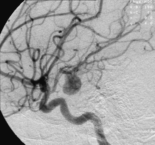
Definition & epidemiology
Secondary ICH is bleeding attributable to a discrete structural, neoplastic, venous, or haemostatic cause rather than to hypertensive or amyloid small-vessel disease. Secondary causes account for roughly 15–20% of spontaneous ICH overall but a far higher proportion in the young and normotensive, in whom a macrovascular cause may be found in a quarter or more of cases. Identifying them changes management entirely — from clot evacuation with lesion resection to endovascular obliteration, anticoagulation for venous thrombosis, or oncological therapy.
Etiology — secondary causes at a glance
| Cause | Suggestive features | Best test |
|---|---|---|
| Arteriovenous malformation (AVM) | Young, lobar, prior seizures/headache; calcification or serpiginous vessels on CT | CTA → DSA (gold standard for nidus/feeders) |
| Dural arteriovenous fistula | Pulsatile tinnitus, cortical venous reflux; near a sinus | DSA |
| Saccular / mycotic aneurysm | Sylvian or medial temporal haematoma, associated SAH; endocarditis (distal mycotic) | CTA/DSA; echocardiography if septic |
| Cavernous malformation | Small lobar/brainstem bleed; "popcorn" lesion with haemosiderin rim, angiographically occult | MRI/SWI (DSA typically negative) |
| Cerebral venous sinus thrombosis | Parasagittal, temporal, or bilateral (deep-venous) haemorrhage; young, peripartum, prothrombotic; disproportionate oedema | CT/MR venography ("empty delta") |
| Tumour (met or primary) | Disproportionate oedema, ring/nodular enhancement, multiple lesions; melanoma, renal cell, thyroid, choriocarcinoma, lung metastases; glioblastoma | Contrast MRI; delayed re-imaging |
| Coagulopathy / anticoagulant | Fluid levels, multicompartment bleeding, drug history, deranged INR/anti-Xa | Coagulation panel, drug-specific assays |
| Sympathomimetic drugs | Cocaine, amphetamines, phenylpropanolamine; young, surge in BP; may unmask AVM/aneurysm | Toxicology; vascular imaging |
| Moyamoya / vasculitis / RCVS | Recurrent events, borderzone pattern, beaded vessels, thunderclap headache | MRA/DSA, vessel-wall MRI |
Diagnostic workup — when to do DSA
CT angiography is the appropriate first-line vascular study in every patient in whom a secondary cause is plausible. Catheter digital subtraction angiography (DSA) remains the gold standard for arteriovenous shunts and small aneurysms and should be pursued when suspicion is high or when CTA is negative but unexplained. The DIAGRAM study formalised the pre-test probability of a macrovascular cause: yield rises sharply with younger age, lobar or purely intraventricular location, absence of a history of hypertension, absence of coagulopathy, and imaging hints such as enlarged vessels, associated subarachnoid blood, or an unusual configuration. Conversely, an older hypertensive patient with a deep basal-ganglia bleed has a very low macrovascular yield and generally does not need DSA.
Reasonable indications for DSA include: lobar ICH in a patient under about 55–65 without a clear hypertensive/amyloid explanation; any ICH with a significant subarachnoid component; isolated or disproportionate intraventricular haemorrhage; imaging features suggesting a shunt or aneurysm; and a negative or equivocal CTA when clinical suspicion persists. When early studies are negative but the picture is atypical, repeat vascular imaging (and contrast MRI) after 6–8 weeks — once the haematoma resolves — can reveal a previously compressed malformation or an underlying tumour.
Clinical pearls
- Bilateral thalamic or deep haemorrhage with prominent oedema should prompt CT/MR venography to exclude internal cerebral vein/straight sinus thrombosis before it is mislabelled hypertensive.
- A haematoma with disproportionate surrounding oedema or nodular enhancement is a tumour until proven otherwise — arrange delayed contrast MRI rather than assuming primary ICH.
- In a febrile patient with a distal lobar bleed, think mycotic aneurysm from infective endocarditis and obtain blood cultures and echocardiography.
- A negative CTA does not exclude a malformation compressed by mass effect; low threshold for DSA and interval re-imaging in the young and normotensive.
Key references: van Asch/Rinkel et al. DIAGRAM prediction score (Lancet Neurol 2013; validation JAMA Neurol 2019); Greenberg et al. AHA/ASA 2022 ICH Guideline (Stroke 2022); Cordonnier et al. ICH secondary causes (Lancet 2018).
Acute Management of ICH (BP Control INTERACT/ATACH-2, Anticoagulation Reversal, ICP, Glucose, Seizure)
Acute management — the bundle concept
Modern ICH care is delivered as a coordinated, time-critical bundle rather than a sequence of isolated interventions. The INTERACT3 stepped-wedge cluster trial (2023, >7,000 patients) showed that a goal-directed care bundle — intensive early systolic BP lowering to below 140 mm Hg within 1 hour, tight glycaemic control (6.1–7.8 mmol/L in non-diabetics; 7.8–10.0 in diabetics), treatment of fever to below 37.5 °C, and rapid reversal of anticoagulation to an INR below 1.5, all maintained for 7 days — improved functional outcome (ordinal modified Rankin common odds ratio 0.86, 95% CI 0.76–0.97, p=0.015). The lesson is that speed and the aggregate of small gains matter more than any single lever.
Acute management — blood-pressure control
INTERACT2 established that early lowering of systolic BP to below 140 mm Hg within 1 hour is safe and shifts the disability distribution favourably (ordinal OR 0.87), though the dichotomous primary endpoint was borderline. ATACH-2 tested more aggressive lowering (target 110–139 vs 140–179 mm Hg) and found no additional functional benefit with more renal adverse events, indicating that below-target overshoot is not helpful and may harm. The AHA/ASA 2022 guideline therefore recommends, for patients presenting with systolic BP 150–220 mm Hg, acute lowering to a target of 140 (range 130–150) mm Hg, delivered smoothly and sustained — minimising peak-to-trough variability, which independently predicts poor outcome.
| Trial / guidance | SBP target | Take-home |
|---|---|---|
| INTERACT2 (2013) | <140 within 1 h vs <180 | Safe; favourable ordinal shift in disability |
| ATACH-2 (2016) | 110–139 vs 140–179 | No added benefit; more renal adverse events with intensive arm |
| INTERACT3 bundle (2023) | <140 within 1 h (bundled) | Improved outcome as part of a care bundle |
| AHA/ASA 2022 | Target ~140 (130–150) for SBP 150–220 | Lower smoothly; avoid large variability and overshoot below 130 |
Acute management — anticoagulation & antithrombotic reversal
| Offending agent | Reversal | Dose / target |
|---|---|---|
| Vitamin K antagonist (warfarin) | 4-factor PCC + IV vitamin K | PCC weight/INR-based; vitamin K 10 mg IV; goal INR <1.3–1.5 (PCC superior to FFP — INCH trial) |
| Dabigatran | Idarucizumab | 5 g IV (two 2.5 g boluses) |
| Factor Xa inhibitors (apixaban, rivaroxaban) | Andexanet alfa (or 4F-PCC if unavailable) | Low/high dose per agent, dose & timing; 4F-PCC 50 U/kg alternative |
| Unfractionated / LMW heparin | Protamine sulfate | 1 mg per 100 U heparin (max 50 mg); partial for LMWH |
| Thrombolytic (tPA) associated | Cryoprecipitate ± tranexamic acid | Cryoprecipitate to replete fibrinogen >150 mg/dL |
| Antiplatelet agents | Platelet transfusion NOT recommended | PATCH trial showed harm; reserve platelets for planned neurosurgery/thrombocytopenia |
ANNEXA-I (2024) confirmed that andexanet alfa reduces anti-Xa activity and improves haemostatic efficacy in factor-Xa-inhibitor-associated ICH (67.0% vs 53.1% with usual care) with less haematoma expansion — but at the cost of more thrombotic events (10.3% vs 5.6%) and ischaemic strokes (6.5% vs 1.5%), so reversal must be weighed against the patient's thrombotic burden. Routine rFVIIa and routine tranexamic acid (TICH-2) do not improve functional outcome and are not recommended for unselected ICH.
Acute management — ICP, glucose, temperature, seizures, VTE
- Intracranial pressure: elevate the head to 30°, provide analgesia/sedation, and treat surges with osmotherapy (mannitol or hypertonic saline); place an external ventricular drain for obstructive hydrocephalus. Avoid prophylactic hyperventilation and prophylactic corticosteroids (harmful).
- Glucose: both hyper- and hypoglycaemia worsen outcome — maintain normoglycaemia and avoid tight-control hypoglycaemia.
- Temperature: treat fever and target normothermia; fever independently predicts poor outcome.
- Seizures: prophylactic antiseizure drugs are not recommended (2022 guideline, no benefit); treat clinical or electrographic seizures, and obtain continuous EEG when consciousness is depressed out of proportion to the lesion.
- Venous thromboembolism: start intermittent pneumatic compression on day 1 (CLOTS-3); pharmacological prophylaxis (LMWH/UFH) may be started 24–48 h after onset once the haematoma is stable.
Clinical pearls
- Smoothness beats aggressiveness — reach ~140 quickly and hold it; large fluctuations and dips below 130 negate benefit.
- Reverse anticoagulation within minutes, not hours; do not wait for the confirmatory INR/anti-Xa to start PCC or a specific antidote in a deteriorating patient.
- Do not transfuse platelets for antiplatelet-associated ICH outside planned surgery — PATCH showed net harm.
- Postpone new "do-not-resuscitate"/care-limitation decisions beyond the first full day; early pessimism is a self-fulfilling prophecy.
Key references: Greenberg et al. AHA/ASA 2022 ICH Guideline (Stroke 2022); Ma et al. INTERACT3 (Lancet 2023); Anderson et al. INTERACT2 (NEJM 2013); Qureshi et al. ATACH-2 (NEJM 2016); Connolly et al. ANNEXA-I (NEJM 2024); Steiner et al. INCH (Lancet Neurol 2016); Baharoglu et al. PATCH (Lancet 2016); Sprigg et al. TICH-2 (Lancet 2018).
Surgical & Minimally-Invasive Treatment of ICH (STICH I/II, MISTIE III, ENRICH, SWITCH; Cerebellar ICH)
Acute management — the evolving surgical rationale
The surgical story in supratentorial ICH is one of a long neutral era followed by a recent, location-specific breakthrough. Open craniotomy removes clot but at the price of surgical tract injury; the field has shifted toward earlier, less-traumatic, minimally invasive evacuation, and toward decompression without evacuation for the most severe deep bleeds. Cerebellar ICH is a separate, long-settled indication.
Acute management — the pivotal trials
| Trial (year) | Intervention / population | Primary result |
|---|---|---|
| STICH (2005) | Early craniotomy vs initial conservative care, supratentorial ICH | No overall benefit; hint of benefit in superficial lobar clots ≤1 cm from surface |
| STICH II (2013) | Early surgery, superficial lobar ICH 10–100 mL, no IVH, ≤1 cm from cortex | No significant benefit; safe, small non-significant favourable trend |
| MISTIE III (2019) | Minimally invasive catheter aspiration + alteplase to residual ≤15 mL | mRS 0–3 at 1 yr 45% vs 41% (NS, p=0.33); mortality 15% vs 23% (HR 0.67); benefit if surgical goal achieved |
| ENRICH (2024) | Trans-sulcal parafascicular evacuation <24 h, 30–80 mL, lobar or anterior basal ganglia | Utility-weighted mRS 0.458 vs 0.374 (favourable); benefit confined to lobar (0.513 vs 0.371); basal-ganglia arm dropped for futility |
| SWITCH (2024) | Decompressive craniectomy (no evacuation) + medical vs medical alone; deep ICH 30–100 mL, age ≤75, GCS 8–13, <66 h | mRS 5–6 at 180 d 44% vs 58% (aRR 0.77, 95% CI 0.59–1.01, p=0.057); mortality 17% vs 27% (trend) |
MISTIE III missed its functional endpoint overall, but a strong dose–response emerged: patients in whom the surgeon actually reached the ≤15 mL residual target did better, and each additional millilitre of clot removed improved outcomes — reframing evacuation extent, not the procedure per se, as the effective ingredient. ENRICH then became the first clearly positive ICH surgery trial by combining very early (median ~17 h) minimally invasive trans-sulcal evacuation with an adaptive design that identified lobar haemorrhage as the responsive phenotype while dropping deep basal-ganglia bleeds for futility. SWITCH tested a different concept — decompressive craniectomy without clot removal for severe deep ICH — and narrowly missed its primary endpoint while showing consistent signals toward reduced death and dependency, leaving craniectomy a reasonable rescue option in selected deteriorating patients.
Acute management — cerebellar ICH
Cerebellar haemorrhage is the clearest surgical indication in all of ICH. Suboccipital craniotomy with clot evacuation is recommended for patients with a cerebellar haematoma greater than about 3 cm in diameter (or roughly ≥15 mL), or with brainstem compression, neurological deterioration, or obstructive hydrocephalus from fourth-ventricular effacement. Crucially, placing an external ventricular drain alone, without evacuating the clot, is insufficient and may precipitate upward transtentorial herniation — the posterior fossa mass must be removed. Surgery should not be delayed until the patient has herniated, because deterioration can be precipitous.
Clinical pearls
- For supratentorial ICH, location now guides the offer: minimally invasive early evacuation is most defensible for accessible lobar clots (ENRICH), whereas deep basal-ganglia bleeds have not shown functional benefit from evacuation.
- Extent of evacuation is the effective dose — aim to reach the residual-volume target, not merely to insert a catheter (MISTIE III).
- Decompressive craniectomy is a life-saving rescue for the deteriorating severe deep ICH patient even though SWITCH was formally borderline.
- A drowsy patient with a >3 cm cerebellar clot needs a surgeon now, not serial scans — evacuate rather than watch, and do not rely on an EVD alone.
Key references: Mendelow et al. STICH (Lancet 2005) & STICH II (Lancet 2013); Hanley et al. MISTIE III (Lancet 2019); Pradilla/Ratcliff et al. ENRICH (NEJM 2024); Beck/Fischer et al. SWITCH (Lancet 2024); Greenberg et al. AHA/ASA 2022 ICH Guideline (Stroke 2022).
Intraventricular Hemorrhage in Adults (CLEAR III, EVD + Thrombolysis, Graeb Score)
Definition & epidemiology
Intraventricular haemorrhage (IVH) is blood within the ventricular system. In adults it is usually secondary — extension from a deep (thalamic or caudate/basal-ganglia) parenchymal ICH — and less often primary, confined to the ventricles (often from a subependymal vascular lesion or the periventricular perforators). IVH complicates 30–50% of spontaneous ICH and is one of the most powerful independent predictors of death and disability: it is a component of the ICH Score, and its presence roughly doubles mortality. The mechanisms of harm are acute obstructive hydrocephalus, direct injury to periventricular structures, and inflammatory/haemodynamic effects of intraventricular clot.
Diagnostic workup — quantifying IVH burden (Graeb & modified Graeb)
The clot burden is graded to prognosticate and to track clearance. The original Graeb score (range 0–12) assigns points to each lateral ventricle (0–4, by degree of blood and dilatation) and to the third and fourth ventricles (0–2 each); higher scores denote greater casting and dilatation. The modified Graeb score (range 0–32) subdivides the ventricular system more granularly (including occipital and temporal horns) and is more sensitive to change, which is why it was used to measure clot resolution in thrombolysis trials. The alternative LeRoux score grades each of the four ventricles 0–4 (range 0–16).
Acute management — EVD and intraventricular thrombolysis
The cornerstone of care for IVH with hydrocephalus or depressed consciousness is cerebrospinal-fluid diversion with an external ventricular drain (EVD), which relieves pressure but drains the casted clot slowly and is prone to catheter occlusion. CLEAR III (2017) tested accelerating clearance by instilling low-dose alteplase (1 mg every 8 hours, up to 12 doses) through the EVD versus saline in 500 patients with IVH and small parenchymal clots (<30 mL):
| Outcome | Alteplase | Saline | Interpretation |
|---|---|---|---|
| mRS 0–3 at 180 d | 48% | 45% | Not significant (RR 1.06) |
| 180-day case fatality | ~18% | ~29% | Significant ~11% absolute mortality reduction (adjusted OR death ~0.50) |
| Severe disability (mRS 5) | Increased | — | Lives saved shifted into severe dependency |
| Safety (ventriculitis, symptomatic bleeding) | No excess | — | Procedure safe |
The net message: intraventricular alteplase safely reduces mortality but does not improve the proportion achieving functional independence, partly because it converts some deaths into severe disability. A pre-specified signal indicated that greater clot removal (reaching a lower end-of-treatment IVH volume, e.g., clearing more than ~80–85% of the clot) was associated with better functional outcomes, suggesting that more complete and faster evacuation — the goal of newer active irrigation/drainage systems — may be where benefit lies.
Prognosis
IVH volume, the Graeb/modified Graeb score, acute hydrocephalus, and the parent ICH volume together drive prognosis. Rapid, complete clot clearance and control of intracranial pressure are the modifiable levers; persistent casting of the third and fourth ventricles portends the worst course.
Clinical pearls
- EVD relieves pressure but clears clot slowly — anticipate catheter occlusion and monitor drainage and ICP closely.
- Intraventricular alteplase is a mortality-reducing, not independence-restoring, intervention; counsel families accordingly given the shift toward severe disability among survivors.
- More clot cleared, faster, tracks with better outcome — the extent-of-removal principle mirrors MISTIE III in the parenchyma.
- Use the modified Graeb score, not just "IVH present/absent," to quantify burden and monitor resolution.
Key references: Hanley et al. CLEAR III (Lancet 2017); Graeb et al. original IVH grading (Radiology 1982); Morgan/Hanley modified Graeb score (Stroke 2013); Greenberg et al. AHA/ASA 2022 ICH Guideline (Stroke 2022).
Hematoma Expansion — Imaging Predictors (Spot Sign, Blend Sign, Black Hole Sign, Swirl, Island)
Definition & epidemiology
Haematoma expansion — conventionally defined as growth of at least 6 mL or 33% (some studies use ≥12.5 mL) on follow-up imaging — occurs in roughly a quarter to a third of patients scanned within a few hours of onset and is the strongest modifiable predictor of death and disability. Most growth happens within the first 6 hours, though anticoagulant-associated bleeding can continue for 24–48 hours. Because expansion is the target of blood-pressure control, reversal, and (in trials) haemostatic therapy, recognising the imaging markers that predict it is a core competency.
Diagnostic workup — CTA and NCCT predictors
| Sign | Modality | Definition |
|---|---|---|
| Spot sign | CTA (source/first-pass) | ≥1 focus of contrast extravasation within the clot, ≥1–2 mm, attenuation ≥120 HU, discontinuous from vessels — marks active bleeding |
| Blend sign | NCCT | Adjacent hypoattenuating and hyperattenuating regions with a clear demarcation and density difference ≥18 HU |
| Black hole sign | NCCT | A hypoattenuating region encapsulated within the hyperattenuating clot, not connected to the edge, with a distinct border (density difference ≥28 HU) |
| Swirl sign | NCCT | A region of hypo- or isoattenuation within the hyperdense clot (unclotted fresh blood — ongoing bleeding) |
| Island sign | NCCT | ≥3 small haematomas scattered and separate from the main clot, or ≥4 small bubble-like/budding foci contiguous with it |
| Satellite sign | NCCT | A small haemorrhage (1–20 mm) separate from but near the main clot |
Additional NCCT predictors include an irregular clot shape and heterogeneous internal density (the Barras shape/density categories), the presence of any intra-haematoma hypodensities, a fluid level (coagulopathy), a large baseline volume, very early presentation, and anticoagulation. The spot sign remains the best-validated single marker of expansion (high specificity/positive predictive value, moderate sensitivity); a spot-sign score and delayed "leakage" on post-contrast or CTV imaging refine risk further.
Pathophysiology
These signs are radiological windows onto the same process: the spot and swirl signs reflect frank contrast/blood extravasation from ruptured vessels; hypodensity-based signs (black hole, blend, heterogeneous density) reflect an admixture of freshly extravasated, not-yet-clotted blood of differing attenuation within an evolving haematoma; and multiplicity/irregularity signs (island, satellite, irregular shape) reflect secondary rupture of surrounding vessels — the "avalanche" model of progressive peripheral bleeding.
Acute management & prognosis
No haemostatic drug has yet converted expansion prediction into improved outcome: spot-sign-guided recombinant factor VIIa (SPOTLIGHT/STOP-IT) and unselected tranexamic acid (TICH-2) reduced growth modestly without improving function. The practical value of the signs is therefore risk stratification — flagging patients for intensive monitoring, rapid and complete reversal, disciplined blood-pressure control, and cautious prognostic counselling — and enrichment of future trials.
Clinical pearls
- A spot sign is an actionable alarm: intensify BP control and reversal and re-image early, even if the initial clot is modest.
- You can predict expansion from NCCT alone when CTA is unavailable — look for blend, black-hole, swirl, island, irregular shape, and heterogeneous density.
- Absence of a spot sign does not guarantee stability, especially in anticoagulated patients; sensitivity is only moderate.
- Expansion markers inform prognosis and monitoring, not a specific proven drug — do not offer rFVIIa or routine tranexamic acid on the basis of a spot sign outside a trial.
Key references: Demchuk et al. PREDICT spot-sign study (Lancet Neurol 2012); Li et al. blend sign (Stroke 2015) & black hole sign (Stroke 2016); Li et al. island sign (Stroke 2017); Barras et al. shape/density (Stroke 2009); Gladstone et al. SPOTLIGHT/STOP-IT (JAMA Neurol 2019); Sprigg et al. TICH-2 (Lancet 2018).
ICH Scores & Prognostication (ICH Score, max-ICH, FUNC) — Components & Mortality
Definition & rationale
Structured prognostic scores standardise communication and risk-adjust cohorts, but in ICH they carry a unique hazard: because early withdrawal of life-sustaining treatment is the commonest proximate cause of death, a pessimistic score can become a self-fulfilling prophecy. Scores should inform, not dictate, and should be applied only after a genuine trial of aggressive care.
Prognosis — the ICH Score (Hemphill 2001)
The ICH Score is the most widely used 30-day mortality predictor, built from five routine variables:
| Variable | Points |
|---|---|
| GCS 3–4 / 5–12 / 13–15 | 2 / 1 / 0 |
| Age ≥80 years | 1 |
| Infratentorial origin | 1 |
| ICH volume ≥30 mL | 1 |
| Intraventricular haemorrhage present | 1 |
The total (0–6) maps to steeply rising 30-day mortality:
| ICH Score | Approx. 30-day mortality |
|---|---|
| 0 | 0% |
| 1 | ~13% |
| 2 | ~26% |
| 3 | ~72% |
| 4 | ~97% |
| 5–6 | ~100% (few patients in derivation) |
Prognosis — max-ICH and FUNC
The max-ICH score (Sembill 2017) was designed explicitly to overcome the self-fulfilling-prophecy bias by deriving prognosis in patients who received maximal treatment (excluding those with early care limitations). It incorporates age, neurological severity (NIHSS), intraventricular haemorrhage, oral anticoagulation, and haematoma volume assessed with different thresholds for lobar versus non-lobar location (total range 0–10), and it calibrates better than the ICH Score in fully-treated patients — a more honest estimate of what aggressive care can achieve.
The FUNC score (Rost 2008) predicts the opposite endpoint — the probability of regaining functional independence at 90 days — which is often the more decision-relevant question:
| Variable | Points |
|---|---|
| ICH volume <30 / 30–60 / >60 mL | 4 / 2 / 0 |
| Age <70 / 70–79 / ≥80 years | 2 / 1 / 0 |
| Location lobar / deep / infratentorial | 2 / 1 / 0 |
| GCS ≥9 / ≤8 | 2 / 0 |
| Pre-ICH cognitive impairment absent / present | 1 / 0 |
The total (0–11) grades the chance of functional independence: patients scoring 11 achieve independence in roughly 80% of cases, whereas those scoring 4 or below essentially never do.
Clinical pearls
- Prognosticate after a committed trial of aggressive care — the AHA/ASA guideline advises postponing new DNR/care-limitation orders beyond the second full hospital day.
- Prefer max-ICH or FUNC when the clinical question is "how good can this get with full treatment?" rather than "what is the average death rate?" — the ICH Score is contaminated by early withdrawals in its derivation cohorts.
- Report the positive framing (probability of independence, FUNC) to families alongside mortality; it reduces premature nihilism.
- No score should override an individual patient's trajectory over the first 24–72 hours.
Key references: Hemphill et al. ICH Score (Stroke 2001); Rost et al. FUNC score (Stroke 2008); Sembill et al. max-ICH score (Neurology 2017); Greenberg et al. AHA/ASA 2022 ICH Guideline (Stroke 2022).
Anticoagulation-Associated ICH & Resumption Timing (When/Whether to Restart)
Definition & epidemiology
Anticoagulant-associated ICH accounts for roughly 15–20% of all ICH and is disproportionately lethal: these haematomas are larger at baseline, expand for longer (up to 24–48 hours with vitamin K antagonists), and carry higher mortality than spontaneous bleeds. Direct oral anticoagulant (DOAC)-associated ICH tends to be somewhat smaller and less severe than warfarin-associated ICH but remains a serious event. Acute management centres on immediate, agent-specific reversal (4-factor PCC and vitamin K for warfarin; idarucizumab for dabigatran; andexanet alfa or 4F-PCC for factor-Xa inhibitors) alongside blood-pressure control.
Prognosis & the resumption dilemma
The hardest decision is whether, and when, to restart anticoagulation in a survivor who had a compelling indication (usually atrial fibrillation). Clinicians must balance the ongoing thromboembolic risk of withholding therapy against the risk of recurrent ICH — a risk that depends heavily on the bleed's mechanism and location. Lobar/CAA-related ICH recurs far more often than deep hypertensive ICH, tilting the balance toward avoidance (or left-atrial-appendage occlusion) in the former and toward resumption in the latter once blood pressure is controlled.
Acute management — evidence on restarting
| Trial / source | Design | Signal |
|---|---|---|
| Observational cohorts/meta-analyses | Resumption vs avoidance after AF-related ICH | Resumption associated with lower thromboembolism and mortality without clear excess recurrence, especially in non-lobar ICH |
| RESTART (2019) | Restart vs avoid antiplatelet after ICH | No increase in recurrent ICH; antiplatelet resumption appears safe |
| SoSTART / APACHE-AF (2021) | Small RCTs, anticoagulation vs avoidance | Underpowered/inconclusive; non-inferiority of avoidance not established |
| PRESTIGE-AF (2025) | RCT, DOAC vs no anticoagulation after ICH with AF (n≈319) | Ischaemic stroke sharply reduced (HR 0.05) but recurrent ICH increased (HR ~10.9); net-benefit HR 0.69 trended in favour of DOAC (NNT ~13, NNH ~24) |
| ENRICH-AF, ASPIRE | Ongoing RCTs (edoxaban; apixaban vs aspirin) | Awaited — will refine the risk–benefit balance |
PRESTIGE-AF (2025) crystallised the trade-off: in ICH survivors with atrial fibrillation, a DOAC almost abolished ischaemic strokes but roughly decupled recurrent ICH, with a composite net clinical benefit that leaned — non-significantly — toward anticoagulation. The decision is therefore genuinely individualised, and the results of ENRICH-AF and ASPIRE are needed before a firm standard emerges.
Acute management — timing & a practical framework
The AHA/ASA 2022 guideline notes that, for patients with non-valvular AF and a spontaneous ICH, avoiding anticoagulation may be reasonable when recurrence risk is high, and that when anticoagulation is chosen, delaying it by at least 4 weeks (in patients without a mechanical valve) may lower recurrence. A workable approach:
- Favour resumption (typically at ~4–8 weeks): deep/hypertensive ICH, well-controlled blood pressure, high CHA₂DS₂-VASc, a reversible bleed trigger, mechanical heart valve (resume earlier, individualised, to prevent valve thrombosis).
- Favour avoidance or LAA occlusion: lobar/CAA-related ICH, cortical superficial siderosis or a high microbleed burden, recurrent ICH, uncontrolled hypertension.
- Antiplatelet therapy can generally be resumed within days when a vascular indication exists (RESTART), as its recurrence risk is low.
Clinical pearls
- Location drives the decision: deep bleeds lean toward restarting, lobar/amyloid bleeds toward withholding or appendage closure.
- DOACs are preferred over warfarin when anticoagulation is resumed for non-valvular AF (lower ICH risk); mechanical valves still require a VKA.
- PRESTIGE-AF shows anticoagulation buys large ischaemic-stroke reduction at the cost of more recurrent ICH — quantify both risks for the individual rather than defaulting.
- Image the brain (GRE/SWI) before deciding: siderosis and lobar microbleeds are red flags for CAA and future haemorrhage.
Key references: Greenberg et al. AHA/ASA 2022 ICH Guideline (Stroke 2022); RESTART Collaboration (Lancet 2019); SoSTART (Lancet Neurol 2021); APACHE-AF (Lancet Neurol 2021); PRESTIGE-AF (Lancet 2025); Kuramatsu/Huttner on anticoagulant-ICH reversal & resumption (JAMA 2015).
Cortical Laminar Necrosis (Imaging, Causes)
Definition & epidemiology
Cortical laminar necrosis (also called pseudolaminar necrosis) is selective death of neurons in specific laminae of the cerebral cortex following a global or severe energy-failure insult. The middle cortical layers — especially the large pyramidal neurons of layer 3 (with layers 5–6 also vulnerable) — are the most metabolically demanding and therefore the first to die when oxygen or glucose delivery fails, producing a characteristic gyriform (ribbon-like) pattern that follows the cortical contour. It is not a haemorrhage and, in most cases, not primarily calcification; recognising it prevents both misdiagnosis as cortical bleeding and false reassurance.
Etiology
- Global hypoxic–ischaemic injury: cardiac arrest, near-drowning, carbon-monoxide or other asphyxial poisoning, prolonged hypotension.
- Status epilepticus / prolonged seizures: excitotoxic energy failure in hyperactive cortex.
- Hypoglycaemia: severe, prolonged neuroglycopenia.
- Metabolic / mitochondrial disease: MELAS and related mitochondrial cytopathies (stroke-like episodes not confined to vascular territories).
- Other: posterior reversible encephalopathy syndrome (PRES), calcineurin-inhibitor/immunosuppressant neurotoxicity, hepatic encephalopathy, and the cortex of maturing large infarcts and watershed (borderzone) ischaemia.
Diagnostic workup — imaging
The signature finding is gyriform cortical T1 hyperintensity on MRI. It is characteristically delayed — typically emerging around 2 weeks after the insult, peaking between roughly 1 and 3 months, and slowly fading over months to a year or more. The T1 shortening is attributed to lipid-laden macrophages and denatured/necrotic protein (and possibly trace mineral deposition), not to methaemoglobin, which is the key point distinguishing it from cortical haemorrhage.
| Sequence / modality | Typical appearance |
|---|---|
| T1-weighted MRI | Gyriform (laminar) cortical hyperintensity, delayed onset (~2 wk+), the hallmark |
| Diffusion-weighted imaging | Cortical restricted diffusion in the acute/subacute phase |
| T2 / FLAIR | Cortical hyperintensity, oedema early, gliosis late |
| GRE / SWI | Usually little or no blooming — helps exclude haemorrhage; mild susceptibility only if mineralisation supervenes |
| Post-contrast T1 | Transient gyriform enhancement possible in the subacute phase |
| CT | Faint cortical (gyriform) hyperdensity; chronic cortical calcification in some cases |
Clinical features & prognosis
Cortical laminar necrosis is a radiological marker of a preceding severe hypoxic-ischaemic or metabolic insult rather than a disease in itself; the clinical picture is that of the underlying cause (e.g., post-arrest encephalopathy, refractory epilepsy). Its distribution and timing help date the injury and infer the mechanism, and — after cardiac arrest — extensive cortical involvement carries adverse prognostic weight. There is no specific treatment beyond addressing the primary cause and preventing recurrence.
Clinical pearls
- Gyriform T1 hyperintensity that appears weeks after an anoxic or seizure event, without GRE blooming, is laminar necrosis — not cortical haemorrhage.
- Timing matters: T1 signal is delayed (peaks 1–3 months), so an early normal T1 does not exclude it — DWI is the acute clue.
- A non-vascular, gyriform cortical distribution crossing arterial territories should prompt consideration of MELAS, hypoglycaemia, PRES, or global anoxia rather than embolic infarction.
- Use GRE/SWI and CT to separate laminar necrosis (minimal susceptibility) from cortical haemorrhage (marked blooming) and from cortical calcification (dense on CT).
Key references: Komiyama et al. cortical laminar necrosis on MRI (AJNR 1998); Siskas et al. laminar necrosis imaging (Neuroradiology 2003); Donaire et al. peri-ictal cortical changes; general neuroradiology references on hypoxic–ischaemic injury.
Subarachnoid Hemorrhage — Definition & Etiology
Definition & epidemiology
Subarachnoid hemorrhage (SAH) is extravasation of blood into the subarachnoid space between the arachnoid and pia mater, where it mixes with cerebrospinal fluid. Overall the commonest cause is head trauma; when clinicians say "SAH" in a stroke context they mean spontaneous (non-traumatic) SAH, which accounts for roughly 5% of all strokes. Incidence is about 6–9 per 100,000 person-years worldwide, with striking geographic variation — approximately twice that in Finland and Japan and lower in Central/South America. Mean age at presentation is the mid-50s (younger than ischemic stroke), and there is a female preponderance (~1.6:1) that emerges after the fifth decade. Despite improvements in care, aneurysmal SAH still carries roughly 30-day case-fatality of 25–35% (including pre-hospital deaths of ~10–15%), and a large share of survivors are left with lasting cognitive impairment.
Etiology — a rupture-risk-driven classification
Of spontaneous SAH, saccular (berry) aneurysm rupture causes about 80–85%, non-aneurysmal perimesencephalic hemorrhage about 10%, and a heterogeneous remainder about 5%. The differential must be worked through deliberately because management diverges sharply.
| Category | Representative causes | Notes |
|---|---|---|
| Saccular aneurysm (80–85%) | Rupture at arterial bifurcations of the circle of Willis — Acom, Pcom/ICA, MCA bifurcation, basilar apex | The lesion that must be excluded first; drives all acute management |
| Non-aneurysmal perimesencephalic (~10%) | Presumed low-pressure venous/capillary bleed around the midbrain | Benign course; diagnosis of exclusion after negative angiography |
| Other vascular (~5%) | AVM, dural arteriovenous fistula, arterial dissection (intradural vertebral), mycotic (infective) aneurysm, spinal AVM/aneurysm, cerebral venous thrombosis, moyamoya, pituitary apoplexy | Often suggested by atypical clot distribution or clinical context |
| Vasculopathy / convexal | Reversible cerebral vasoconstriction syndrome (RCVS), cerebral amyloid angiopathy (CAA), vasculitis, PRES | Typically produce isolated cortical (convexal) SAH — see dedicated topic |
| Other | Sympathomimetic drugs (cocaine, amphetamines), coagulopathy/anticoagulants, sickle cell disease, tumor (metastasis) | History and toxicology essential |
Perimesencephalic (non-aneurysmal) SAH
Perimesencephalic SAH is a distinct, largely benign entity in which blood is centered immediately anterior to the midbrain or pons (interpeduncular, prepontine, ambient cisterns), without extension into the lateral Sylvian or anterior interhemispheric fissures and without significant intraventricular blood. Patients are typically neurologically intact with a less explosive headache. The presumed source is venous or perforator/capillary rather than arterial, which explains the very low rates of rebleeding and delayed cerebral ischemia and the excellent prognosis. The essential caveat: a ruptured posterior circulation aneurysm (basilar or vertebral) can produce an identical CT pattern, so the "perimesencephalic" label may only be applied after high-quality vascular imaging excludes an aneurysm. Most centers require a negative CT angiogram; policy on whether a single negative CTA suffices or whether digital subtraction angiography (DSA) is still needed remains debated, though contemporary evidence supports that a good-quality negative CTA with a classic perimesencephalic pattern makes DSA of low yield.
Risk factors for aneurysm formation & rupture
Modifiable factors include hypertension, cigarette smoking (the strongest modifiable factor, roughly doubling risk), heavy alcohol use and sympathomimetic drugs. Non-modifiable/constitutional factors include female sex, a first-degree family history of aneurysm or SAH, autosomal dominant polycystic kidney disease (ADPKD), and heritable connective-tissue disorders (e.g., vascular Ehlers–Danlos, Loeys–Dietz). Aneurysm formation reflects hemodynamic wall stress at bifurcations superimposed on degenerative/inflammatory remodeling of the internal elastic lamina and media.
Clinical pearls
- "Spontaneous SAH is aneurysmal until proven otherwise" — do not anchor on a benign perimesencephalic pattern until a posterior-circulation aneurysm has been excluded by angiography.
- The single most useful discriminator between aneurysmal and perimesencephalic bleeding is the distribution of blood on the first CT, not headache severity.
- Isolated blood confined to the cortical convexity (sparing the basal cisterns) points away from aneurysm and toward CAA (older) or RCVS (younger) — a different work-up entirely.
Key references: Hoh et al. 2023 AHA/ASA Guideline for the Management of Patients With Aneurysmal Subarachnoid Hemorrhage (Stroke 2023); Steiner et al. ESO Guidelines for the Management of Intracranial Aneurysms and SAH (Cerebrovasc Dis 2013); Rinkel & Wijdicks — perimesencephalic SAH literature; Vlak et al. prevalence of unruptured aneurysms (Lancet Neurol 2011).
SAH — Clinical Presentation & Complications
Clinical features
The cardinal symptom is thunderclap headache: a sudden, severe, "worst headache of my life" that reaches maximal intensity within seconds to a minute. Approximately 10–15% of aneurysmal SAH is heralded days to weeks earlier by a sentinel (warning) headache from a minor leak — a critical, frequently missed opportunity, since these patients often present again with catastrophic rupture. Associated features include nausea/vomiting, neck stiffness and photophobia (chemical meningismus, evolving over hours), transient or persistent loss of consciousness (from an abrupt rise in intracranial pressure that transiently exceeds cerebral perfusion pressure), and seizures at onset. Focal signs may localize the aneurysm — a painful third-nerve palsy with pupillary involvement suggests a posterior communicating artery aneurysm, and abulia/paraparesis suggests anterior communicating artery rupture. Terson syndrome (intraocular — vitreous, subhyaloid or retinal — hemorrhage) accompanies severe cases and predicts worse outcome. A substantial minority present in coma or with sudden death.
Early complications
Rebleeding is the most feared early event: risk is highest in the first 24 hours (and disproportionately within the first 2–12 hours), reported at up to ~4–15% in the first day if the aneurysm is unsecured, and it roughly doubles mortality. This hazard is the entire rationale for urgent aneurysm securing and for controlling extreme hypertension before treatment. Acute hydrocephalus occurs in ~15–30%, from intraventricular blood obstructing CSF flow or impaired arachnoid resorption; it manifests as declining consciousness and is treated with an external ventricular drain (EVD). Seizures occur in a minority.
Systemic & neurogenic complications
A catecholamine surge at ictus drives extracranial organ dysfunction. Neurogenic stress cardiomyopathy (including Takotsubo/apical ballooning) produces troponin elevation, regional wall-motion abnormalities, reduced ejection fraction and, on ECG, deep symmetric T-wave inversions ("cerebral T waves"), QT prolongation, ST changes and arrhythmia — usually reversible over days. Neurogenic pulmonary edema can develop abruptly. These may mimic acute coronary syndrome; recognizing the neurologic cause avoids inappropriate antithrombotic therapy in a patient with intracranial hemorrhage.
Hyponatremia — cerebral salt wasting vs SIADH
Hyponatremia is common (up to ~30–50%) and clinically important because it can worsen cerebral edema and, if managed with fluid restriction in a volume-depleted patient, may precipitate delayed cerebral ischemia. The key distinction is volume status: cerebral salt wasting (CSW) is a hypovolemic natriuresis, whereas SIADH is euvolemic/hypervolemic water retention. In SAH the therapeutic bias is toward maintaining euvolemia and replacing sodium — fluid restriction is generally avoided.
| Feature | Cerebral salt wasting (CSW) | SIADH |
|---|---|---|
| Volume status | Hypovolemic (net salt & water loss) | Euvolemic / mildly hypervolemic |
| Primary mechanism | Renal Na⁺ wasting (natriuretic peptides, sympathetic) | Inappropriate ADH → water retention |
| Urine sodium | High | High |
| Urine output | High (polyuria) | Normal/low |
| Serum uric acid / BUN:Cr | May be elevated (dehydration) | Low uric acid, low-normal BUN |
| Central venous pressure / clinical volume | Low | Normal/high |
| Treatment | Volume + sodium repletion (isotonic/hypertonic saline; fludrocortisone) | Fluid restriction risky in SAH — prefer hypertonic saline; consider vaptan cautiously |
Distinguishing the two at the bedside is notoriously difficult, and both may coexist; in practice, because inducing hypovolemia is hazardous, the pragmatic default in SAH is to keep the patient euvolemic and give sodium (hypertonic saline for symptomatic or severe hyponatremia) rather than to restrict fluids.
Clinical pearls
- A "sentinel headache" is a diagnosis you make in retrospect at your peril — treat every first thunderclap headache as possible SAH.
- Troponin rise and ECG changes after SAH usually reflect neurogenic cardiac stunning, not coronary occlusion; do not reflexively anticoagulate.
- Never fluid-restrict a hyponatremic SAH patient reflexively — hypovolemia is a trigger for delayed cerebral ischemia. Assess volume and give salt.
- Sudden neurologic decline after admission has three usual explanations: rebleeding, acute hydrocephalus, or (after day 3) delayed cerebral ischemia — image and act quickly.
Key references: Hoh et al. 2023 AHA/ASA aSAH Guideline (Stroke 2023); Connolly et al. 2012 AHA/ASA aSAH Guideline (Stroke 2012); Sherlock et al. hyponatremia after SAH; Naidech et al. rebleeding predictors.
Diagnosis of SAH — NCCT, Lumbar Puncture & Angiography
Non-contrast CT — sensitivity is time-dependent
Non-contrast CT (NCCT) of the head is the first test in any suspected SAH. Its sensitivity is exquisite when performed early and declines predictably as subarachnoid blood is cleared and becomes isodense to CSF. With a modern multidetector scanner and a competent reader, sensitivity approaches 100% within the first 6 hours of headache onset, then falls over subsequent days.
| Time from ictus | Approx. NCCT sensitivity for SAH |
|---|---|
| <6 hours | ~98–100% (modern scanner, expert read) |
| Within 24 hours | ~93% |
| Day 3 | ~85% |
| Day 5–7 | ~50% |
| >1 week | ~30% and falling |
The "6-hour rule"
Prospective data (Perry et al., BMJ 2011, and subsequent validations) established that a NCCT performed within 6 hours of headache onset, interpreted by a qualified radiologist on a modern (≥16-slice) scanner, has a sensitivity for aneurysmal SAH near 98.7–100% and a very high negative predictive value. In a neurologically intact patient meeting all of these conditions, a normal early CT may reasonably obviate lumbar puncture. Important caveats: the rule requires onset (not arrival) within 6 hours, a competent reader and current-generation hardware, and profound anemia (hematocrit roughly <30%) can render blood isodense and lower sensitivity. Beyond 6 hours, a normal CT no longer excludes SAH and further testing is required.
Lumbar puncture — timing & xanthochromia
When suspicion persists after a negative/late CT, lumbar puncture (LP) is the traditional confirmatory test. The key concept is xanthochromia — the yellow discoloration of CSF supernatant produced by enzymatic breakdown of oxyhemoglobin to bilirubin. Because this conversion occurs in vivo over time, xanthochromia may be absent very early and is most reliable when the LP is performed at least 12 hours after ictus; the UK standard is spectrophotometric quantification of bilirubin (more sensitive/specific than the naked eye), whereas many other centers rely on visual inspection. A rising or persistent red-cell count that does not clear from CSF tube 1 to tube 4 favors true SAH over a traumatic tap, but red-cell clearance alone is unreliable; elevated opening pressure and xanthochromia are more dependable. A completely acellular, non-xanthochromic CSF obtained ≥12 hours from onset argues strongly against SAH.
The CT-negative headache — CTA vs LP
In patients presenting after 6 hours, or when any doubt remains, the choice between LP and CT angiography (CTA) is individualized. CTA can directly demonstrate an aneurysm but detects incidental unruptured aneurysms (~3% of adults) that may not be the culprit, risking overdiagnosis; LP directly interrogates for blood in the subarachnoid space. Many pathways use LP to confirm/exclude hemorrhage and reserve angiography for confirmed SAH.
Vascular imaging to find the source
Once SAH is confirmed, the aneurysm must be localized. CTA is the usual first vascular study (sensitivity ~95–98%), rapidly available and often sufficient to plan treatment, but it may miss aneurysms <3 mm. Digital subtraction angiography (DSA) with 3D rotational acquisition remains the gold standard (highest spatial resolution, defines neck morphology and perforators, allows treatment in the same session). If initial angiography is negative in a non-perimesencephalic (aneurysmal-pattern) SAH, a repeat DSA in ~1–2 weeks is recommended because the yield of a delayed study is ~10–20% (an aneurysm initially obscured by thrombus or vasospasm); MRI of brain and spine should be considered to seek an alternative source.
Clinical pearls
- The 6-hour rule only holds with onset within 6 hours, a modern scanner and an expert reader — miss any element and a normal CT does not exclude SAH.
- Do not perform the LP too early: give xanthochromia time to develop (≥12 hours) or you may get a false-negative.
- A negative first angiogram in a diffuse aneurysmal-pattern SAH mandates a repeat study; a classic perimesencephalic pattern with a good-quality negative CTA usually does not.
- An incidental aneurysm on CTA is not automatically the bleeding source — correlate its location with the clot distribution.
Key references: Perry et al. Sensitivity of CT within 6 hours (BMJ 2011); Perry et al. non-academic validation (Neurology 2015); Backes et al. early CT sensitivity (Stroke 2016); Hoh et al. 2023 AHA/ASA aSAH Guideline (Stroke 2023); UK National External Quality Assessment CSF spectrophotometry standard.
Management of Aneurysmal SAH
Initial stabilization & system of care
Aneurysmal SAH is a neurologic emergency best managed in a high-volume center with neurosurgical, endovascular and neurocritical-care capability — higher case volume is associated with better outcomes (a class I emphasis of the 2023 guideline). Priorities are airway/breathing/circulation, treatment of acute hydrocephalus with an EVD when consciousness is depressed, analgesia, and control of extreme blood pressure to limit rebleeding while the aneurysm is unsecured.
Securing the aneurysm — timing & modality
Because early rebleeding is the dominant early threat, the ruptured aneurysm should be secured as early as feasible, preferably within 24 hours of presentation. The two modalities are microsurgical clipping and endovascular coiling. The pivotal ISAT trial (Molyneux et al.) randomized 2,143 patients with aneurysms judged suitable for either treatment and found a significant absolute reduction in death or dependency at one year with coiling (23.5% vs 30.9% with clipping). Long-term follow-up confirmed a durable survival/independence advantage for coiling, at the cost of a small excess of late target-aneurysm rebleeding and higher retreatment rates. The complementary lesson from BRAT and clinical experience is that modality should be chosen by an experienced multidisciplinary team based on aneurysm and patient factors — coiling is favored when both are feasible and for posterior-circulation and poor-grade patients, whereas clipping may be preferred for wide-neck MCA aneurysms, those with a large space-occupying hematoma requiring evacuation, and some very young patients where durability matters.
Blood pressure before & after securing
Before the aneurysm is secured, extreme hypertension should be controlled to reduce rebleeding while avoiding hypotension that could compromise cerebral perfusion; a commonly used practical target is systolic pressure below ~160 mm Hg (titrated agents such as labetalol, nicardipine or clevidipine), individualized to baseline pressure and clinical status. After the aneurysm is secured, blood-pressure goals liberalize, and if delayed cerebral ischemia develops, pressure is deliberately raised rather than lowered. The 2023 guideline emphasizes minimizing blood-pressure variability and maintaining euvolemia.
Nimodipine
Oral/enteral nimodipine 60 mg every 4 hours for 21 days is a class I intervention for every aneurysmal SAH patient who can tolerate it. Crucially, nimodipine improves neurologic outcome and reduces delayed-cerebral-ischemia–related poor outcome without consistently reducing angiographic vessel narrowing — its benefit is best understood as neuroprotection rather than reversal of large-artery spasm. If the full dose provokes hypotension, a split schedule of 30 mg every 2 hours is used; the enteral route is strongly preferred, and intravenous nimodipine has been associated with fatal medication errors (an explicit safety warning). Other calcium antagonists and, notably, routine statins and intravenous magnesium are not recommended, having failed to improve outcome in trials.
Antifibrinolytics
Short-course antifibrinolytic therapy (e.g., tranexamic acid) to prevent pre-treatment rebleeding is not routinely recommended. The randomized ULTRA trial found that ultra-early, short-course tranexamic acid did not improve functional outcome at six months, and the 2023 guideline concludes that routine antifibrinolytic use does not improve outcomes. Any use is at most a brief bridge in settings where aneurysm securing must be delayed, weighed against thrombotic risk.
Supportive neurocritical care
- Euvolemia, not prophylactic hypervolemia — the old "triple-H" prophylaxis has been abandoned in favor of maintaining normal volume and treating ischemia when it occurs.
- Sodium/glucose/temperature: avoid hyponatremia and hypovolemia; control hyperglycemia (avoiding hypoglycemia) and fever.
- Seizures: routine prophylaxis is not recommended; treat clinical seizures and give a short course after a documented seizure. Phenytoin specifically is discouraged given cognitive/outcome concerns.
- Hydrocephalus/ICP: EVD for acute obstructive/communicating hydrocephalus; a minority require permanent shunting.
- VTE prophylaxis: mechanical compression until the aneurysm is secured, then pharmacologic prophylaxis can be added.
Clinical pearls
- The two interventions with the strongest outcome evidence are early aneurysm securing and enteral nimodipine — everything else is supportive.
- Nimodipine protects the brain but will not fix the angiogram; do not stop it because the TCDs still show spasm, and do not expect it to substitute for treating symptomatic DCI.
- "Triple-H" prophylaxis is obsolete — keep patients euvolemic and reserve induced hypertension for actual clinical deterioration.
- Give nimodipine enterally; intravenous administration has caused deaths and carries a boxed safety warning.
Key references: Hoh et al. 2023 AHA/ASA aSAH Guideline (Stroke 2023); Molyneux et al. ISAT (Lancet 2002; long-term follow-up Lancet 2015); Spetzler et al. Barrow Ruptured Aneurysm Trial (BRAT); Post et al. ULTRA trial (Lancet 2021); Dorhout Mees et al. calcium antagonists Cochrane review.
Cerebral Vasospasm & Delayed Cerebral Ischemia
Definitions — vasospasm is not the same as DCI
Two overlapping but distinct concepts must be separated. Angiographic (or sonographic) vasospasm is objective narrowing of large intracranial arteries. Delayed cerebral ischemia (DCI) is a clinical syndrome — new focal deficit or a decline of ≥2 points on the Glasgow Coma Scale lasting ≥1 hour, not attributable to another cause (rebleeding, hydrocephalus, seizure, metabolic derangement) — and/or new infarction on imaging. The distinction matters because up to two-thirds of patients have some arterial narrowing but only ~30% develop DCI, and DCI is driven by more than large-vessel spasm alone: microcirculatory dysfunction, microthrombosis, cortical spreading depolarizations and impaired autoregulation all contribute. This is why treatments that relax large arteries do not reliably prevent DCI.
Timing & risk stratification
DCI characteristically begins around day 3–4, peaks at day 7–10, and resolves by roughly day 14–21. The strongest predictor is the volume and thickness of cisternal/ventricular blood, captured by the modified Fisher scale; poor clinical grade adds risk. The VASOGRADE tool combines the WFNS grade and modified Fisher score into a simple green/yellow/red stratification of DCI risk.
Detection & monitoring
In good-grade patients, serial neurologic examination is the most sensitive monitor. In poor-grade or sedated patients, adjuncts are needed. Transcranial Doppler (TCD) tracks flow velocities; because velocity rises with both vasospasm and hyperemia, the Lindegaard ratio (mean MCA velocity ÷ mean extracranial ICA velocity) is used to separate the two.
| Severity | MCA mean flow velocity | Lindegaard ratio (MCA/extracranial ICA) |
|---|---|---|
| Normal | <120 cm/s | <3 |
| Mild vasospasm | 120–150 cm/s | 3–4.5 |
| Moderate vasospasm | 150–200 cm/s | 4.5–6 |
| Severe vasospasm | >200 cm/s | >6 |
A rapid day-to-day rise in velocity (>50 cm/s/day) is worrisome. TCD has high specificity but modest sensitivity, and is unreliable for the anterior and posterior cerebral arteries. CT angiography with CT perfusion confirms narrowing and, importantly, tissue-level hypoperfusion; DSA is definitive and enables treatment in the same setting.
Prevention
The only pharmacologic agent proven to improve outcome is enteral nimodipine (see Management topic). Maintenance of euvolemia is standard. Prophylactic hypervolemia/"triple-H," statins and intravenous magnesium are not recommended. The endothelin-receptor antagonist clazosentan reduces angiographic vasospasm but has not consistently improved functional outcome in Western trials (it is approved in Japan) — a paradigmatic example of the vasospasm–DCI dissociation.
Treatment of symptomatic DCI
Once DCI is diagnosed and the aneurysm is secured, the first-line intervention is induced hypertension: raise blood pressure with vasopressors while maintaining euvolemia, titrating stepwise to reversal of the deficit rather than to a fixed number (the isolated hemodilution and prophylactic-hypervolemia components of old triple-H are no longer pursued). If deterioration is refractory to hemodynamic augmentation, endovascular rescue is indicated: intra-arterial vasodilators (verapamil, nicardipine, milrinone) for diffuse/distal spasm and balloon angioplasty for accessible, focal proximal large-vessel spasm. These measures are effective for large-artery narrowing but do not address the microvascular contributors to DCI.
| Tier | Intervention | Comment |
|---|---|---|
| Baseline (all patients) | Enteral nimodipine 60 mg q4h ×21 days; euvolemia; serial exams ± TCD | Only nimodipine has proven outcome benefit |
| Symptomatic DCI, first-line | Induced hypertension (vasopressors, euvolemia), titrated to neuro exam | Avoid in unsecured aneurysm; watch cardiac/pulmonary tolerance |
| Refractory DCI | Intra-arterial vasodilators (verapamil/nicardipine/milrinone); balloon angioplasty for proximal focal spasm | Angioplasty durable but risk of vessel rupture; vasodilators transient |
| Not recommended | Prophylactic triple-H, routine statins, IV magnesium, prophylactic angioplasty | No outcome benefit ± harm |
Clinical pearls
- Treat the patient, not the TCD number — asymptomatic sonographic spasm does not by itself warrant induced hypertension, and DCI can occur with unimpressive velocities.
- The Lindegaard ratio exists to stop you from calling hyperemia "spasm" — always index MCA velocity to the extracranial ICA.
- Modified Fisher (blood burden) is your best early flag for who will develop DCI — front-load surveillance in these patients.
- Reversing a large-artery narrowing on DSA does not guarantee neurologic recovery, because microcirculatory failure and spreading depolarizations also drive DCI.
Key references: Hoh et al. 2023 AHA/ASA aSAH Guideline (Stroke 2023); Vergouwen et al. DCI definition consensus (Stroke 2010); Lindegaard et al. TCD ratio; Frontera et al. modified Fisher and DCI; de Oliveira Manoel et al. VASOGRADE; Macdonald et al. CONSCIOUS/clazosentan program.
Unruptured Intracranial Aneurysm — Management
The clinical problem
Unruptured intracranial aneurysms (UIAs) are present in roughly 3% of the adult population (meta-analytic prevalence ~3.2%), yet the great majority never rupture. Management therefore hinges on weighing a often-small annual rupture risk against the upfront, one-time risk of preventive treatment, framed by the patient's age, life expectancy and values. Overtreatment of low-risk lesions can cause more harm than the aneurysm.
Natural history — ISUIA
The International Study of Unruptured Intracranial Aneurysms (ISUIA, 2003) provided the foundational natural-history data, showing that rupture risk rises steeply with size and is higher for posterior-circulation and posterior-communicating lesions. Representative 5-year cumulative rupture rates in patients without prior SAH:
| Aneurysm size | Anterior circulation (excl. Pcom) | Posterior circulation & Pcom |
|---|---|---|
| <7 mm | ~0% | ~2.5% |
| 7–12 mm | ~2.6% | ~14.5% |
| 13–24 mm | ~14.5% | ~18.4% |
| ≥25 mm | ~40% | ~50% |
ISUIA is critiqued for selection bias (higher-risk aneurysms were preferentially treated and removed from observation), so it likely underestimates the risk of small anterior lesions; the Japanese UCAS cohort broadly corroborated the size/location gradient and highlighted daughter-sac/irregular morphology as an independent risk marker.
Rupture-risk scoring — PHASES
The PHASES score (pooled analysis of six prospective cohorts, Greving et al. 2014) estimates 5-year rupture risk from readily available variables.
| Component | Category | Points |
|---|---|---|
| (P) Population | North American/European (non-Finnish) / Japanese / Finnish | 0 / 3 / 5 |
| (H) Hypertension | No / Yes | 0 / 1 |
| (A) Age | <70 / ≥70 years | 0 / 1 |
| (S) Size | <7.0 / 7.0–9.9 / 10.0–19.9 / ≥20 mm | 0 / 3 / 6 / 10 |
| (E) Earlier SAH | No / Yes (from a different aneurysm) | 0 / 1 |
| (S) Site | ICA / MCA / ACA·Pcom·posterior | 0 / 2 / 4 |
| PHASES total | Approx. 5-year rupture risk |
|---|---|
| ≤2 | 0.4% |
| 3 | 0.7% |
| 4 | 0.9% |
| 5 | 1.3% |
| 6 | 1.7% |
| 7 | 2.4% |
| 8 | 3.2% |
| 9 | 4.3% |
| 10 | 5.3% |
| 11 | 7.2% |
| ≥12 | 17.8% |
PHASES is useful for structuring discussion but has recognized limitations: it was derived cross-sectionally, does not incorporate aneurysm morphology or growth, and external validations show only modest discrimination — it should inform, not dictate, decisions.
Growth-risk scoring — ELAPSS
Aneurysm growth is itself a strong predictor of eventual rupture, and the ELAPSS score estimates 3- and 5-year growth risk to guide surveillance intensity (see the dedicated tables in the Grading/Scores context). Its components are Earlier SAH, aneurysm Location, Age, Population, aneurysm Size and Shape (regular vs irregular).
| Component | Category | Points |
|---|---|---|
| Earlier SAH | Yes / No | 0 / 1 |
| Location | ICA·ACA·Acom / MCA / Pcom·posterior | 0 / 3 / 5 |
| Age (per band) | ≤60 up to >95 years | 0 up to 8 (≈1 per 5 yrs) |
| Population | N.America·China·Europe / Japan / Finland | 0 / 1 / 7 |
| Size | 1.0–2.9 / 3.0–4.9 / 5.0–6.9 / 7.0–9.9 / ≥10 mm | 0 / 4 / 10 / 13 / 22 |
| Shape | Regular / Irregular | 0 / 4 |
Totals stratify into low (0–9), intermediate (10–19) and high (≥20) growth risk, with 5-year growth probability rising from roughly single digits to well over one-third across the range.
Treat or observe?
There is no single threshold; decisions integrate rupture-risk features (size ≥7 mm, posterior/Pcom location, irregular shape/daughter sac, documented growth, symptomatic mass effect, prior SAH from another aneurysm, family history, active smoking/hypertension) against treatment risk (which rises with age, size, posterior location and comorbidity) and remaining life expectancy. Very small (<7 mm) incidental anterior-circulation aneurysms in patients without additional risk factors are frequently observed. All patients are counseled on smoking cessation and blood-pressure control, the two modifiable levers on both formation and rupture.
Surveillance
Observed aneurysms are followed with non-invasive imaging (MRA or CTA), commonly at ~6–12 months after discovery and then at lengthening intervals if stable; documented interval growth shifts the balance toward treatment.
Clinical pearls
- PHASES and ELAPSS are conversation aids, not verdicts — a low score in a young patient with a 50-year horizon still warrants individualized judgment.
- Growth on surveillance is a hard indicator to intervene, largely independent of the baseline score.
- Posterior-circulation and posterior-communicating location punches above its size for rupture risk — treat these more assertively than a same-size anterior lesion.
- The most reliably beneficial "treatment" for many small UIAs is smoking cessation and antihypertensive therapy.
Key references: Wiebers et al. ISUIA (Lancet 2003); Greving et al. PHASES (Lancet Neurol 2014); Backes et al. ELAPSS (Neurology 2017); UCAS Japan Investigators (N Engl J Med 2012); Thompson et al. AHA/ASA Guidelines for Management of Unruptured Intracranial Aneurysms (Stroke 2015).
Brain Aneurysm — Diagnosis, Imaging & Screening
Imaging modalities
Three modalities detect and characterize intracranial aneurysms, each with a role.
| Modality | Strengths | Limitations |
|---|---|---|
| CT angiography (CTA) | Fast, widely available; sensitivity ~95–98%; good for acute triage and treatment planning | Ionizing radiation, iodinated contrast; may miss aneurysms <3 mm; bony artifact near skull base |
| MR angiography (MRA) | Time-of-flight sequence needs no contrast — ideal for repeated screening/surveillance; no radiation | Lower resolution for very small aneurysms; flow artifacts; slower, less accessible acutely |
| Digital subtraction angiography (DSA) | Gold standard; highest spatial resolution; 3D rotational imaging defines neck/perforators; enables treatment | Invasive; small neurologic complication risk; resource-intensive |
In acute SAH, CTA usually comes first and DSA follows when needed. For asymptomatic screening and surveillance, contrast-free 3D time-of-flight MRA is generally preferred to avoid cumulative radiation and contrast exposure.
Who should be screened?
Population-wide screening is not justified given the ~3% prevalence and the low absolute rupture risk of most small aneurysms. Screening is targeted to groups with meaningfully elevated risk:
| Indication | Rationale / notes |
|---|---|
| ≥2 first-degree relatives with intracranial aneurysm or aSAH (familial intracranial aneurysm) | Familial clustering substantially raises risk; screen first-degree relatives with MRA |
| Autosomal dominant polycystic kidney disease (ADPKD) | Aneurysm prevalence several-fold higher, especially with a family history of aneurysm/SAH; screen such patients |
| Heritable connective-tissue disease | Vascular Ehlers–Danlos (type IV), Loeys–Dietz and related disorders; individualized screening and caution with invasive angiography (arterial fragility) |
| Coarctation of the aorta; other associations | Reported higher aneurysm prevalence |
| Prior aSAH survivor | Surveillance for de novo and residual/recurrent aneurysms (see long-term care) |
The strength of the familial signal scales with the number of affected relatives; a single affected first-degree relative confers a more modest increase, and shared decision-making about whether to screen is appropriate. When screening is performed and negative, periodic re-imaging over years is often advised because aneurysms can form later, particularly in smokers.
Interpreting the study
Report should capture size (maximum diameter), location, neck width and dome-to-neck ratio, morphology (regular vs irregular, daughter sac/bleb), multiplicity, and relation to perforators/parent vessel — all of which feed both rupture-risk estimation (PHASES/ELAPSS) and treatment planning. In multiple aneurysms after SAH, identifying the culprit relies on correlating each aneurysm's location with the clot distribution and looking for a focal irregularity or Murphy's teat.
Clinical pearls
- Use non-contrast MRA — not repeated CTA — for surveillance and screening to spare cumulative radiation and contrast.
- Screen ADPKD patients with a family history of aneurysm/SAH; the yield is meaningfully higher than in sporadic ADPKD.
- In vascular Ehlers–Danlos, weigh catheter angiography carefully — arterial fragility raises procedural risk; prefer non-invasive imaging when possible.
- A negative screen is not forever — aneurysms can develop later, so plan interval re-imaging in high-risk groups, especially ongoing smokers.
Key references: Thompson et al. AHA/ASA Unruptured Intracranial Aneurysm Management Guideline (Stroke 2015); Vlak et al. prevalence and risk factors (Lancet Neurol 2011); Rinkel — familial aneurysm screening literature; ADPKD screening consensus statements.
Endovascular Treatment of Cerebral Aneurysm
Principles
Endovascular therapy aims to exclude the aneurysm from the circulation from within the vessel, promoting intra-aneurysmal thrombosis and, ultimately, neointimal healing across the neck. Since ISAT demonstrated better outcomes with coiling for aneurysms suitable for either treatment, the endovascular armamentarium has expanded well beyond simple coils, enabling treatment of increasingly complex morphologies. A central trade-off across techniques is durability versus the need for antiplatelet therapy — a particular concern in the acute ruptured setting where an external drain or surgery may be required.
Techniques
| Technique | Best-suited aneurysms | Key considerations |
|---|---|---|
| Primary (bare-platinum) coiling | Narrow-neck saccular aneurysms; workhorse for ruptured aneurysms | No antiplatelet requirement; recurrence/recanalization risk rises with size and wide neck; needs follow-up imaging |
| Balloon-assisted coiling (remodeling) | Moderately wide-neck aneurysms | Temporary balloon supports coil mass; avoids permanent implant/antiplatelets |
| Stent-assisted coiling | Wide-neck aneurysms needing a scaffold | Requires dual antiplatelet therapy (DAPT) — problematic in acute rupture (bleeding, EVD/surgery) |
| Flow diverter (e.g., Pipeline and successors) | Large/giant, wide-neck, fusiform, blister and cavernous/paraophthalmic ICA aneurysms | Redirects flow along parent vessel; progressive occlusion over weeks–months; needs DAPT; not first-line for ruptured aneurysms; perforator/branch-jailing considerations |
| Intrasaccular flow disruptor — WEB (Woven EndoBridge) | Wide-neck bifurcation aneurysms (MCA bifurcation, basilar apex, Acom) | Self-expanding mesh deployed inside the sac; generally no DAPT — an advantage in ruptured wide-neck bifurcation aneurysms |
| Adjuncts (e.g., PulseRider, coated/hydrogel coils) | Selected wide-neck bifurcation lesions; improving packing/healing | Niche/emerging roles |
Durability & follow-up
The principal limitation of coiling is recanalization — coil compaction or continued inflow, most frequent in large and wide-neck aneurysms — which can require retreatment. This is why endovascularly treated aneurysms are followed with imaging (MRA and/or DSA) over time, and why flow diverters and intrasaccular devices were developed to improve durable exclusion of morphologies that coil poorly. Flow diverters achieve high long-term occlusion rates but heal gradually, so the aneurysm is not immediately protected.
Complications
- Thromboembolic events — the commonest complication; mitigated by antiplatelet therapy with stents/flow diverters (which in turn adds hemorrhagic risk).
- Intraprocedural rupture — uncommon but serious.
- Delayed complications — in-stent stenosis, delayed aneurysm rupture after flow diversion of very large aneurysms, and, rarely, delayed ipsilateral parenchymal hemorrhage.
- Recanalization/retreatment — as above.
Clinical pearls
- The DAPT requirement is the pivotal issue in ruptured aneurysms — it steers you away from stents/flow diverters and toward primary coiling or a WEB device, which avoid dual antiplatelets.
- A flow diverter does not seal the aneurysm on the table — occlusion matures over weeks to months, so it is generally unsuitable when immediate protection from rebleeding is needed.
- Wide-neck bifurcation aneurysms are the classic problem child; the WEB device was designed for exactly this geometry.
- Plan durable follow-up imaging after coiling — recanalization is the price of the technique's lower upfront morbidity.
Key references: Molyneux et al. ISAT (Lancet 2002; 2015); Becske et al. PUFS/Pipeline for uncoilable large aneurysms; WEB-IT and WEBCAST studies (WEB device); Hoh et al. 2023 AHA/ASA aSAH Guideline (Stroke 2023); Brinjikji et al. flow-diverter and coiling outcome meta-analyses.
SAH Grading Scales
Why grade?
Grading scales serve two purposes: clinical grade at presentation (Hunt–Hess, WFNS) prognosticates survival and functional outcome, while radiographic grade (modified Fisher, Graeb) predicts the risk of delayed cerebral ischemia and hydrocephalus. Documenting both at admission standardizes communication and risk assessment.
Hunt & Hess scale (clinical)
| Grade | Clinical features |
|---|---|
| 1 | Asymptomatic or mild headache and slight nuchal rigidity |
| 2 | Moderate–severe headache, nuchal rigidity; no deficit other than cranial-nerve palsy |
| 3 | Drowsiness, confusion, or mild focal deficit |
| 4 | Stupor, moderate–severe hemiparesis, early decerebrate rigidity |
| 5 | Deep coma, decerebrate posturing, moribund appearance |
Serious systemic disease or severe vasospasm adds one grade. Higher grades correlate with progressively worse survival.
World Federation of Neurosurgical Societies (WFNS) scale
The WFNS scale is more reproducible because it anchors on the Glasgow Coma Scale plus the presence of a major motor deficit.
| Grade | GCS | Motor deficit |
|---|---|---|
| 1 | 15 | Absent |
| 2 | 13–14 | Absent |
| 3 | 13–14 | Present |
| 4 | 7–12 | Present or absent |
| 5 | 3–6 | Present or absent |
Fisher and modified Fisher scales (radiographic)
The original Fisher scale (1980) grades the pattern of blood on the initial CT; the modified Fisher scale (Frontera 2006) improved prediction of symptomatic vasospasm/DCI by separately accounting for the thickness of subarachnoid blood and the presence of intraventricular hemorrhage (IVH).
| Original Fisher grade | CT appearance |
|---|---|
| 1 | No subarachnoid blood detected |
| 2 | Diffuse or thin (<1 mm) subarachnoid layer |
| 3 | Localized clot and/or thick (≥1 mm) subarachnoid layer |
| 4 | Intracerebral or intraventricular hemorrhage with diffuse or no SAH |
| Modified Fisher grade | SAH thickness | IVH | Approx. symptomatic DCI risk |
|---|---|---|---|
| 0 | None | None | Very low (~0–5%) |
| 1 | Thin | Absent | ~12–24% |
| 2 | Thin | Present | ~25–33% |
| 3 | Thick | Absent | ~33–35% |
| 4 | Thick | Present | ~40% |
The core teaching: thick cisternal blood and the presence of IVH are what drive DCI risk — the modified scale makes this explicit.
Graeb score (intraventricular hemorrhage burden)
When SAH is accompanied by IVH, the Graeb score quantifies ventricular blood burden and helps predict hydrocephalus and outcome. The original score (0–12) grades each lateral ventricle (0–4) and the third and fourth ventricles (0–2 each) by amount of blood and degree of distension; the modified Graeb score (0–32) is more granular and is used to track IVH resolution (e.g., with intraventricular thrombolysis).
| Region | Scoring basis | Max points |
|---|---|---|
| Each lateral ventricle | Trace/partial/full blood, with extra point for expansion | 4 (×2 = 8) |
| Third ventricle | Blood present ± distension | 2 |
| Fourth ventricle | Blood present ± distension | 2 |
| Total | 12 |
Clinical pearls
- Use WFNS (or GCS) for reproducible clinical grading and prognosis; use modified Fisher for DCI risk — they answer different questions.
- VASOGRADE combines WFNS with modified Fisher into a single green/yellow/red DCI-risk category for triage.
- Record the grade after resuscitation and EVD placement — a poor initial GCS from hydrocephalus can improve dramatically once CSF is diverted, changing prognosis.
- Thick cisternal clot plus IVH is the highest-risk radiographic combination for both vasospasm/DCI and hydrocephalus.
Key references: Hunt & Hess (J Neurosurg 1968); Report of the WFNS Committee on a Universal SAH Grading Scale (1988); Fisher et al. (Neurosurgery 1980); Frontera et al. modified Fisher scale (Neurosurgery 2006); Graeb et al. (Radiology 1982); Morgan et al. modified Graeb score.
Nontraumatic Convexal (Cortical) SAH
Definition & why it is different
Convexal (cortical) SAH is bleeding confined to one or a few cortical sulci over the cerebral convexity, characteristically sparing the basal cisterns, Sylvian and interhemispheric fissures and ventricles. This distribution is the antithesis of aneurysmal SAH and reliably points away from a ruptured circle-of-Willis aneurysm toward a small-vessel or cortical-surface source. It is often an incidental or subtle finding and is best appreciated on CT or, especially, on MRI susceptibility sequences.
An age-stratified differential
The two dominant causes split cleanly by age, and recognizing this dichotomy directs the entire work-up and management.
| Age group | Leading cause | Clinical clues |
|---|---|---|
| Older (≥~60 years) | Cerebral amyloid angiopathy (CAA) | Transient focal neurologic episodes ("amyloid spells", often spreading/migratory sensorimotor symptoms mimicking TIA/seizure); MRI shows lobar microbleeds and cortical superficial siderosis; heralds future lobar ICH |
| Younger (<~60 years) | Reversible cerebral vasoconstriction syndrome (RCVS) | Recurrent thunderclap headaches, triggers (postpartum, vasoactive/serotonergic drugs, exertion, sexual activity); segmental "string-of-beads" vasoconstriction that reverses within ~3 months |
Other causes across ages include cortical/cerebral venous sinus thrombosis, dural arteriovenous fistula, posterior reversible encephalopathy syndrome (PRES), infective (mycotic) or inflammatory vasculopathy/vasculitis, endocarditis, moyamoya, severe carotid stenosis/occlusion with hemodynamic failure, and coagulopathy. Notably, a ruptured aneurysm is an uncommon cause of a purely convexal pattern.
Diagnostic work-up
- MRI with GRE/SWI is the key test — cortical superficial siderosis and lobar cerebral microbleeds support CAA (and the modified Boston criteria); it also detects underlying infarction, PRES or venous congestion.
- Vascular imaging (CTA/MRA, and DSA where needed) to seek RCVS-type vasoconstriction, dural AV fistula or venous thrombosis; MR/CT venography for suspected sinus thrombosis.
- In RCVS the initial vascular study can be normal, so repeat vessel imaging at ~1–3 weeks may be required to capture the evolving vasoconstriction.
Management implications
Management is cause-specific and diverges from aneurysmal SAH. In suspected/confirmed CAA, the priority is to avoid or minimize antithrombotic and anticoagulant therapy given the high future lobar-hemorrhage risk, and to control blood pressure. In RCVS, care is supportive with removal of precipitating vasoactive drugs, symptom control, and consideration of calcium-channel blockers (e.g., nimodipine) for headache — while recognizing the evidence base is observational. Venous thrombosis requires anticoagulation, and a dural AV fistula requires targeted (often endovascular) treatment — underscoring why establishing the mechanism is essential before choosing therapy.
Clinical pearls
- Blood in the sulci over the convexity but not in the basal cisterns is not an aneurysm pattern — pivot from "find the aneurysm" to the CAA-vs-RCVS age-based differential.
- Transient, spreading sensorimotor "amyloid spells" are frequently mistaken for TIA — but antithrombotics can be harmful in the CAA patient who is actually having them.
- In RCVS, a normal first angiogram does not exclude the diagnosis; the vasoconstriction may only appear on repeat imaging days to weeks later.
- Always get an SWI/GRE sequence — superficial siderosis and lobar microbleeds are the fingerprint of CAA.
Key references: Kumar et al. atraumatic convexal SAH — etiologies (Neurology 2010); Ducros — RCVS reviews (Lancet Neurol 2012); Charidimou et al. cortical superficial siderosis and CAA; Greenberg et al. Boston criteria v2.0 for CAA (Lancet Neurol 2022).
Aneurysm Wall Enhancement — Vessel-Wall MRI as an Instability Marker
Concept & technique
Conventional luminal imaging (CTA, MRA, DSA) shows the aneurysm's shape but not the biology of its wall. High-resolution vessel-wall MRI (VWI) uses black-blood (blood- and CSF-suppressed) sequences before and after gadolinium to image the aneurysm wall directly. Aneurysm wall enhancement (AWE) — focal or circumferential post-contrast enhancement of the wall — has emerged as a candidate imaging biomarker of an unstable, biologically active aneurysm.
Biological basis
AWE is thought to reflect the pathologic features of a vulnerable wall: inflammatory cell (macrophage) infiltration, adventitial vasa vasorum proliferation and neovascularization, endothelial dysfunction and wall remodeling. Histopathologic studies correlating enhancing walls with resected/autopsy specimens support an association between AWE and inflammatory, degenerative wall changes rather than a simple, quiescent wall.
Clinical associations
- AWE is more frequent in ruptured than unruptured aneurysms, and among unruptured aneurysms it is associated with symptomatic lesions, larger size, irregular morphology and documented growth — i.e., features already linked to rupture.
- Conversely, the absence of wall enhancement has a high negative predictive value for stability, which is arguably its most useful attribute for reassurance and surveillance decisions.
- AWE is being studied as an adjunct that could refine risk beyond size/location scores (PHASES/ELAPSS), potentially flagging small aneurysms that are nonetheless biologically active.
Caveats & current role
VWI/AWE is not yet a validated, standalone trigger for treatment. Enhancement is a qualitative finding with variability in acquisition and interpretation across centers, potential confounding by slow flow and adjacent structures, and a lack of prospective outcome data linking AWE to future rupture in treatment-naïve patients. At present it is best positioned as a supplementary marker — most helpful when it corroborates other high-risk features or, by its absence, supports conservative surveillance — pending standardization and longitudinal validation.
Clinical pearls
- Think of AWE as a window on wall biology rather than geometry — it complements, not replaces, size/morphology and PHASES/ELAPSS.
- A non-enhancing wall is reassuring (high negative predictive value for stability); a newly or strongly enhancing wall should raise your index of suspicion, especially with concurrent growth or irregularity.
- Do not treat on AWE alone — it is not yet a validated independent indication for intervention; integrate it with the whole clinical picture.
Key references: Edjlali et al. wall enhancement and unstable aneurysms (Stroke 2014); Larsen et al. AWE histopathologic correlation (JAHA 2018); Hartman et al. VWI as surrogate marker of aneurysm instability (Stroke: Vascular and Interventional Neurology 2023); Texakalidis et al. AWE meta-analyses.
Post-Discharge & Long-Term Care after SAH
The survivor's trajectory
Surviving the acute hemorrhage is only the first phase. Even patients classified as having a "good" functional outcome (independent on the modified Rankin Scale) frequently harbor substantial cognitive and psychosocial morbidity that undermines return to prior life. The 2023 AHA/ASA guideline elevates structured, multidisciplinary follow-up to a formal recommendation, including early screening for these deficits with validated tools.
Cognitive, psychological & physical sequelae
- Cognition — deficits in executive function, memory, attention and processing speed are common and may persist for years; anterior communicating artery aneurysms are classically associated with amnestic/executive syndromes.
- Mood & behavior — depression, anxiety, and post-traumatic stress symptoms are frequent; fatigue is one of the most disabling and under-recognized complaints.
- Physical — headache, focal deficits from infarction, and, in a subset, epilepsy.
- Chronic hydrocephalus — a minority of survivors (roughly 1 in 5) develop shunt-dependent hydrocephalus; new or progressive cognitive decline, gait disturbance or incontinence after discharge should prompt evaluation.
Management is multidisciplinary rehabilitation addressing physical, cognitive, behavioral and quality-of-life domains, with neuropsychological assessment, mood screening and treatment, fatigue management, and vocational/driving counseling.
Surveillance for recurrent & de novo aneurysms
SAH survivors remain at risk from residual/recurrent aneurysm (especially after coiling, given recanalization) and from the formation of entirely new (de novo) aneurysms, estimated at roughly 1–2% per year cumulatively. Consequently, long-term vascular surveillance with non-invasive imaging (MRA preferred to avoid cumulative radiation/contrast) is advised, with interval and duration individualized to the treatment modality, completeness of occlusion, number of aneurysms, family history and age. Treated aneurysms are followed for durability of occlusion; a common practice is periodic imaging (for example, re-screening for de novo aneurysms at intervals of several years in appropriate patients).
Modifiable risk-factor control
The two interventions that most reduce future aneurysm formation, growth and rupture are smoking cessation and blood-pressure control — both warrant sustained emphasis at every follow-up, alongside moderation of alcohol and avoidance of sympathomimetic drugs. These are as important to the long-term prognosis as any imaging-surveillance schedule.
Clinical pearls
- A good mRS is not the same as a good life — screen every survivor for cognitive impairment, mood disorder and fatigue, which routinely persist after "successful" recovery.
- New cognitive decline, gait disorder or incontinence weeks–months after SAH should trigger evaluation for chronic (often normal-pressure-type) hydrocephalus requiring a shunt.
- Smoking cessation is the single most impactful long-term intervention — it lowers de novo aneurysm formation and rupture risk; make it a recurring conversation, not a one-time mention.
- Plan durable vascular surveillance — coiled aneurysms recanalize and new aneurysms form over years — and use MRA to limit cumulative radiation and contrast.
Key references: Hoh et al. 2023 AHA/ASA aSAH Guideline (Stroke 2023); Al-Khindi et al. cognitive and functional outcome after aSAH (Stroke 2010); Wermer et al. de novo aneurysm formation after SAH; Molyneux et al. ISAT long-term recurrence/rebleeding (Lancet 2015).
CVST — Clinical Presentation & Etiology (Risk Factors, Syndromes by Sinus)
Definition & epidemiology
Cerebral venous and dural sinus thrombosis (CVST) is occlusion of a cerebral vein or dural venous sinus by thrombus, producing a distinctive combination of raised intracranial pressure, venous congestion, and, in a minority, venous (often haemorrhagic) infarction that does not respect arterial territories. Once considered rare and lethal, CVST is now recognised as a treatable cause of stroke with an incidence of roughly 1.3–1.6 per 100,000 per year overall, and considerably higher in young women (up to 12 per million during pregnancy/puerperium). It accounts for perhaps 0.5–1% of all strokes but a disproportionate share of stroke in patients under 50. The International Study on Cerebral Vein and Dural Sinus Thrombosis (ISCVT, 624 patients) remains the reference cohort: median age 37, roughly 75% female, reflecting the influence of sex-specific prothrombotic states.
Pathophysiology
Two pathophysiological consequences flow from venous outflow obstruction. First, occlusion of a cortical vein or sinus raises local venous and capillary pressure, producing vasogenic then cytotoxic oedema and, if pressure exceeds the tolerance of the capillary bed, diapedetic haemorrhage — hence the characteristic haemorrhagic venous infarct that crosses arterial boundaries (classically the parasagittal frontoparietal cortex with superior sagittal sinus thrombosis, or the temporal lobe with vein of Labbé/transverse sinus thrombosis). Second, thrombosis of the major sinuses (superior sagittal, transverse) impairs cerebrospinal fluid resorption at the arachnoid granulations, generating a communicating hydrocephalus-like rise in intracranial pressure that may present as isolated intracranial hypertension without parenchymal lesion. The two mechanisms frequently coexist.
Clinical features — four canonical syndromes
Presentation is protean and subacute in most (days to weeks), which is the principal reason for diagnostic delay. Four overlapping syndromes should be memorised:
- Isolated intracranial hypertension — headache (present in ~90% overall and often the only symptom), papilloedema, transient visual obscurations, pulsatile tinnitus, sixth-nerve palsy. Mimics idiopathic intracranial hypertension; any "atypical IIH" (older, male, no obesity) warrants venous imaging.
- Focal syndrome — seizures (in up to 40%, far commoner than in arterial stroke), focal deficits, and haemorrhage from venous infarction.
- Encephalopathy — depressed consciousness, delirium, often with bilateral deep lesions.
- Cavernous sinus syndrome — painful ophthalmoplegia, chemosis, proptosis (see cavernous sinus topic).
Thunderclap headache can occur and CVST enters the differential of sudden severe headache. Seizure at onset, thalamic/basal-ganglia signs (deep venous system), and rapidly declining consciousness are markers of severity.
Localisation — syndromes by sinus
| Site | Frequency (approx.) | Characteristic features |
|---|---|---|
| Superior sagittal sinus | ~60% | Bilateral or parasagittal motor deficits, seizures, raised ICP; parasagittal haemorrhagic infarcts |
| Transverse / sigmoid sinus | ~40–70% (often with SSS) | Isolated headache/raised ICP; temporal-lobe infarct via vein of Labbé; pulsatile tinnitus; may cause aphasia (dominant) |
| Deep venous system (internal cerebral veins, vein of Galen, straight sinus) | ~10% | Bilateral thalamic/basal-ganglia oedema, encephalopathy, coma; worst prognosis; behavioural/memory sequelae |
| Cortical vein (isolated) | ~5% | Focal deficit or seizure without raised ICP; easily missed; SWI/T2* essential |
| Cavernous sinus | <5% | Painful ophthalmoplegia (III, IV, VI), proptosis, chemosis; often septic (facial/sphenoid source) |
Etiology & risk factors
CVST is almost always multifactorial; a provoking factor is identified in ~85% and more than one in nearly half. Categorise systematically:
- Hormonal/obstetric — combined oral contraceptives (the single commonest risk factor in young women, OR ~6), pregnancy and especially the puerperium.
- Inherited thrombophilia — factor V Leiden, prothrombin G20210A, protein C/S and antithrombin deficiency; hyperhomocysteinaemia. Present in ~20–35%.
- Acquired thrombophilia — antiphospholipid syndrome, nephrotic syndrome, malignancy (solid tumour, and haematologic disease including paroxysmal nocturnal haemoglobinuria, essential thrombocythaemia, polycythaemia vera with JAK2), inflammatory bowel disease, Behçet disease, sarcoidosis.
- Local causes — otomastoiditis and sinusitis (septic thrombosis, classically transverse/sigmoid or cavernous), meningitis, head trauma, neurosurgery, jugular catheterisation, dural puncture (post-LP low-CSF states predispose).
- Systemic/drug — dehydration, sepsis, L-asparaginase and other chemotherapies, androgens/erythropoietin, COVID-19, and vaccine-induced immune thrombotic thrombocytopenia (VITT, see dedicated topic).
Clinical pearls
- Headache that is new, progressive, worse supine or on Valsalva, or accompanied by a seizure demands venous imaging even with a normal non-contrast CT.
- Seizures are common in CVST — a first seizure with a parasagittal or temporal haemorrhage should trigger MRV, not merely an EEG.
- "Atypical IIH" and post-partum headache with any focal sign are CVST until proven otherwise.
- A bilateral cortical/subcortical haemorrhage, or a haemorrhage that ignores arterial territory, is venous until the sinuses are cleared.
- Absence of a classic risk factor does not exclude CVST; conversely, finding one cause should not stop the search for others, as combinations drive risk.
Key references: Ferro JM, et al. ISCVT (Stroke 2004); Saposnik G, et al. AHA/ASA Scientific Statement on Cerebral Venous Thrombosis (Stroke 2011); Ferro JM, et al. ESO Guideline for CVST (Eur J Neurol 2017); Bousser MG, Ferro JM. Cerebral venous thrombosis (Lancet Neurol 2007).
Anatomy of Cerebral Veins & Dural Sinuses
Organising principle
The cerebral venous system is valveless, allowing bidirectional flow and rich collateralisation — the anatomical basis both for the variable clinical expression of CVST and for the frequent recovery when collaterals open. It is divided into a superficial (cortical) system draining the cortex and subcortical white matter, and a deep system draining the periventricular white matter, basal ganglia, thalami, and choroid plexus. Both ultimately converge on the dural venous sinuses, which lie between the periosteal and meningeal layers of the dura and drain to the internal jugular veins.
Superficial (cortical) venous drainage
- Superior cerebral veins (8–12) drain the superolateral and medial hemispheric surfaces upward into the superior sagittal sinus (SSS), entering against the direction of sinus flow as "bridging veins" — vulnerable to shearing (subdural haematoma) and to congestion in SSS thrombosis.
- Superficial middle cerebral (Sylvian) vein runs along the lateral sulcus and drains to the sphenoparietal sinus/cavernous sinus.
- Vein of Trolard — the dominant superior anastomotic vein linking the Sylvian vein to the SSS.
- Vein of Labbé — the inferior anastomotic vein linking the Sylvian vein to the transverse sinus; its occlusion (or sacrifice during temporal surgery) produces temporal-lobe venous infarction with aphasia if dominant.
Deep venous drainage
Paired internal cerebral veins (formed near the foramen of Monro from the thalamostriate, septal, and choroidal veins) run posteriorly through the velum interpositum and unite with the paired basal veins of Rosenthal (draining the medial temporal lobe, insula, and midbrain) to form the single midline great cerebral vein of Galen. The vein of Galen joins the inferior sagittal sinus to form the straight sinus, which runs in the tentorial attachment to the confluence of sinuses (torcular Herophili). Deep system thrombosis floods the thalami and basal ganglia bilaterally — the imaging signature of a poor-prognosis encephalopathic CVST.
Dural sinus confluence and outflow
At the internal occipital protuberance the SSS, straight sinus, and occipital sinus meet at the torcular. Flow is typically asymmetric: the SSS drains predominantly into the (usually dominant) right transverse sinus, while the straight sinus drains preferentially into the left. Each transverse sinus curves forward as the sigmoid sinus, exits the skull at the jugular foramen, and becomes the internal jugular vein. Asymmetry and hypoplasia of a transverse sinus are common normal variants and a major source of MRV misinterpretation (see Diagnosis topic).
Cavernous sinus — a special case
The paired cavernous sinuses flank the sella and sphenoid. Uniquely, structures traverse the sinus itself rather than merely its wall:
| Location | Structures |
|---|---|
| Within the sinus (through the blood) | Internal carotid artery (cavernous segment) with peri-arterial sympathetic plexus; abducens nerve (VI) lying inferolateral to the ICA |
| In the lateral wall (superior to inferior) | Oculomotor (III), trochlear (IV), ophthalmic V1, maxillary V2 |
Because VI runs free within the sinus adjacent to the ICA, it is often the first nerve affected in cavernous sinus pathology (thrombosis, fistula, tumour), and an isolated abducens palsy with orbital congestion is an important localising clue. Venous inflow is via the superior and inferior ophthalmic veins (linking the face to the sinus — the anatomic route of septic thrombosis from a "danger triangle" facial infection) and the sphenoparietal sinus; outflow is via the superior and inferior petrosal sinuses to the transverse/sigmoid sinus and jugular bulb. The two cavernous sinuses communicate through intercavernous sinuses, permitting bilateral spread.
Clinical pearls
- The valveless system means facial and orbital infection can ascend to the cavernous sinus — the anatomic rationale for never squeezing a mid-face furuncle.
- A dominant transverse sinus (usually right) and a hypoplastic contralateral one is a normal variant; do not over-call it thrombosis without corroborating SWI/parenchymal findings.
- Vein of Labbé anatomy explains why transverse sinus thrombosis and dominant-hemisphere temporal surgery both threaten language.
- Bilateral thalamic abnormality should always prompt review of the internal cerebral veins, vein of Galen, and straight sinus (deep CVST) as well as the artery of Percheron.
Key references: Kiliç T, Akakin A. Anatomy of cerebral veins and sinuses (Front Neurol Neurosci 2008); Rhoton AL. The cerebral veins (Neurosurgery 2002); Standring S. Gray's Anatomy, 42nd ed.
Diagnosis of CVST (CT/CTV, MRV, Empty Delta, D-dimer, Pitfalls)
Diagnostic strategy
No single test is definitive; diagnosis rests on demonstrating thrombus within a vein/sinus and the absence of flow, corroborated by a compatible parenchymal pattern. The practical algorithm is: non-contrast CT to screen and exclude mimics, followed by a dedicated venographic study — CT venography (CTV) or MR venography (MRV) with parenchymal MRI including a blood-sensitive sequence. Digital subtraction angiography is reserved for equivocal cases (e.g. isolated cortical vein thrombosis) or planned endovascular therapy.
Non-contrast CT
Normal in 25–30% and therefore never sufficient to exclude CVST. Direct signs are hyperdensity of a thrombosed sinus (dense triangle sign in the posterior SSS) or a hyperdense cord sign of a thrombosed cortical vein. Indirect signs include diffuse oedema, bilateral or territory-crossing (often haemorrhagic) infarcts, and small juxtacortical haemorrhages. Beware that a high haematocrit or dehydration can render normal sinuses spuriously dense.
The empty delta sign
On contrast-enhanced CT (or CTV), the empty delta (empty triangle) sign is enhancement of the dural walls of the posterior SSS surrounding a non-enhancing central thrombus — the venous counterpart to a filling defect. It is specific but appears only after ~5 days and before ~2 months (the enhancing collateral dura develops over time), is absent very early and in chronic organised thrombus, and can be mimicked by a high-splitting SSS.
CT venography
CTV gives excellent bony/sinus detail, is fast and widely available, and is superior for the skull-base sinuses. Thrombus appears as a filling defect. Limitations are iodinated contrast and radiation, and difficulty distinguishing chronic thrombus from a hypoplastic sinus.
MRI/MRV
MRI with MRV is the reference standard, adding parenchymal detail and thrombus signal evolution. Two venographic techniques are used: time-of-flight (TOF) MRV (no contrast, but prone to flow-related artefact and signal loss where flow is slow or in-plane) and contrast-enhanced MRV (more reliable for slow flow and small/hypoplastic sinuses — generally preferred). Crucially, the diagnosis hinges on direct visualisation of thrombus on parenchymal sequences, whose signal changes with clot age:
| Clot age | T1 | T2 | Comment |
|---|---|---|---|
| Acute (0–5 days) | Iso | Hypo (deoxyhaemoglobin) | Isointense T1 with T2 hypointensity mimics a normal flow void — a classic early-miss pitfall |
| Subacute (6–15 days) | Hyper | Hyper (methaemoglobin) | Easiest to see; bright thrombus on T1 |
| Chronic (>15 days) | Iso/variable | Iso/variable, with enhancement | Organised, may partially recanalise; enhancing |
Susceptibility-weighted imaging (SWI)/T2* is the single most valuable sequence for isolated cortical vein thrombosis, showing the clot as a blooming hypointense cord when MRV is normal, and for detecting the small juxtacortical haemorrhages of venous congestion.
D-dimer
A raised D-dimer supports the diagnosis, but a normal value does not exclude CVST — sensitivity falls in patients presenting with isolated headache, with a long symptom duration (subacute/chronic organised thrombus), or with limited-extent thrombosis. D-dimer should therefore never gate imaging in a patient with a compelling clinical picture; when pre-test probability is high, proceed directly to venography regardless of D-dimer.
Imaging pitfalls (the crux of CVST diagnosis)
- Hypoplastic/atretic transverse sinus — a very common normal variant (usually the left), producing a flow gap on TOF-MRV that mimics thrombosis. Clues to normality: a correspondingly small bony jugular foramen/groove on CT, smooth tapering, absence of thrombus signal on SWI, and no parenchymal congestion. Contrast-enhanced MRV or CTV resolves most cases.
- Arachnoid (Pacchionian) granulations — well-defined, rounded CSF-density/CSF-signal filling defects, typically in the transverse sinus and lateral SSS, that must not be read as thrombus; they follow CSF on all sequences and lack blooming on SWI.
- Normal flow gaps and in-plane flow artefact on TOF-MRV, and slow flow in the anterior SSS near the crista galli.
- Sinus septations, prominent arachnoid granulations, and a high sinus confluence can create pseudo-empty-delta appearances.
- Chronic thrombus may enhance and mimic patency; correlate with clinical course and prior imaging.
Clinical pearls
- The commonest reason to miss acute CVST on MRI is mistaking T2-hypointense acute thrombus for a normal flow void — always add SWI and a contrast study.
- Match the venogram to the parenchyma: a "flow gap" without congestion, without SWI clot, and with a small bony jugular foramen is a hypoplastic sinus, not thrombosis.
- Isolated cortical vein thrombosis is a SWI diagnosis; a normal MRV does not exclude it.
- Do not let a normal D-dimer overrule a strong clinical suspicion.
Key references: Saposnik G, et al. AHA/ASA Statement (Stroke 2011); Ferro JM, et al. ESO Guideline (Eur J Neurol 2017); Leach JL, et al. Imaging of cerebral venous thrombosis (RadioGraphics 2006); Dentali F, et al. D-dimer in CVST (Blood 2012).
Acute Management of CVST (Anticoagulation Despite Haemorrhage, Endovascular Therapy, Decompression)
Principles
Acute treatment has three aims: arrest thrombus propagation and promote recanalisation with anticoagulation; control the complications of raised intracranial pressure and seizures; and treat the underlying cause. The counter-intuitive cornerstone — anticoagulate even when the venous infarct has bled — reflects that the haemorrhage is a consequence of venous outflow obstruction, and relieving that obstruction protects the brain more than the (small) added bleeding risk.
Anticoagulation — including in the presence of haemorrhage
Full-dose parenteral anticoagulation is indicated for essentially all patients with acute CVST, including those with a venous haemorrhagic infarct or intracerebral haemorrhage at presentation. Both AHA/ASA (2011) and ESO (2017) endorse this, extrapolating from two small randomised trials (Einhäupl 1991, using intravenous heparin; de Bruijn/CVST 1999, using nadroparin) and consistent observational data. Low-molecular-weight heparin (LMWH) is preferred over unfractionated heparin (UFH) based on a more favourable efficacy/safety signal (including in patients with baseline haemorrhage) and easier dosing; UFH is reserved for patients who may need rapid reversal, procedures, or who have renal failure or haemodynamic instability. There is no role for correcting the coagulopathy of a venous ICH by withholding anticoagulation — the presence of haemorrhage is not a contraindication.
Endovascular therapy
Endovascular treatment (mechanical thrombectomy with or without local thrombolysis) is not routinely beneficial. The randomised TO-ACT trial (Coutinho et al., JAMA Neurol 2020) enrolled patients with severe CVST (coma, mental-status disturbance, deep venous or large-lesion thrombosis) and was stopped early for futility: endovascular therapy plus standard care did not improve 12-month mRS 0–1 compared with standard care alone, and there was no mortality benefit. Endovascular therapy is therefore reserved as rescue for the individual patient who deteriorates despite adequate anticoagulation, delivered at experienced centres — not offered as first-line even for severe presentations. Systemic thrombolysis is not recommended.
Decompressive surgery
In contrast to the pharmacological and endovascular data, decompressive craniectomy (or haematoma evacuation) is life-saving in the subset with a large parenchymal lesion causing herniation. The DECOMPRESS registries and cohort data show that among patients with impending or actual transtentorial herniation from a large venous infarct/haemorrhage, decompression can achieve survival with functional independence in a substantial proportion — an outcome unusually favourable for such a dire presentation, reflecting the reversibility of venous (as opposed to arterial) injury. It should be offered without delay when a unilateral large lesion produces a declining level of consciousness or pupillary signs; a guideline Class I/IIa recommendation. Anticoagulation is generally continued/resumed around surgery.
Managing raised intracranial pressure and seizures
- Raised ICP — head-of-bed elevation and treatment of the thrombosis are primary; for threatened vision from severe papilloedema, acetazolamide, serial lumbar puncture (safe once no herniation risk and imaging shows no large mass lesion), CSF shunting, or optic-nerve-sheath fenestration may be needed. Routine osmotic therapy and steroids are not recommended (steroids may worsen a prothrombotic state and did not help in ISCVT).
- Seizures — treat clinical seizures with antiseizure medication. Prophylactic ASM is reasonable for patients with a supratentorial lesion (parenchymal haemorrhage/infarct) who have had a seizure, and is often extended in that group given high early-seizure risk; primary prophylaxis in lesion-free patients is not routinely advised.
Clinical pearls
- A haemorrhagic venous infarct is an indication for, not a contraindication to, anticoagulation — treat the cause, not the CT appearance.
- Choose LMWH over UFH unless rapid reversibility or a procedure is anticipated.
- Do not reach for endovascular thrombectomy for severity alone — TO-ACT showed no benefit; reserve it for deterioration despite anticoagulation.
- In a herniating patient with a large venous lesion, decompressive craniectomy can produce good functional survival — act early; the "futile" instinct from arterial stroke does not transfer.
- Sight-threatening papilloedema is an emergency of its own and may need CSF diversion even as the sinus recanalises.
Key references: Coutinho JM, et al. TO-ACT (JAMA Neurol 2020); Einhäupl KM, et al. Heparin in sinus-venous thrombosis (Lancet 1991); de Bruijn SFTM, Stam J. Nadroparin RCT (Stroke 1999); Ferro JM, et al. Decompressive surgery in CVT — a registry (Stroke 2011); ESO Guideline (Eur J Neurol 2017).
Subacute & Long-term Management of CVST (DOAC vs Warfarin, Duration, ACTION-CVT/SECRET)
Transition to oral anticoagulation
Once the patient is stabilised on parenteral heparin, treatment transitions to oral anticoagulation to prevent recurrent venous thromboembolism (cerebral or systemic) and to encourage continued recanalisation. The historical standard was a vitamin-K antagonist (warfarin) targeting INR 2.0–3.0, bridged from heparin. The central modern question is whether direct oral anticoagulants (DOACs) can replace warfarin, and for how long anticoagulation should continue.
DOAC vs warfarin — the evidence
- RE-SPECT CVT (Ferro et al., JAMA Neurol 2019) — a small randomised trial (n=120) of dabigatran 150 mg twice daily versus dose-adjusted warfarin. No recurrent VTE in either arm and comparable, low bleeding rates; the trial established feasibility and safety but was underpowered for efficacy.
- ACTION-CVT (Yaghi et al., Stroke 2022) — a large multicentre real-world cohort (~845 patients) comparing DOACs (predominantly apixaban and rivaroxaban) with warfarin. Rates of recurrent venous thrombosis were similar between groups, while DOAC-treated patients had numerically lower major haemorrhage and similar recanalisation, supporting DOACs as a reasonable alternative in routine practice.
- SECRET (Field et al., Stroke 2023) — a Canadian randomised feasibility trial of rivaroxaban versus standard-of-care anticoagulation begun in the acute phase; it confirmed feasibility and safety (no symptomatic ICH attributable to rivaroxaban; comparable functional and recanalisation outcomes), again underpowered for definitive efficacy but reassuring for early DOAC use.
Taken together, DOACs (apixaban, rivaroxaban, dabigatran) are now a widely accepted alternative to warfarin for most patients with CVST and are increasingly first-choice given fixed dosing and no monitoring. Important exceptions where warfarin (or continued LMWH) remains preferred: antiphospholipid syndrome (especially triple-positive — DOACs inferior in TRAPS), pregnancy and lactation, active malignancy (LMWH often favoured), severe renal impairment, and severe thrombocytopenia. DOACs must not be used in VITT-associated CVST during the acute thrombocytopenic phase without haematology input.
Duration of anticoagulation
Duration is individualised by the provoking context, mirroring venous thromboembolism principles:
| Context | Suggested duration |
|---|---|
| Provoked by a transient/reversible factor (e.g. oral contraceptive, pregnancy, infection, trauma) | 3–6 months |
| Unprovoked | 6–12 months |
| Recurrent CVST/VTE, or severe ("high-risk") thrombophilia (e.g. antithrombin deficiency, antiphospholipid syndrome, homozygous factor V Leiden, combined defects) | Indefinite/lifelong |
| Active cancer | Continue while cancer active (LMWH or DOAC per oncologic context) |
Extent of recanalisation on follow-up MRV does not by itself dictate duration; many patients do well with persistent partial occlusion, and continued flow gaps in the chronic phase are common. A thrombophilia screen is reasonable but should be timed to avoid the acute phase and anticoagulation-related assays (e.g. protein C/S, antithrombin), and its results rarely change acute management.
Contraception, pregnancy counselling, and recurrence
Combined hormonal contraception must be stopped permanently after CVST. Long-term recurrence of CVST is low (~2–4% per patient over years) with a somewhat higher rate of extracranial VTE; prior CVST is not a contraindication to future pregnancy but warrants prophylactic LMWH during pregnancy and the puerperium (see pregnancy topic).
Prognosis
Overall outcome is favourable relative to arterial stroke: roughly 80% achieve functional independence (mRS 0–1), acute mortality is ~4%, and long-term death/dependency ~10–15% (ISCVT). Independent predictors of poor outcome, captured in the ISCVT risk score, include malignancy, coma/depressed consciousness, deep venous system thrombosis, mental-status disturbance, intracerebral haemorrhage, and male sex. Residual symptoms — chronic headache, cognitive complaints, and depression — are common even in the "recovered" and merit active follow-up.
Clinical pearls
- DOACs are a reasonable default for oral-phase CVST in most patients (ACTION-CVT, RE-SPECT CVT, SECRET) — but not in antiphospholipid syndrome, pregnancy, or the acute VITT window.
- Match duration to provoking context, not to the follow-up MRV; incomplete recanalisation alone is not a reason to anticoagulate indefinitely.
- Time thrombophilia testing carefully; acute-phase and on-anticoagulation results are unreliable and seldom alter early care.
- Counsel every patient to stop combined hormonal contraception permanently and to receive prophylaxis in any future pregnancy.
Key references: Yaghi S, et al. ACTION-CVT (Stroke 2022); Ferro JM, et al. RE-SPECT CVT (JAMA Neurol 2019); Field TS, et al. SECRET (Stroke 2023); Ferro JM, et al. ESO Guideline (Eur J Neurol 2017); Ferro JM, et al. ISCVT prognosis/risk score (Stroke 2004/2009).
CVST in Pregnancy & VITT-associated CVST
CVST in pregnancy and the puerperium
Pregnancy and especially the puerperium are among the strongest acquired risk states for CVST, reflecting the physiological hypercoagulability of gestation (rising factors, falling protein S, reduced fibrinolysis), venous stasis, dehydration (hyperemesis, labour), and post-partum volume/hormonal shifts. Risk is highest in the third trimester and the first weeks post-partum. CVST is a leading cause of pregnancy-associated stroke and must be considered in any peripartum woman with new headache, seizure, focal deficit, or altered consciousness — a presentation that overlaps with, and must be distinguished from, (pre)eclampsia and posterior reversible encephalopathy syndrome.
Diagnosis in pregnancy
MRI/MRV without gadolinium is the imaging modality of choice, avoiding ionising radiation; non-contrast time-of-flight MRV plus SWI usually suffices, and gadolinium is avoided in pregnancy unless essential. CT/CTV can be used when MRI is unavailable, with abdominal shielding. D-dimer is physiologically elevated in pregnancy and is unhelpful for exclusion.
Management in pregnancy
- Anticoagulation — full-dose LMWH is the treatment of choice throughout pregnancy; it does not cross the placenta. Warfarin is teratogenic (especially weeks 6–12) and is avoided; DOACs are contraindicated in pregnancy and lactation (inadequate safety data, placental transfer). Around delivery, LMWH is converted to a plan permitting neuraxial anaesthesia and managing peripartum bleeding (typically switching to UFH or holding LMWH per timing protocols).
- Postpartum — anticoagulation continues for at least 6 weeks post-partum and to complete a minimum total course (commonly 3–6 months and at least 6 weeks post-partum); warfarin and LMWH are both compatible with breastfeeding, whereas DOACs are not.
- Delivery — CVST is not in itself an indication for caesarean section; mode of delivery is obstetric, with anticoagulation timing coordinated for neuraxial anaesthesia.
Future pregnancies are not contraindicated after CVST; the absolute recurrence risk in subsequent pregnancy is low. Prophylactic LMWH is recommended antepartum and for 6 weeks post-partum in women with prior CVST.
VITT-associated CVST
Vaccine-induced immune thrombotic thrombocytopenia (VITT), also termed thrombosis with thrombocytopenia syndrome, is a rare, severe prothrombotic disorder that emerged after adenoviral-vector COVID-19 vaccines (ChAdOx1 nCoV-19/AstraZeneca and Ad26.COV2.S/Janssen), typically 5–30 days after the first dose. It is mechanistically a spontaneous HIT-like syndrome: pathogenic IgG antibodies against platelet factor 4 (PF4) activate platelets via FcγRIIa, causing simultaneous thrombosis and consumptive thrombocytopenia. CVST is the signature manifestation and carries high mortality, often occurring alongside splanchnic-vein thrombosis.
Recognising and confirming VITT
Suspect VITT in anyone presenting with thrombosis (especially CVST or splanchnic) 4–42 days after an adenoviral-vector vaccine, with the triad of thrombocytopenia, markedly elevated D-dimer (out of proportion, often >4× upper limit), and hypofibrinogenaemia. Confirmation is by a PF4 ELISA (the rapid/automated chemiluminescence HIT assays are frequently falsely negative in VITT and must not be used to exclude it); functional platelet-activation assays support the diagnosis.
Treatment of VITT-associated CVST
Management diverges sharply from ordinary CVST and is guided by haematology (ASH/ISTH/expert guidance):
- Avoid all heparin and heparin-based products; use non-heparin anticoagulants — argatroban, bivalirudin, danaparoid, fondaparinux, or a DOAC — with dosing adjusted to platelet count and bleeding risk (anticoagulation is given even in the presence of CVST-related haemorrhage, as in other CVST, once platelet-directed therapy is under way).
- High-dose intravenous immunoglobulin (IVIG), 1 g/kg/day for 2 days, to block FcγRIIa-mediated platelet activation and raise the platelet count — a cornerstone that improves outcome.
- Avoid platelet transfusion unless there is life-threatening haemorrhage or emergent surgery, as it may fuel further thrombosis.
- Consider corticosteroids and, in refractory cases, plasma exchange to remove pathogenic antibody.
Because the adenoviral-vector vaccines have been largely withdrawn in high-income settings, incident VITT is now rare, but the syndrome remains the archetype of an immune, PF4-driven CVST and its management template applies to rare "VITT-like" spontaneous cases.
Clinical pearls
- New peripartum headache/seizure is CVST until MRV clears it — and must be separated from eclampsia/PRES.
- In pregnancy and lactation use LMWH; never warfarin (teratogenic) or DOACs (contraindicated) during gestation, and no DOACs while breastfeeding.
- In post-vaccine thrombosis, thrombocytopenia + very high D-dimer + low fibrinogen = VITT: confirm with PF4 ELISA, give IVIG, use a non-heparin anticoagulant, and do not transfuse platelets.
- A negative rapid HIT immunoassay does not exclude VITT — insist on the ELISA.
Key references: Greinacher A, et al. Thrombotic thrombocytopenia after ChAdOx1 (NEJM 2021); Pavord S, et al. Clinical features of VITT (NEJM 2021); Furie KL, et al. AHA/ASA guidance on CVST in the setting of VITT (Stroke 2021); ISTH/ASH interim guidance on VITT (2021–2022); ESO Guideline on CVST (Eur J Neurol 2017).
Cerebral Arteriovenous Malformation (Spetzler-Martin, Rupture Risk, ARUBA)
Definition & epidemiology
A brain arteriovenous malformation (AVM) is a congenital tangle (nidus) of dysplastic vessels in which feeding arteries connect directly to draining veins through a high-flow, low-resistance shunt without an intervening capillary bed. Prevalence is roughly 10–18 per 100,000; detection is rising with imaging. AVMs are the commonest cause of spontaneous intracerebral haemorrhage in young adults and a frequent cause of new-onset focal seizures.
Pathophysiology & anatomy
The absent capillary bed produces arterialised draining veins, venous hypertension, and a "steal" that may render adjacent brain chronically ischaemic. The nidus is fed by pial (and sometimes dural) arteries and may harbour flow-related aneurysms (on feeding arteries) and intranidal aneurysms, which raise haemorrhage risk. Haemorrhage is usually parenchymal but can be intraventricular or subarachnoid.
Clinical features
- Haemorrhage (~50% of presentations) — the most consequential event; case-fatality per bleed ~10–15% and morbidity higher, though generally less devastating than aneurysmal SAH.
- Seizures (~25–35%) — focal or secondarily generalised.
- Headache and progressive focal deficits (from steal, mass effect, or venous hypertension); increasingly, incidental discovery.
Spetzler-Martin grading
The Spetzler-Martin (S-M) scale predicts operative risk by summing three anatomical features:
| Feature | Points |
|---|---|
| Size of nidus: <3 cm | 1 |
| Size of nidus: 3–6 cm | 2 |
| Size of nidus: >6 cm | 3 |
| Eloquent adjacent cortex (sensorimotor, language, visual, thalamus/hypothalamus, internal capsule, brainstem, cerebellar nuclei) | 1 (vs 0) |
| Deep venous drainage | 1 (vs 0) |
Grades run I–V (sum 1–5); a separate Grade VI denotes an inoperable lesion. Surgical morbidity/mortality rises steeply with grade (low for I–II, high for IV–V). The Supplementary (Lawton-Young) grade adds patient age, unruptured status, and nidus diffuseness to sharpen risk stratification and is now widely combined with S-M for surgical decision-making.
Rupture risk
The overall annual haemorrhage risk of an unruptured AVM is approximately 2–4%, but this is strongly modified by lesion features. A useful shorthand (Spetzler/Ondra-derived): baseline ~1% per year, rising with each of the high-risk features:
- Prior haemorrhage — the strongest predictor; annual re-rupture risk rises to ~5–10%, highest in the first year.
- Exclusively deep venous drainage.
- Deep/infratentorial (e.g. basal ganglia, thalamus, brainstem, posterior fossa) location.
- Associated aneurysm (feeding-artery or intranidal).
An AVM with all high-risk features may exceed 8–10% annual rupture risk, whereas a superficial, small, previously unruptured lesion with superficial drainage may bleed at <1% per year. Cumulative lifetime risk is substantial in young patients, which underlies the perennial tension over treating unruptured lesions.
Management & the ARUBA controversy
Treatment options are microsurgical resection (definitive, best for low-grade lesions), stereotactic radiosurgery (for small, deep, or eloquent lesions, with a 2–3 year latency to obliteration during which rupture risk persists), and endovascular embolisation (usually adjunctive — to reduce flow before surgery/radiosurgery — and rarely curative alone). Ruptured AVMs are generally treated, as re-bleeding risk is high.
For unruptured AVMs, the randomised ARUBA trial (Mohr et al., Lancet 2014) compared medical management alone with intervention (any modality) and was halted early: the intervention arm had a markedly higher rate of the primary outcome (death or symptomatic stroke), and the final long-term follow-up (Lancet Neurology 2020) confirmed that medical management remained superior over extended observation. ARUBA has been criticised for short follow-up relative to a lifelong disease, heterogeneous/low surgical representation, and inclusion of high-grade lesions, but it decisively shifted practice toward conservative management of most unruptured AVMs, reserving intervention for selected low-grade, surgically favourable lesions or those with high-risk features (e.g. associated aneurysm), ideally within multidisciplinary and trial settings. Ruptured AVMs were not addressed by ARUBA and continue to warrant treatment.
Clinical pearls
- Deep venous drainage, deep location, prior haemorrhage, and associated aneurysm are the features that turn a "low-risk" AVM into a high-risk one.
- ARUBA supports observation for most unruptured AVMs — but does not apply to ruptured lesions, which should be treated.
- Combine Spetzler-Martin with the Supplementary (Lawton-Young) grade for surgical decisions; a low combined score identifies genuinely low-risk surgical candidates.
- Radiosurgery does not protect against haemorrhage during its multi-year latency — counsel accordingly.
- Always hunt for a flow-related or intranidal aneurysm on angiography; its presence changes risk and target selection.
Key references: Spetzler RF, Martin NA (J Neurosurg 1986); Lawton MT, et al. Supplementary grading (Neurosurgery 2010); Mohr JP, et al. ARUBA (Lancet 2014); Mohr JP, et al. ARUBA final follow-up (Lancet Neurol 2020); Ondra SL, et al. Natural history (J Neurosurg 1990).
Cerebral Cavernous Malformation (Familial Genes, Imaging, Haemorrhage Risk)
Definition & epidemiology
A cerebral cavernous malformation (CCM, cavernoma, or cavernous angioma) is a well-circumscribed cluster of thin-walled, endothelium-lined sinusoidal "caverns" without intervening brain parenchyma and, critically, without a large arterial feeder — a low-flow (angiographically occult) lesion. Prevalence is ~0.4–0.8% of the population; CCMs account for 5–15% of vascular malformations and are a common incidental MRI finding in the SWI era.
Genetics — sporadic vs familial
Most CCMs are sporadic and solitary, frequently associated with a developmental venous anomaly (DVA). Familial CCM is autosomal dominant with incomplete penetrance and characteristically multiple lesions that accrue over life. Three genes are implicated, all encoding components of a common cytoskeletal/endothelial-junction signalling complex:
| Gene | Protein | Notes |
|---|---|---|
| CCM1 | KRIT1 | Commonest; founder mutation in Hispanic-American families |
| CCM2 | Malcavernin (MGC4607) | — |
| CCM3 | PDCD10 | Most aggressive phenotype — earlier/more frequent haemorrhage, more lesions, associated meningiomas and scoliosis |
Lesion genesis follows a "two-hit" model (germline plus somatic mutation), explaining multiplicity in familial disease. Genetic testing and screening MRI of first-degree relatives are appropriate when familial disease is suspected (multiple lesions, family history). Radiotherapy can induce de-novo CCMs, especially in children.
Imaging
MRI is the imaging modality of choice; CCMs are occult on catheter angiography. The classic appearance is a "popcorn" or "mulberry" lesion with a mixed-signal core of blood products of differing ages surrounded by a complete hypointense haemosiderin rim that blooms on T2*/SWI. The Zabramski classification stages lesions by MRI signal:
| Type | MRI | Correlate |
|---|---|---|
| I | Hyperintense T1 core | Subacute haemorrhage |
| II | Mixed "popcorn" core with haemosiderin rim | Classic; haemorrhages/thromboses of varying age |
| III | Hypointense on T2/T2* | Chronic resolved haemorrhage |
| IV | Punctate, visible only on SWI/GRE | Tiny lesions; common in familial disease |
SWI dramatically increases lesion detection and is essential for counting lesions in familial disease. Always look for an accompanying DVA in sporadic solitary CCM.
Clinical features & haemorrhage risk
CCMs present with seizures (the commonest symptomatic manifestation, especially supratentorial/temporal lesions), focal deficits, headache, or symptomatic haemorrhage; many are incidental. Bleeds are typically small and low-pressure (unlike AVM), so outcomes are often better, but brainstem lesions can be disproportionately symptomatic. Annual symptomatic haemorrhage risk depends heavily on prior bleeding and location:
- No prior haemorrhage — low, roughly 0.2–1% per lesion per year.
- After a symptomatic haemorrhage — substantially higher, on the order of ~5% or more per year (higher still in the first 1–2 years and for brainstem lesions), before risk tends to decline again ("temporal clustering" of re-bleeds).
- Brainstem and deep location — higher haemorrhage rates and morbidity.
Management
Management is individualised. Incidental, asymptomatic, surgically inaccessible lesions are observed. Surgical resection is considered for accessible symptomatic lesions — a single accessible bleed, medically refractory epilepsy attributable to the lesion, or a brainstem/deep lesion after a second symptomatic haemorrhage where the cumulative risk justifies the operative hazard. Complete resection (including the haemosiderin rim in epilepsy cases) is curative for that lesion; radiosurgery is controversial, of unproven benefit, and generally reserved for select inoperable symptomatic lesions. Anticoagulation and antiplatelet therapy are not absolutely contraindicated and observational data suggest they do not increase (and may reduce) haemorrhage risk, allowing their cautious use when otherwise indicated. There is emerging interest in molecular therapies targeting the CCM signalling pathway (e.g. Rho-kinase inhibition) but none is established.
Clinical pearls
- A "popcorn" lesion with a blooming haemosiderin rim on SWI and a normal angiogram is a CCM.
- Multiple lesions imply familial disease — test genetically, screen relatives, and note CCM3 (PDCD10) is the aggressive subtype.
- Do not resect the associated DVA when removing a CCM — sacrificing the DVA risks venous infarction.
- Re-bleeding clusters temporally and is highest after a first symptomatic bleed and in brainstem lesions — this, not incidental discovery, drives surgical timing.
- SWI is mandatory for lesion counting; standard sequences undercount tiny type-IV cavernomas.
Key references: Zabramski JM, et al. Familial cavernous malformations (J Neurosurg 1994); Akers A, et al. Synopsis of Guidelines for Clinical Management of CCM — Angioma Alliance (Neurosurgery 2017); Flemming KD, et al. Cavernous malformation natural history (Neurology/JAMA Neurol); Al-Shahi Salman R, et al. Prospective CCM cohort (Lancet Neurol).
Developmental Venous Anomaly (DVA)
Definition & epidemiology
A developmental venous anomaly (DVA; venous angioma, cerebral venous developmental venous malformation) is the commonest intracranial vascular malformation, found in perhaps 2–3% of the population and often incidentally. It represents an anatomical variant of normal venous drainage rather than a true neoplastic or high-flow malformation: a radially arranged cluster of dilated medullary (white-matter) veins converging on a single enlarged transcortical or subependymal collecting vein — the classic "caput medusae" feeding a "palm-tree" trunk.
Pathophysiology & anatomy
The key concept is that a DVA is functional tissue's only venous drainage for that territory; the intervening brain is normal and relies on the anomaly. This is why a DVA must never be resected or occluded — doing so causes venous infarction/haemorrhage of the drained region. DVAs are low-flow and low-pressure. They are frequently associated with a cerebral cavernous malformation in the same territory (the presumed source when a "DVA" appears to have bled — the haemorrhage almost always arises from an associated CCM, not the DVA itself). Rarely, outflow restriction of the collecting vein produces symptomatic congestion.
Imaging
On contrast-enhanced CT or MRI, the DVA enhances as a stellate collection of small veins draining into a dilated collecting vein (caput medusae) — pathognomonic. On susceptibility sequences the collecting vein blooms. On catheter angiography the arterial phase is normal and the anomaly appears only in the venous phase, distinguishing it from an AVM (which shows early arteriovenous shunting). Always scrutinise the adjacent parenchyma on SWI for a co-existing cavernoma.
Clinical features & management
The overwhelming majority are asymptomatic and require no treatment — the correct management is recognition and reassurance. When a DVA is found near a symptomatic haemorrhage or seizure focus, the cause is nearly always an associated CCM, which is what is treated (leaving the DVA intact). Uncommonly, DVAs are implicated in venous congestion, restricted-outflow symptoms, or (with an associated arterialised/"atypical" DVA) shunting; these are exceptional. Anticoagulation is not contraindicated by the presence of a DVA.
Clinical pearls
- A DVA is a normal variant and the sole venous drainage of that brain — never occlude or resect it.
- "Caput medusae" on the venous phase with a normal arterial phase = DVA, not AVM.
- If a DVA territory bleeds, look hard for an associated cavernoma on SWI — that is the culprit and the treatment target.
- Report DVAs as incidental and benign to avoid unnecessary anxiety and intervention.
Key references: Lasjaunias P, et al. Developmental venous anomalies — a review (Neurosurgery 1986); San Millán Ruíz D, et al. DVA imaging and physiology (AJNR); Angioma Alliance CCM guidelines (Neurosurgery 2017).
Dural Arteriovenous Fistula (Borden/Cognard, Cortical Venous Reflux)
Definition & epidemiology
A dural arteriovenous fistula (dAVF) is an abnormal shunt between meningeal arteries and a dural venous sinus or cortical/leptomeningeal vein, located within the dura (unlike a pial AVM, which has a parenchymal nidus). dAVFs are usually acquired in adults, often after sinus thrombosis, trauma, infection, or surgery, and account for ~10–15% of intracranial vascular malformations. The transverse/sigmoid sinus and cavernous sinus are the commonest locations.
Pathophysiology — the primacy of venous drainage
Clinical behaviour is dictated almost entirely by the pattern of venous drainage, specifically the presence of cortical venous reflux (CVR) — retrograde arterialised flow into leptomeningeal veins. CVR raises cortical venous pressure and drives the "aggressive" complications: intracranial haemorrhage, venous infarction, and progressive non-haemorrhagic neurological deficits (including a dementia-like or parkinsonian picture from venous congestion). Fistulas that drain antegrade into a sinus without reflux are "benign" and present, if at all, with pulsatile tinnitus, bruit, or ophthalmic symptoms. Two classification systems formalise this and are essential to reporting.
Borden classification
| Type | Venous drainage | Behaviour |
|---|---|---|
| I | Into a dural sinus (or meningeal vein), antegrade, no cortical venous reflux | Benign |
| II | Into a dural sinus with reflux into cortical veins | Aggressive |
| III | Directly into cortical/leptomeningeal veins (no sinus), i.e. pure CVR | Aggressive |
Cognard classification
The Cognard system refines this, incorporating flow direction within the sinus and venous ectasia:
| Type | Features |
|---|---|
| I | Sinus drainage, antegrade flow, no CVR — benign |
| IIa | Sinus drainage with retrograde flow in the sinus (no cortical reflux) |
| IIb | Sinus drainage with reflux into cortical veins |
| IIa+b | Both retrograde sinus flow and cortical venous reflux |
| III | Direct cortical venous drainage without venous ectasia |
| IV | Direct cortical venous drainage with venous ectasia (aneurysmal dilatation) — highest haemorrhage risk |
| V | Drainage into spinal perimedullary veins — progressive myelopathy |
Any lesion with cortical venous reflux (Borden II–III; Cognard IIb and above) carries a materially elevated annual risk of haemorrhage or non-haemorrhagic neurological deficit — on the order of ~10–15% per year for symptomatic CVR, and lower but non-trivial for asymptomatic CVR — and generally warrants treatment. Type V (Cognard) can masquerade as a progressive myelopathy years before diagnosis.
Clinical features
- Benign (no CVR): pulsatile tinnitus, audible bruit, headache, and (for anterior/cavernous lesions) ophthalmic symptoms.
- Aggressive (CVR): intracranial haemorrhage, seizures, focal deficits, raised intracranial pressure/papilloedema (from impaired venous outflow), rapidly progressive cognitive decline or parkinsonism (bithalamic/deep venous congestion), and — for spinal drainage — progressive myelopathy.
Diagnostic workup & management
Digital subtraction angiography is the gold standard, defining feeders, the fistulous point, and — most importantly — the venous drainage pattern for classification. MRI/MRA and time-resolved (4D) MRA/CTA can screen and show engorged cortical veins, flow voids, and venous congestion (T2 hyperintensity). Treatment is guided by grade: benign lesions without CVR may be observed or treated for intolerable symptoms; lesions with CVR are treated to eliminate reflux, primarily by endovascular embolisation (transarterial with liquid embolics such as Onyx, or transvenous), with surgical disconnection or stereotactic radiosurgery for selected cases. The therapeutic goal is obliteration of the CVR/fistulous point, not merely flow reduction.
Clinical pearls
- Cortical venous reflux is the pivotal feature — its presence (Borden II–III; Cognard IIb+) defines an aggressive lesion that needs treatment.
- Consider a dAVF in any adult with new pulsatile tinnitus, or with unexplained rapidly progressive cognitive decline/parkinsonism and bithalamic venous congestion on MRI.
- A spinal-draining (Cognard V) cranial dAVF can present as a slowly progressive myelopathy — image the whole neuraxis.
- Prior sinus thrombosis predisposes to dAVF; new symptoms after CVST warrant angiographic reassessment.
- DSA is required — cross-sectional imaging can miss a fistula or under-grade the venous drainage.
Key references: Borden JA, et al. Classification of spinal and cranial dural AVFs (J Neurosurg 1995); Cognard C, et al. Cerebral dural AVFs — venous drainage and clinical course (Radiology 1995); Zipfel GJ, et al. Modified classification (Neurosurg Focus 2009); Reynolds MR, et al. Intracranial dAVF (Stroke 2017).
Carotid-Cavernous Fistula (Direct vs Indirect, Presentation)
Definition
A carotid-cavernous fistula (CCF) is an abnormal shunt between the carotid arterial circulation and the cavernous sinus. Arterialisation of the cavernous sinus and its tributaries — chiefly the superior and inferior ophthalmic veins — produces the characteristic orbital and ocular syndrome. CCFs are classified physiologically (high- vs low-flow) and by the Barrow classification according to arterial supply.
Barrow classification
| Type | Supply | Flow | Typical setting |
|---|---|---|---|
| A (direct) | Direct communication between the internal carotid artery and cavernous sinus | High-flow | Trauma (commonest), or ruptured cavernous ICA aneurysm; young men or elderly with aneurysm |
| B (indirect/dural) | Meningeal branches of the ICA | Low-flow | Spontaneous, dural |
| C (indirect/dural) | Meningeal branches of the ECA | Low-flow | Spontaneous, dural |
| D (indirect/dural) | Meningeal branches of both ICA and ECA | Low-flow | Commonest indirect type; older women, hypertension |
Type A (direct) CCFs are typically post-traumatic high-flow shunts with abrupt, florid symptoms. Types B–D (indirect/dural CCFs) are effectively dural arteriovenous fistulas of the cavernous sinus — low-flow, often spontaneous (classically in post-menopausal, hypertensive women), and more insidious.
Clinical features
The clinical picture reflects arterialised venous pressure transmitted to the orbit and the cranial nerves within the cavernous sinus:
- Classic triad of pulsatile exophthalmos, chemosis (conjunctival injection/"corkscrew" arterialised episcleral vessels), and an orbital/cranial bruit (audible to the patient and on auscultation).
- Ophthalmoplegia — from involvement of cranial nerves III, IV, and VI; the abducens nerve (VI), running free within the sinus, is often affected early, producing horizontal diplopia.
- Raised intraocular pressure and secondary glaucoma, proptosis, orbital pain, and reduced visual acuity; vision loss can occur from venous congestion, ischaemia, or glaucoma.
- Direct (high-flow) fistulas present acutely and dramatically after trauma; indirect (low-flow) fistulas present subacutely with a "red eye" often misdiagnosed as conjunctivitis, thyroid eye disease, or orbital inflammation.
A dangerous variant drains posteriorly and/or into cortical veins rather than anteriorly to the orbit — such cortical venous drainage raises the risk of haemorrhage and may present with few orbital signs ("white-eyed" fistula), demanding a high index of suspicion.
Diagnostic workup
Orbital ultrasound/Doppler shows a dilated, arterialised superior ophthalmic vein with reversed flow; CT/MRI show proptosis, an enlarged superior ophthalmic vein, extraocular muscle engorgement, and a bulging cavernous sinus. Catheter angiography (DSA) is definitive, characterising the arterial supply (Barrow type), the fistulous point, and the venous drainage (particularly any cortical venous reflux) — the information that drives treatment.
Management
Direct (Type A) CCFs generally require treatment, most often endovascular occlusion (detachable coils/balloons or covered stents; transarterial or transvenous), because of high flow and threat to vision. Indirect/dural (Types B–D) CCFs may resolve spontaneously — a significant fraction thrombose, and intermittent manual carotid compression is sometimes used to promote closure of low-flow lesions with intact vision. Treatment (usually transvenous embolisation via the inferior petrosal sinus or superior ophthalmic vein) is indicated for progressive visual loss, intolerable symptoms, raised intraocular pressure refractory to medical therapy, or — urgently — any cortical venous drainage.
Clinical pearls
- Pulsatile proptosis + chemosis + orbital bruit is a CCF until proven otherwise — auscultate the orbit.
- A red, congested eye with an early abducens palsy after head trauma is a direct CCF; think of it before "conjunctivitis."
- The "white-eyed" CCF with posterior/cortical drainage lacks orbital signs but carries haemorrhage risk — image the venous outflow.
- Cortical venous drainage on DSA converts a nuisance into an emergency and mandates treatment.
- Rising intraocular pressure or falling acuity is the trigger to intervene in an otherwise indolent dural CCF.
Key references: Barrow DL, et al. Classification and treatment of spontaneous carotid-cavernous fistulas (J Neurosurg 1985); Ellis JA, et al. CCF review (Neurosurg Focus 2012); Miller NR. Diagnosis and management of dural CCFs (Neurosurg Focus 2007).
Cerebral Amyloid Angiopathy (Boston Criteria v2.0, Lobar Microbleeds, cSS, ARIA)
Definition & epidemiology
Cerebral amyloid angiopathy (CAA) is deposition of amyloid-β in the walls of small-to-medium cortical and leptomeningeal arteries and arterioles (sparing deep perforators and the systemic vasculature), producing a distinctive lobar/corticosubcortical pattern of haemorrhagic and ischaemic injury. It is a disease of the elderly, strongly age-dependent, and is the second commonest cause of spontaneous intracerebral haemorrhage after hypertensive arteriopathy — the leading cause of lobar ICH in older adults. CAA shares the amyloid-β substrate with Alzheimer disease and frequently coexists with it, contributing independently to cognitive decline.
Pathophysiology
Amyloid-β (predominantly Aβ40) accumulates in vessel walls, causing loss of smooth muscle, wall fragility, microaneurysm formation, fibrinoid necrosis, and impaired vasoreactivity and perivascular clearance. The consequences seen on imaging are: lobar intracerebral haemorrhage, lobar cerebral microbleeds, cortical superficial siderosis (cSS) and convexity subarachnoid haemorrhage, plus ischaemic markers — white-matter hyperintensities, cortical microinfarcts, and enlarged perivascular spaces in the centrum semiovale (reflecting impaired perivascular drainage, in contrast to the basal-ganglia perivascular spaces of hypertensive disease). The strictly lobar (cortical-subcortical) distribution — sparing deep grey nuclei, brainstem, and cerebellar deep structures — is the diagnostic fingerprint.
Clinical features
- Lobar ICH — often recurrent, in older normotensive or treated patients; occipital and parietal predilection.
- Transient focal neurological episodes ("amyloid spells") — stereotyped, spreading paraesthesiae or positive/negative phenomena, usually related to cSS/convexity SAH; important because they mimic TIA yet anticoagulation/antiplatelet therapy is hazardous, and they predict future lobar ICH.
- Cognitive impairment/dementia — via microbleeds, microinfarcts, white-matter injury, and coexisting Alzheimer pathology.
Boston criteria version 2.0
The Boston criteria v2.0 (Charidimou et al., Lancet Neurology 2022) updated the diagnosis by incorporating non-haemorrhagic MRI markers alongside the haemorrhagic ones, improving sensitivity without sacrificing specificity, and applying to patients aged ≥50 presenting with spontaneous ICH, transient focal neurological episodes, or cognitive impairment/dementia — with no deep haemorrhagic lesions and no other cause. The two qualifying white-matter (non-haemorrhagic) features are: (1) severe visible perivascular spaces in the centrum semiovale (>20 in a hemisphere), and (2) multispot white-matter hyperintensities pattern.
| Category | MRI requirement (in the absence of any deep haemorrhagic lesion or other cause) |
|---|---|
| Probable CAA | ≥2 strictly lobar haemorrhagic lesions (any combination of lobar ICH, cerebral microbleeds, or foci of cortical superficial siderosis / convexity SAH), OR 1 strictly lobar haemorrhagic lesion plus ≥1 white-matter feature (severe centrum-semiovale perivascular spaces or multispot WMH) |
| Possible CAA | 1 strictly lobar haemorrhagic lesion, OR 1 white-matter feature (severe centrum-semiovale perivascular spaces or multispot WMH), with no lobar haemorrhagic lesion |
| Probable CAA with supporting pathology / Definite CAA | Requires tissue (biopsy or autopsy) demonstrating CAA — definite CAA needs full post-mortem examination |
Cortical superficial siderosis (especially disseminated) is now weighted heavily as it is the strongest imaging predictor of future symptomatic ICH. SWI/T2* is essential for detecting microbleeds and siderosis.
Management & the ARIA connection
There is no disease-modifying therapy; management centres on minimising haemorrhage risk: strict blood-pressure control, and careful avoidance of antithrombotic and — especially — anticoagulant drugs unless a compelling indication exists, since CAA markedly raises ICH risk (a high microbleed burden and any cSS are relative contraindications to anticoagulation; left-atrial-appendage occlusion is an alternative to consider in atrial fibrillation). CAA is highly relevant to amyloid-related imaging abnormalities (ARIA) seen with anti-amyloid monoclonal antibodies (lecanemab, donanemab, aducanumab) in Alzheimer disease: removing vascular amyloid destabilises the vessel wall, producing ARIA-E (vasogenic oedema/effusion) and ARIA-H (microhaemorrhages and siderosis). Baseline CAA burden, a high microbleed count, cSS, and ApoE ε4 homozygosity substantially increase ARIA risk — so CAA imaging markers now directly govern eligibility for and monitoring during anti-amyloid immunotherapy, and pre-existing CAA/cSS is a contraindication in current dosing guidance.
Clinical pearls
- Strictly lobar microbleeds, cSS, and centrum-semiovale (not basal-ganglia) perivascular spaces distinguish CAA from hypertensive small-vessel disease.
- Cortical superficial siderosis is the single strongest predictor of future lobar ICH — treat any "amyloid spell" as a red flag, not a TIA to be antiplatelet-treated.
- Boston v2.0 lets you reach "probable CAA" using one haemorrhagic lesion plus a white-matter feature — learn the two non-haemorrhagic markers.
- Avoid anticoagulation where possible in established CAA; a high microbleed burden or any cSS should prompt LAA occlusion or a very cautious risk-benefit discussion.
- Screen for CAA markers before anti-amyloid immunotherapy — cSS, >4 microbleeds, and ApoE ε4/ε4 flag high ARIA risk.
Key references: Charidimou A, et al. Boston criteria v2.0 (Lancet Neurol 2022); Greenberg SM, Charidimou A. Boston criteria 2.0 diagnosis (Stroke); Wilson D, et al. cSS and ICH risk (Lancet Neurol 2019); Sperling RA, et al. ARIA recommendations (Alzheimers Dement 2011); Cummings J, et al. Lecanemab appropriate-use recommendations (2023).
CAA-Related Inflammation (ABRA / CAA-ri)
Definition & concept
CAA-related inflammation (CAA-ri) is an inflammatory response to vascular amyloid-β, occupying a spectrum from a perivascular, non-destructive infiltrate (CAA-ri proper, often with associated meningeal reaction) to a transmural, destructive angiodestructive vasculitis termed amyloid-β-related angiitis (ABRA). It matters disproportionately because, unlike CAA itself, it is treatable and often reversible with immunosuppression — so recognising it changes outcome. It is regarded as the spontaneous, human correlate of the ARIA-E reaction provoked by anti-amyloid immunotherapy.
Clinical features
CAA-ri typically presents subacutely (days to weeks) in an older adult with a combination of:
- Subacute cognitive decline or encephalopathy (behavioural change, confusion) — the commonest presentation.
- Seizures and headache.
- Focal neurological deficits, sometimes stroke-like.
The tempo (subacute, sometimes relapsing) contrasts with the acute apoplexy of a CAA-related lobar ICH and with the chronic course of degenerative dementia.
Imaging & diagnosis
The imaging hallmark is asymmetric, patchy or confluent white-matter hyperintensity (vasogenic oedema), often extending to involve the subcortical U-fibres and cortex, sometimes with leptomeningeal enhancement and mass effect — superimposed on the background haemorrhagic markers of CAA: multiple lobar microbleeds and/or cortical superficial siderosis on SWI. The coexistence of an inflammatory white-matter lesion with disseminated lobar microbleeds/siderosis in an older patient is the key pattern. Clinicoradiological diagnostic criteria (Chung 2011; Auriel/Greenberg 2016) allow a probable or possible CAA-ri diagnosis without biopsy when the characteristic syndrome, the asymmetric white-matter oedema, and the requisite CAA haemorrhagic markers are present and other causes are excluded; CSF may show elevated protein and anti-Aβ autoantibodies. Brain-and-meningeal biopsy remains the definitive test and is pursued when the picture is atypical or fails to respond, showing perivascular/transmural inflammation with amyloid-laden vessels (and giant cells/granulomas in ABRA).
Management & prognosis
Most patients respond to immunosuppression: high-dose corticosteroids (e.g. pulse methylprednisolone followed by an oral taper) are first-line, with clinical and radiological improvement often within weeks. Steroid-refractory or relapsing cases are treated with additional immunosuppressants (cyclophosphamide, mycophenolate, or others). Prognosis with treatment is generally favourable — a marked contrast to untreated cases — though relapses occur and residual deficits are common; the underlying CAA (and its ICH risk) persists. Because of the tight parallel with ARIA-E, the CAA-ri experience informs corticosteroid management of severe symptomatic ARIA during anti-amyloid antibody therapy.
Clinical pearls
- Subacute encephalopathy or seizures + asymmetric white-matter oedema + multiple lobar microbleeds/cSS in an older adult = CAA-ri until proven otherwise — and it is treatable.
- You can often diagnose probable CAA-ri on clinicoradiological criteria and start steroids; reserve biopsy for atypical or refractory cases.
- Always run SWI/T2* — the diagnosis hinges on finding the underlying haemorrhagic CAA markers beneath the inflammatory oedema.
- CAA-ri is the spontaneous mirror of ARIA-E; both reflect an immune reaction to vascular amyloid and both respond to corticosteroids.
- Do not mistake it for a tumour, PRES, or leukoencephalopathy — the microbleed/siderosis backdrop is the tell.
Key references: Auriel E, et al. Validation of clinicoradiological criteria for CAA-related inflammation (JAMA Neurol 2016); Chung KK, et al. CAA-ri clinicoradiological syndrome (J Neurol Neurosurg Psychiatry 2011); Salvarani C, et al. Aβ-related angiitis (ABRA) series (Medicine/Neurology); Antolini L, et al. CAA-ri outcomes (Neurology 2021).
Cerebral Microbleeds (Interpretation, Anticoagulation Implications)
Definition & detection
Cerebral microbleeds (CMBs) are small (generally ≤5–10 mm), rounded foci of signal loss ("blooming") on T2*-gradient-echo or, more sensitively, susceptibility-weighted imaging (SWI), corresponding pathologically to perivascular deposits of haemosiderin-laden macrophages — the footprints of prior microhaemorrhage from a diseased small vessel. They are invisible on CT and on conventional spin-echo MRI. Detection depends heavily on sequence, field strength, and echo time; SWI at 3 T detects substantially more than T2* at 1.5 T, which matters when a "count" drives clinical decisions. Standardised rating scales (MARS, BOMBS) improve reproducibility.
Interpretation — location is mechanism
The anatomical distribution of CMBs points to the underlying arteriopathy, exactly as for macrohaemorrhage:
| Distribution | Likely arteriopathy |
|---|---|
| Strictly lobar (cortical-subcortical), often with cSS | Cerebral amyloid angiopathy |
| Deep (basal ganglia, thalamus, brainstem) or mixed deep+lobar | Hypertensive/arteriolosclerotic small-vessel disease |
Mimics that must not be counted as CMBs include cavernomas, calcification, diffuse axonal injury microhaemorrhages, haemorrhagic metastases, and flow voids of small vessels. Rarer causes of numerous CMBs include radiation vasculopathy, infective endocarditis, fat/air embolism, CADASIL and other genetic small-vessel diseases, and critical-illness/high-altitude microbleeds.
Clinical significance
CMBs are markers of a bleeding-prone small-vessel arteriopathy and are independently associated with increased risk of both future intracerebral haemorrhage and ischaemic stroke, as well as cognitive decline — reflecting that the same vasculopathy causes ischaemic and haemorrhagic injury. Higher burden confers higher risk, and lobar/CAA-pattern bleeds and cSS carry the strongest haemorrhage signal.
Anticoagulation implications
The pivotal clinical question is whether CMBs should modify antithrombotic decisions. Key principles:
- CMBs increase both ischaemic and haemorrhagic risk, so their presence rarely overturns a strong indication for antithrombotic therapy (e.g. atrial fibrillation) — the ischaemic benefit usually still outweighs bleeding risk, and this was reinforced by pooled analyses (e.g. the microbleeds-and-stroke collaboration) showing that most patients with CMBs still net-benefit from secondary prevention.
- High burden matters: a large number of CMBs (commonly cited threshold ≥5, and especially very high counts) is associated with a disproportionately elevated ICH risk that can approach or exceed ischaemic benefit — warranting individualised caution.
- Pattern matters more than count: a strictly lobar/CAA pattern, and above all cortical superficial siderosis, signals the highest ICH risk and should prompt reconsideration of anticoagulation — with left-atrial-appendage occlusion a reasonable alternative in atrial fibrillation.
- CMBs alone are not a contraindication to intravenous thrombolysis or to indicated anticoagulation; a very high burden modestly raises post-thrombolysis ICH risk but does not by itself preclude treatment in the acute setting.
In practice, integrate CMB burden and pattern with the competing ischaemic risk, blood-pressure control, and the specific indication, rather than reacting to their mere presence.
Clinical pearls
- Read the pattern: strictly lobar (± cSS) means CAA; deep/mixed means hypertensive disease — this dictates ICH risk more than the raw count.
- A few CMBs should not deprive an AF patient of anticoagulation; the net benefit usually holds.
- Cortical superficial siderosis and a very high lobar CMB burden are the findings that genuinely shift the anticoagulation calculus toward LAA occlusion or avoidance.
- Sequence dependence is real — specify SWI vs T2* and field strength when a count is being used for decisions, and beware CMB mimics.
- CMBs are a reason to intensify blood-pressure control, not merely to withhold drugs.
Key references: Greenberg SM, et al. Cerebral microbleeds — a field guide (Lancet Neurol 2009); Wardlaw JM, et al. STRIVE (Lancet Neurol 2013); Charidimou A, et al. Microbleeds and antithrombotic therapy (Lancet Neurol / Stroke); Wilson D, et al. Microbleeds and recurrent stroke risk — pooled analysis (Lancet Neurol 2019).
Superficial Siderosis (Cortical vs Infratentorial)
Definition
Superficial siderosis is deposition of haemosiderin along the pial/subpial surfaces of the central nervous system following chronic or repeated subarachnoid bleeding, seen as characteristic gyriform/curvilinear hypointensity (blooming) on T2*/SWI coating the brain surface. Two clinically and topographically distinct entities share this substrate and must be separated, because their causes, evaluation, and prognosis differ sharply: classical (infratentorial) superficial siderosis and cortical superficial siderosis (cSS).
Classical (infratentorial) superficial siderosis
Classical superficial siderosis reflects chronic, low-grade bleeding into the subarachnoid space, with haemosiderin preferentially coating the infratentorial structures — cerebellum (especially the vermis and folia, the "bat-wing" cerebellar atrophy), brainstem, cranial nerves (notably the vestibulocochlear nerve, which has a long glia-covered central segment), and the spinal cord. The clinical triad is:
- Progressive sensorineural hearing loss (the most characteristic feature, from VIII-nerve/cochlear-nucleus siderosis).
- Cerebellar ataxia.
- Myelopathy (and sometimes anosmia).
The essential task is to find and stop the chronic bleeding source: a dural defect with a fluid-filled collection (often a ventral spinal CSF-venous or dural abnormality), a prior trauma/surgery cavity, a nerve-root avulsion pseudomeningocele, an ependymoma or other CNS tumour, or a vascular malformation. Whole-neuraxis MRI (brain and entire spine) with dedicated sequences, and sometimes CT myelography/dynamic imaging, is required to localise the dural leak. Treatment is surgical repair of the source; iron chelation (e.g. deferiprone) has been tried with limited, inconsistent benefit.
Cortical superficial siderosis (cSS)
Cortical superficial siderosis is siderosis confined to the supratentorial convexity cortical sulci and is, in older adults, a hallmark of cerebral amyloid angiopathy — the residue of repeated convexity (non-aneurysmal) subarachnoid haemorrhages and bleeding from fragile superficial amyloid-laden vessels. It is graded as focal (restricted to ≤3 sulci) or disseminated (≥4 sulci); disseminated cSS is the strongest imaging predictor of future symptomatic lobar intracerebral haemorrhage in CAA. Clinically, cSS underlies the stereotyped transient focal neurological episodes ("amyloid spells") that mimic TIA or seizure. In younger patients, focal cSS/convexity SAH has a broader differential (reversible cerebral vasoconstriction syndrome, dural AVF, endocarditis, PRES, coagulopathy).
Why the distinction matters
| Feature | Classical (infratentorial) | Cortical (cSS) |
|---|---|---|
| Distribution | Cerebellum, brainstem, cranial nerves, cord | Supratentorial convexity sulci |
| Typical cause | Chronic dural/CSF leak, tumour, prior trauma/surgery | Cerebral amyloid angiopathy (in the elderly) |
| Hallmark syndrome | Sensorineural deafness, ataxia, myelopathy | Transient focal neurological episodes; predicts lobar ICH |
| Key action | Hunt for and repair the bleeding source (whole-spine imaging) | Diagnose CAA (Boston v2.0); avoid antithrombotics where possible |
Clinical pearls
- Progressive sensorineural hearing loss + ataxia + myelopathy with gyriform brainstem/cerebellar hypointensity on SWI is classical superficial siderosis — image the entire spine to find the dural leak.
- Convexity sulcal siderosis in an older adult is cortical superficial siderosis and effectively a diagnosis of CAA — and disseminated cSS is the strongest predictor of future lobar ICH.
- Do not antiplatelet/anticoagulate an "amyloid spell" from cSS as if it were a TIA — you risk precipitating lobar haemorrhage.
- SWI/T2* is mandatory; siderosis is invisible on CT and on routine spin-echo MRI.
- In classical siderosis, treating the source (not chelation) is what alters the course.
Key references: Kumar N. Superficial siderosis — a clinical review (Neurology); Charidimou A, et al. Cortical superficial siderosis in CAA (Brain 2015 / Neurology); Wilson D, et al. cSS and ICH risk (Lancet Neurol 2019); Charidimou A, et al. Boston criteria v2.0 (Lancet Neurol 2022).
Cerebral Small Vessel Disease — Overview (WMH, Lacunes, PVS, Atrophy; STRIVE)
Definition & burden
Cerebral small vessel disease (SVD) is a group of pathological processes affecting the brain's small perforating arteries, arterioles, capillaries, and venules, producing a characteristic constellation of imaging and clinical features. It is enormously important at the population level: SVD causes ~25% of ischaemic strokes (lacunar), the majority of vascular cognitive impairment, a large share of spontaneous ICH, and contributes to gait disorder, late-life depression, and mortality — yet is frequently under-recognised because much of its damage is clinically "covert."
Pathophysiology & classification
The two commonest types are arteriolosclerosis (age- and hypertension-related; lipohyalinosis, fibrinoid necrosis, and microatheroma of deep perforators — the cause of most lacunar strokes and deep ICH) and cerebral amyloid angiopathy (amyloid-β in cortical/leptomeningeal vessels — lobar bleeding). Rarer monogenic forms (CADASIL/NOTCH3, CARASIL/HTRA1, COL4A1/A2, Fabry, and others) present younger and inform mechanism. The final common pathways are impaired autoregulation and vasoreactivity, endothelial/blood-brain-barrier dysfunction, and perivascular clearance failure, producing both ischaemic (incomplete/complete infarction, demyelination) and haemorrhagic injury.
STRIVE — the standardised imaging lexicon
The STandards for ReportIng Vascular changes on nEuroimaging (STRIVE) consensus (Wardlaw et al., Lancet Neurology 2013; updated as STRIVE-2, Duering et al., Lancet Neurology 2023) standardised the terminology and imaging definitions of SVD markers, which every neurologist should use precisely:
| Marker | Definition / appearance |
|---|---|
| Recent small subcortical infarct | Recent infarct in a perforator territory, ≤~20 mm, DWI-positive; evolves to a lacune or disappears |
| Lacune (of presumed vascular origin) | Round/ovoid CSF-filled cavity 3–15 mm in a perforator territory, often with a hyperintense FLAIR rim |
| White-matter hyperintensity (WMH) of presumed vascular origin | Bilateral, largely symmetric FLAIR/T2 hyperintensity in periventricular/deep white matter (leukoaraiosis) |
| Enlarged perivascular spaces (PVS) | CSF-signal spaces along penetrating vessels; basal-ganglia PVS ↔ hypertensive SVD, centrum-semiovale PVS ↔ CAA |
| Cerebral microbleed | Small blooming focus on T2*/SWI (see microbleeds topic) |
| Brain atrophy | Volume loss not attributable to a specific macroscopic focal injury |
| Cortical superficial siderosis / (in STRIVE-2) cortical microinfarcts, incidental DWI-positive lesions | Added/expanded markers reflecting refined MRI capability |
STRIVE-2 additionally emphasises a total SVD burden approach — summing markers into a composite score that predicts stroke, cognitive, and functional outcomes better than any single feature.
Clinical features
- Lacunar syndromes — pure motor, pure sensory, sensorimotor, ataxic hemiparesis, dysarthria-clumsy hand — from a single perforator infarct.
- Vascular cognitive impairment — classically a dysexecutive, slowed-processing profile with relatively preserved episodic memory early.
- Gait apraxia, imbalance and falls, urinary urgency, and late-life depression/apathy — the "subcortical" syndrome of confluent white-matter disease.
- Intracerebral haemorrhage — deep (hypertensive) or lobar (CAA).
Diagnostic workup & management
MRI (FLAIR, DWI, T2*/SWI) characterises the markers; consider monogenic SVD when onset is young, there is a strong family history, migraine with aura, or atypical distribution (e.g. anterior temporal/external-capsule WMH in CADASIL). Management is vascular risk-factor control — above all blood pressure (the dominant modifiable driver), plus smoking cessation, glycaemic and lipid management, and physical activity; antiplatelet therapy for ischaemic lacunar stroke (but not long-term dual antiplatelet — SPS3 showed harm), with attention to the raised bleeding risk of advanced SVD. No specific disease-modifying therapy yet exists; trials of endothelial-targeted agents are ongoing.
Clinical pearls
- Use STRIVE terms precisely — "lacune," "WMH of presumed vascular origin," "enlarged PVS" — and consider total SVD burden rather than isolated findings.
- Basal-ganglia perivascular spaces track hypertensive SVD; centrum-semiovale PVS track CAA — a free localiser of mechanism.
- Blood-pressure control is the single most effective intervention across the SVD spectrum.
- Avoid long-term dual antiplatelet therapy in lacunar/small-vessel stroke (SPS3) — bleeding risk outweighs benefit.
- Young-onset, familial, migrainous, or anterior-temporal-predominant WMH should trigger a search for CADASIL and other monogenic causes.
Key references: Wardlaw JM, et al. STRIVE (Lancet Neurol 2013); Duering M, et al. STRIVE-2 (Lancet Neurol 2023); Pantoni L. Cerebral small vessel disease (Lancet Neurol 2010); Benavente OR, et al. SPS3 (NEJM/Lancet 2012–2013).
WMH Rating & Binswanger Disease (Fazekas, ARWMC); Covert SVD & Silent Infarcts
Rating white-matter hyperintensities
White-matter hyperintensities (WMH; leukoaraiosis) of presumed vascular origin are quantified with visual rating scales for clinical and research use. The two most used are the Fazekas scale and the Age-Related White Matter Changes (ARWMC / Wahlund) scale; automated volumetric segmentation is increasingly used but visual scales remain the practical bedside standard.
Fazekas scale
The Fazekas scale rates periventricular and deep white-matter hyperintensity separately on FLAIR/T2, each 0–3:
| Grade | Periventricular (PVH) | Deep white matter (DWMH) |
|---|---|---|
| 0 | Absent | Absent |
| 1 | Caps or thin pencil-line lining | Punctate foci |
| 2 | Smooth halo | Beginning confluence of foci |
| 3 | Irregular PVH extending into the deep white matter | Large confluent areas |
Confluent change (grade 2–3, particularly deep confluent WMH) correlates with clinical impact — gait disorder, cognitive slowing, falls, and stroke/dementia risk — whereas a few punctate foci (grade 1) are common and often age-appropriate. The ARWMC (Wahlund) scale extends rating to five regions per hemisphere (frontal, parieto-occipital, temporal, infratentorial, basal ganglia) on a 0–3 scale and captures basal-ganglia lesions, useful across CT and MRI.
Binswanger disease
Binswanger disease (subcortical arteriosclerotic encephalopathy) is the severe end of hypertensive small-vessel white-matter injury: extensive confluent (Fazekas 3) periventricular and deep WMH with multiple lacunes, in a patient with a subcortical vascular cognitive/behavioural syndrome. Clinically it produces an insidious, sometimes stepwise, subcortical dementia — psychomotor slowing, dysexecutive and attentional impairment, apathy/depression, early gait disturbance (small-stepped, "lower-body parkinsonism"/gait apraxia), pseudobulbar affect, and urinary urgency — on a background of long-standing, often poorly controlled hypertension. It is a clinicoradiological diagnosis (there is no single confirmatory test) and overlaps with the broader construct of subcortical ischaemic vascular dementia; management is aggressive vascular risk-factor control, especially blood pressure.
Covert SVD and silent infarcts
Much SVD is "covert" — detected on imaging without a corresponding recognised clinical event — yet it is far from benign:
- Silent (covert) brain infarcts — most are small subcortical/lacunar infarcts found incidentally; they are several times more common than symptomatic stroke and roughly double the risk of subsequent stroke and dementia. The term "silent" is a misnomer: careful assessment often reveals subtle cognitive or gait effects.
- Covert WMH progression and incident lacunes/microbleeds accumulate subclinically and predict future clinical stroke, cognitive decline, disability, and death.
- The discovery of covert SVD is a legitimate trigger to intensify vascular prevention (blood-pressure control foremost), even absent a clinical event.
Incidental DWI-positive lesions (asymptomatic acute microinfarcts) and cortical microinfarcts, newly emphasised in STRIVE-2, further underline that ongoing covert injury is common and prognostically meaningful.
Clinical pearls
- Report WMH with a named scale (Fazekas grade for PVH and DWMH); confluent grade 2–3 disease is the clinically meaningful threshold.
- Confluent WMH + multiple lacunes + subcortical dementia + hypertension = Binswanger disease; the treatment lever is blood pressure.
- "Silent" infarcts are not silent — each roughly doubles future stroke and dementia risk and should intensify prevention.
- Covert SVD found incidentally is an opportunity for prevention, not an artefact to ignore.
- Distinguish vascular confluent WMH from demyelinating/inflammatory patterns (see leukoencephalopathy DDx) before attributing all white-matter change to SVD.
Key references: Fazekas F, et al. MR signal abnormalities in ageing (AJR 1987); Wahlund LO, et al. ARWMC scale (Stroke 2001); Vermeer SE, et al. Silent brain infarcts — Rotterdam Study (NEJM 2003; Lancet Neurol 2007); Duering M, et al. STRIVE-2 (Lancet Neurol 2023); Caplan LR. Binswanger disease revisited (Neurology).
Differential Diagnosis of Leukoencephalopathies; Thalamic & Corpus Callosum Lesion DDx
Approaching a leukoencephalopathy
"Leukoencephalopathy" describes diffuse or multifocal white-matter signal abnormality with a broad differential spanning vascular, inflammatory, infective, toxic-metabolic, and genetic causes. The clinical value lies in reading the pattern, symmetry, distribution, enhancement, restriction, and accompanying features rather than the mere presence of white-matter change. Age of onset, tempo (acute vs chronic/progressive), family history, and systemic clues narrow the field rapidly.
A pattern-based differential
| Category | Representative causes & clues |
|---|---|
| Vascular / genetic SVD | CADASIL (NOTCH3; anterior temporal pole & external capsule WMH, migraine with aura, lacunes, microbleeds); CARASIL (HTRA1; alopecia, spondylosis); COL4A1/A2; hypertensive Binswanger pattern (symmetric confluent periventricular WMH) |
| Inflammatory / demyelinating | Multiple sclerosis (ovoid periventricular/Dawson fingers, juxtacortical, infratentorial, callosal; open-ring enhancement); ADEM (large, fluffy, monophasic in children); Susac syndrome (central callosal "snowball/icicle" lesions, branch retinal artery occlusions, hearing loss); neuromyelitis optica; CLIPPERS |
| Infective | PML (JC virus; asymmetric, subcortical U-fibre involvement, no/minimal enhancement, immunosuppression); HIV encephalopathy; subacute sclerosing panencephalitis; Lyme; Whipple |
| Toxic / metabolic / iatrogenic | Posterior reversible encephalopathy syndrome (PRES; parieto-occipital vasogenic oedema); osmotic demyelination; chemotherapy/radiation leukoencephalopathy; toxic (heroin "chasing the dragon," carbon monoxide — globus pallidus); vitamin B12/folate; metabolic leukodystrophies |
| Metabolic / genetic leukodystrophies (adult-onset) | Metachromatic leukodystrophy, X-linked adrenoleukodystrophy (parieto-occipital, splenium, enhancing leading edge), Alexander disease (frontal-predominant), vanishing white matter, CTX; mitochondrial (MELAS — non-vascular-territory cortical lesions), Fabry, Susac as above |
| Neoplastic / other | Lymphomatosis cerebri / CNS lymphoma; gliomatosis pattern; CAA-related inflammation (asymmetric oedema + lobar microbleeds/cSS) |
Useful discriminators: DWI restriction (acute ischaemia, active demyelination edge, PML edge, CJD cortical ribboning), enhancement pattern (open-ring in demyelination; leading-edge in X-ALD; absent in PML), U-fibre involvement (spared in vascular disease, involved in PML), and symmetry (metabolic/toxic tend symmetric; MS and PML asymmetric).
Bilateral thalamic lesion differential
Bilateral thalamic signal change is a high-yield pattern with a focused differential — the neurologist's task is to separate vascular from non-vascular causes quickly:
- Deep cerebral venous thrombosis — internal cerebral veins/vein of Galen/straight sinus occlusion → bithalamic oedema ± haemorrhage (always exclude with venography — see CVST topics).
- Artery of Percheron infarction — a single dominant perforator supplying both paramedian thalami (± rostral midbrain), producing bithalamic infarcts with altered consciousness, vertical gaze palsy, and memory disturbance.
- Top-of-the-basilar embolism (thalami + midbrain + occipital).
- Wernicke encephalopathy — medial thalami, mammillary bodies, periaqueductal grey, tectal plate (thiamine deficiency; may enhance).
- Metabolic/toxic/infective — osmotic demyelination, hypoxic-ischaemic injury, Leigh and other mitochondrial disease, viral encephalitides (Japanese encephalitis, flaviviruses, influenza-associated acute necrotising encephalopathy), Creutzfeldt-Jakob disease (pulvinar/hockey-stick sign in variant CJD), and deep bilateral gliomas/lymphoma.
Corpus callosum lesion differential
The corpus callosum is relatively resistant to ischaemia (dual blood supply), so callosal lesions are diagnostically informative:
- Multiple sclerosis — callosal-septal interface lesions, Dawson fingers (the commonest cause of chronic callosal lesions).
- Susac syndrome — central callosal "snowball" lesions and "spokes/icicles" with characteristic central callosal holes on follow-up; triad with retinopathy and hearing loss.
- Cytotoxic lesion of the corpus callosum (CLOCC) / transient splenial lesion — a reversible, DWI-bright splenial lesion associated with seizures/antiseizure-drug withdrawal, infections, metabolic derangement, and high-altitude illness.
- Diffuse axonal injury (trauma), CNS lymphoma and high-grade glioma ("butterfly" across the splenium/genu), Marchiafava-Bignami disease (alcohol; callosal necrosis, classically the body), PRES, and infarction of the pericallosal territory.
Clinical pearls
- Anterior temporal-pole and external-capsule WMH with migraine and lacunes in a middle-aged patient = CADASIL — send NOTCH3 testing.
- Central callosal snowball lesions + branch retinal artery occlusions + sensorineural hearing loss = Susac syndrome, not MS.
- Bilateral paramedian thalami: think artery of Percheron and deep cerebral venous thrombosis first — one needs thrombolysis-era stroke care, the other anticoagulation.
- Asymmetric subcortical white matter with U-fibre involvement and no enhancement in an immunosuppressed patient = PML; a reversible DWI-bright splenial lesion is usually a benign CLOCC.
- Always add SWI: lobar microbleeds/cSS beneath asymmetric white-matter oedema reveal CAA-related inflammation masquerading as a leukoencephalopathy.
Key references: Sarbu N, et al. White-matter diseases — pattern-based MRI approach (RadioGraphics 2016); Ayrignac X, et al. Adult-onset genetic leukoencephalopathies (Brain); Chen JJ, Susac JO. Susac syndrome (Lancet Neurol); Renard D, et al. Bilateral thalamic lesions — differential; Blaauw J, Meiners LC. The splenium of the corpus callosum (review).
Intracranial Atherosclerotic Disease (ICAD) & Management of Intracranial Stenosis
Definition & epidemiology
Intracranial atherosclerotic disease (ICAD) is stenosis of the major intracranial arteries — intracranial internal carotid (ICA), middle cerebral (MCA, especially the M1 segment), intracranial vertebral, and basilar arteries — from atherosclerotic plaque. It is one of the most common causes of stroke worldwide and is disproportionately prevalent in East Asian, Black, and Hispanic populations, in whom it may account for 30–50% of ischemic strokes versus roughly 8–10% in populations of European ancestry. The dominant risk factors are hypertension, diabetes, metabolic syndrome, and smoking. Symptomatic high-grade ICAD carries one of the highest recurrent-stroke risks of any stroke mechanism: in the medical arm of WASID, patients with 70–99% stenosis had a roughly 18% risk of ipsilateral stroke at one year despite antithrombotic therapy.
Pathophysiology & anatomy
ICAD produces ischemia by four non-exclusive mechanisms, which matter because they dictate therapy: (1) artery-to-artery embolism from plaque rupture; (2) branch/perforator occlusion when plaque overgrows the ostium of a lenticulostriate or basilar perforator (plaque covering the perforator origin — "branch atheromatous disease"); (3) hemodynamic/borderzone hypoperfusion distal to a flow-limiting stenosis, especially with impaired collateral or autoregulatory reserve; and (4) in-situ thrombotic occlusion. Borderzone infarct patterns and impaired vasomotor reactivity identify hemodynamically vulnerable patients. Basilar and MCA perforator territory lesions respond poorly to revascularization because the mechanism is small-vessel ostial occlusion, not the trunk stenosis itself.
Diagnostic workup
Non-invasive screening uses CTA or contrast MRA (both tend to overestimate stenosis relative to catheter angiography), transcranial Doppler (elevated velocities, sensitive for MCA), and increasingly high-resolution vessel-wall MRI, which distinguishes atherosclerotic plaque (eccentric, T2-heterogeneous, may enhance) from vasculitis (concentric, smooth wall enhancement), dissection, or RCVS. Digital subtraction angiography remains the reference standard and uses the WASID method: percent stenosis = (1 − [diameter at stenosis / diameter of normal proximal parent artery]) × 100. Physiologic imaging (perfusion, quantitative MRA distal flow, CO2 reactivity) stratifies hemodynamic risk.
Management (medical/surgical/endovascular)
Aggressive medical therapy is first-line and was validated by SAMMPRIS. Percutaneous transluminal angioplasty and stenting (Wingspan) is reserved for the minority who fail maximal medical therapy, and even then remains controversial given periprocedural risk.
| Trial | Population | Design | Key result |
|---|---|---|---|
| WASID (2005) | Symptomatic 50–99% intracranial stenosis | Warfarin vs aspirin | No benefit of warfarin; more hemorrhage/death. Aspirin preferred. Highest recurrence with ≥70% stenosis, recent event, women |
| SAMMPRIS (2011) | Symptomatic 70–99%, recent TIA/stroke (n=451) | Wingspan stent + aggressive medical vs aggressive medical alone | Stopped early — 30-day stroke/death 14.7% (stent) vs 5.8% (medical); 1-yr primary endpoint 20% vs 12.2%. Benefit of medical therapy durable at long-term follow-up |
| VISSIT (2015) | Symptomatic 70–99% (n=112) | Balloon-expandable stent vs medical | Negative — more 30-day and 1-yr stroke/TIA with stenting |
| WEAVE (2019) | On-label Wingspan use (n=152) | Prospective safety registry | Periprocedural (72-h) stroke/bleed/death only 2.6% with strict on-label selection; off-label use within 7 days of stroke 23.9% |
| WOVEN (2020) | 1-yr follow-up of on-label WEAVE patients | Registry | Favorable durable 1-year stroke/death rate (~8.5%), supporting careful selection |
The SAMMPRIS "aggressive medical therapy" regimen is the durable takeaway: dual antiplatelet therapy (aspirin 325 mg + clopidogrel 75 mg) for 90 days then single antiplatelet, systolic BP target <140 mm Hg (<130 if diabetic), LDL-C <70 mg/dL with high-intensity statin, plus a structured lifestyle-modification program. WEAVE/WOVEN show that when stenting is used, on-label selection (70–99% stenosis, ≥2 prior events, >7 days after the last stroke, mRS ≤3, age 22–80) sharply reduces the periprocedural hazard that doomed SAMMPRIS.
Prognosis & clinical pearls
- Do not stent early after an event — periprocedural risk is highest in the first days and drives the negative trials. Stabilize medically first.
- Basilar/perforator-territory ICAD does especially poorly with stenting; the mechanism is ostial branch occlusion, which a trunk stent cannot fix.
- Consider permissive/cautious blood-pressure management in the acute hemodynamic phase of a flow-limiting stenosis before shifting to aggressive long-term control.
- Reserve angioplasty/stenting for genuine medical failure (recurrent events on optimal DAPT + risk-factor control) with hemodynamic compromise, at high-volume centers.
Key references: Chimowitz et al. SAMMPRIS (NEJM 2011); Derdeyn et al. SAMMPRIS long-term (Lancet 2014); Chimowitz et al. WASID (NEJM 2005); Alexander et al. WEAVE (Stroke 2019); WOVEN (J Neurointerv Surg 2021); Zaidat et al. VISSIT (JAMA 2015); AHA/ASA Secondary Prevention Guideline 2021.
Symptomatic Carotid Artery Stenosis (NASCET/ECST, Timing of Revascularization)
Definition & epidemiology
Symptomatic carotid stenosis is atherosclerotic narrowing of the extracranial ICA that has caused an ipsilateral ischemic event — retinal (amaurosis fugax, retinal artery occlusion) or hemispheric (TIA or non-disabling stroke) — within the preceding 6 months. Extracranial large-artery atherosclerosis causes roughly 10–15% of ischemic strokes. The recurrent-stroke risk after a symptomatic high-grade stenosis is front-loaded and steep: without revascularization, the risk of ipsilateral stroke approaches 15–20% within 2 weeks and continues to climb, which is the entire rationale for urgent intervention.
Diagnostic workup — measuring stenosis (NASCET vs ECST)
Degree of stenosis is the master variable for treatment decisions, and how it is measured matters. The two landmark trials used different denominators:
| Method | Formula | Effect |
|---|---|---|
| NASCET (North American) | (1 − [minimal residual lumen / diameter of normal distal ICA beyond the bulb]) × 100 | Reference standard used by most guidelines; yields a lower percentage for a given lesion |
| ECST (European) | (1 − [minimal residual lumen / estimated original diameter at the bulb]) × 100 | Larger denominator → higher percentage for the same lesion |
Approximate conversion: NASCET 70% ≈ ECST 82%; NASCET 50% ≈ ECST 65–70%. Always specify the method. Duplex ultrasound is first-line screening; consensus velocity criteria classify ≥70% stenosis by peak systolic velocity (PSV) >230 cm/s, end-diastolic velocity >100 cm/s, and ICA/CCA PSV ratio >4; the 50–69% band corresponds to PSV 125–230 cm/s. CTA or contrast MRA confirms the degree, defines arch and tandem intracranial disease, and identifies near-occlusion; catheter angiography is reserved for discordant non-invasive studies.
Management — thresholds and timing
Pooled NASCET/ECST data (Rothwell) define who benefits. Revascularization is clearly indicated for 70–99% symptomatic stenosis, of modest and selective benefit for 50–69% (greater in men, older patients, and hemispheric rather than retinal events), and of no benefit for <50% or for near-occlusion.
| Symptomatic stenosis (NASCET) | Benefit of CEA vs medical | Comment |
|---|---|---|
| 70–99% (not near-occlusion) | ~16–17% absolute 5-yr risk reduction; NNT ~6 | Strong indication (Class I) |
| 50–69% | ~4.6% absolute reduction; NNT ~20 | Selective — favor men, age ≥75, hemispheric symptoms, <2-week timing |
| <50% | No benefit | Medical therapy |
| Near-occlusion | No clear benefit | See separate topic |
Timing is decisive. The pooled analysis showed the number needed to treat to prevent one ipsilateral stroke at 5 years was about 5 when surgery was done within 2 weeks of the event, rising to ~125 when delayed beyond 12 weeks — because most preventable strokes occur early. Current AHA/ASA and ESO guidance therefore recommends revascularization within 2 weeks of a non-disabling ischemic event, provided there is no large infarct or hemorrhagic risk that mandates delay. Very early surgery (<48 h) after a completed stroke may carry higher perioperative risk; individualize. Carotid endarterectomy (CEA) is generally first-line; carotid artery stenting (CAS) is an alternative, favored in younger patients and in surgically hostile necks (see CAS/TCAR topic). All patients receive antiplatelet therapy, high-intensity statin, and aggressive risk-factor control regardless of revascularization.
Clinical pearls
- "Symptomatic" is time-limited to 6 months and side-specific — a right-hemisphere event does not make a left carotid stenosis symptomatic.
- Do not delay for a "cooling-off" period; the evidence favors revascularization within 2 weeks. The old teaching of waiting 4–6 weeks is obsolete for non-disabling events.
- Crescendo TIAs or stroke-in-evolution with a tight stenosis warrant urgent (even emergent) evaluation for revascularization.
- Reassess apparent "near-occlusion" carefully — it changes both the benefit calculus and the surgical plan.
Key references: NASCET (NEJM 1991); ECST (Lancet 1998); Rothwell et al. pooled analysis and timing (Lancet 2004); AHA/ASA 2021 Secondary Prevention Guideline; ESO Carotid Guideline 2021; SVS Clinical Practice Guidelines 2022.
Asymptomatic Carotid Stenosis (Modern Medical Therapy vs Intervention; ACST-2, CREST-2, ECST-2)
Definition & the central controversy
Asymptomatic carotid stenosis is a hemodynamically significant ICA narrowing (conventionally ≥60–70%) in a patient with no ipsilateral ischemic event in the prior 6 months. The core question — whether to revascularize — has shifted dramatically because the natural history has improved. The foundational trials (ACAS 1995, ACST-1 2004) showed CEA halved the 5-year stroke risk (roughly 11% down to 5–6%), but that absolute benefit (~1% per year) was established against an antithrombotic-and-lifestyle standard that predates statins, modern antihypertensives, and smoking-cessation programs. Under contemporary intensive medical therapy the annual ipsilateral stroke risk in asymptomatic patients has fallen to roughly 0.5–1%, narrowing or erasing the surgical advantage for average-risk patients.
Diagnostic workup & high-risk features
Duplex ultrasound is the screening and surveillance workhorse (velocity criteria as for symptomatic disease), with CTA/MRA to confirm and to identify "high-risk plaque" features that may still justify intervention: silent embolic infarcts on MRI, microembolic signals on transcranial Doppler, plaque echolucency, intraplaque hemorrhage on vessel-wall MRI, rapid stenosis progression, and contralateral occlusion. Routine population screening of asymptomatic individuals is not recommended (USPSTF).
Management — the evidence, including 2021–2025 data
| Trial | Question | Result |
|---|---|---|
| ACAS (1995) / ACST-1 (2004) | CEA vs medical (older era) | ~5–6% absolute 5-yr stroke reduction; benefit ~1%/yr; required perioperative risk <3% |
| ACST-2 (2021) | CAS vs CEA in asymptomatic (n=3625) | Equivalent — 30-day death/stroke/MI 3.9% (CAS) vs 3.2% (CEA); 5-yr non-procedural stroke and fatal/disabling stroke similar. Either is reasonable with expert operators |
| ECST-2 (2025, 2-yr interim) | Optimized medical therapy ± revascularization in low-to-intermediate-risk (CAR score <20%), symptomatic or asymptomatic (n=429) | No benefit of adding revascularization — primary composite ~10% in both arms (HR 0.96). Supports medical therapy alone in selected lower-risk patients |
| CREST-2 (2025) | Two parallel trials in asymptomatic ≥70%: CAS + intensive medical management (IMM) vs IMM alone; CEA + IMM vs IMM alone | Stenting trial (n=1245): CAS+IMM 2.8% vs IMM 6.0% (P=0.02, NNT ~31). CEA trial (n=1240): CEA+IMM 3.7% vs IMM 5.3% (P=0.24, not significant). Periprocedural stroke/death very low (CAS 1.3%, CEA 1.5%) |
Interpreting CREST-2 (presented Nov 2025, SVIN; NEJM). This long-awaited trial reframes asymptomatic carotid management. Under genuinely intensive modern medical management the event rate was low (~1.4%/yr), confirming that best medical therapy alone is a legitimate strategy. Adding stenting produced a modest but statistically significant reduction; adding endarterectomy did not reach significance. Crucially, the two trials were separate and were not powered to compare CAS with CEA head-to-head, so CREST-2 does not establish stenting as superior to surgery — it establishes that (a) intensive medical therapy sets a high bar, and (b) periprocedural risk with contemporary operators is much lower than in the historical trials. Guidelines had not yet incorporated these results at the time of writing; management should individualize on plaque risk features, life expectancy, patient preference, and local procedural outcomes.
Prognosis & clinical pearls
- The decision hinges on whether the patient's projected life expectancy exceeds ~3–5 years and whether "high-risk plaque" features raise the individual event rate above the low modern baseline.
- Intensive medical therapy (statin to LDL <70, BP control, antiplatelet, smoking cessation) is mandatory for everyone and is now a competitive standalone strategy — the "IMM arm" of CREST-2 did well.
- If intervening, insist on documented low periprocedural risk (<3% for asymptomatic). A high-risk operator erases any benefit.
- ECST-2's CAR (Carotid Artery Risk) score and similar tools help identify low-risk patients in whom revascularization can be safely deferred.
Key references: ACAS (JAMA 1995); Halliday et al. ACST-1 (Lancet 2004) & ACST-2 (Lancet 2021); ECST-2 2-yr interim results (Lancet Neurol 2025); CREST-2 primary results (NEJM 2025, presented SVIN Nov 2025); AHA/ASA 2021 Guideline; SVS 2022 Guidelines.
Carotid Endarterectomy (CEA) — Indications, Risks, Perioperative Care
Definition & indications
Carotid endarterectomy is surgical removal of the atheromatous plaque from the carotid bifurcation and proximal ICA, restoring luminal caliber and eliminating an embolic source. It remains the reference revascularization procedure against which stenting is measured. Evidence-based indications: symptomatic 70–99% stenosis (strong), symptomatic 50–69% (selective), and asymptomatic ≥70% in patients with adequate life expectancy where documented perioperative risk is low — a threshold now being reconsidered in light of CREST-2 and modern medical therapy. Guideline thresholds require perioperative stroke/death rates below 6% for symptomatic and below 3% for asymptomatic disease; above these, the operation does more harm than good.
Technique & perioperative management
CEA is performed under general or cervical-block anesthesia through a longitudinal arteriotomy (conventional, usually with patch angioplasty to reduce restenosis) or as eversion endarterectomy. Intraoperative cerebral monitoring (EEG, somatosensory evoked potentials, stump pressure, or transcranial Doppler) guides selective shunting during cross-clamp. Antiplatelet therapy (aspirin) is continued perioperatively; a high-intensity statin reduces perioperative stroke and death and should be started preoperatively. Meticulous blood-pressure control is central: avoid hypotension during clamping and hypertension postoperatively.
Risks & complications
| Complication | Notes |
|---|---|
| Perioperative stroke/death | ~2–3% asymptomatic, up to ~6% symptomatic (guideline ceiling); embolic or hemodynamic |
| Myocardial infarction | Higher with CEA than CAS (CREST: 2.3% vs 1.1%) — reflects surgical/anesthetic stress; a CEA-specific hazard |
| Cranial nerve injury | ~5% (often transient) — hypoglossal (tongue deviation), recurrent/superior laryngeal (hoarseness), marginal mandibular (lip droop), rarely glossopharyngeal |
| Cerebral hyperperfusion syndrome | Ipsilateral headache, seizures, focal deficit, risk of ICH days after surgery in tight stenosis with impaired autoregulation; treat by strict BP control |
| Neck hematoma | Airway emergency; requires rapid evacuation |
| Restenosis | Lower with patch angioplasty; early restenosis usually myointimal hyperplasia, late is recurrent atheroma |
CEA vs CAS & clinical pearls
CREST (n=2502) found no difference in the composite of periprocedural stroke/MI/death plus 4-year ipsilateral stroke between CEA and CAS, but the components differed: more periprocedural stroke with CAS, more MI with CEA, with an age interaction favoring CEA in patients older than ~70 and CAS in younger patients. This drives modern selection.
- Favor CEA over CAS in older patients (>70), heavily calcified/tortuous arch anatomy, and severe aortic arch atheroma.
- Hyperperfusion syndrome is under-recognized — new ipsilateral headache or seizure after CEA is not "normal," and permissive hypertension is dangerous.
- The MI signal is real; optimize cardiac risk preoperatively but remember most peri-CEA MIs are managed medically and are less disabling than stroke.
- Timing rules from the symptomatic-stenosis topic apply: operate within 2 weeks of a non-disabling event.
Key references: Brott et al. CREST (NEJM 2010) & 10-yr follow-up (NEJM 2016); NASCET (NEJM 1991); AHA/ASA 2021 Guideline; SVS Clinical Practice Guidelines 2022; ESO/ESVS Carotid Guidelines 2021–2023.
Carotid Artery Stenting (CAS) & Transcarotid Artery Revascularization (TCAR)
Definition & rationale
Carotid artery stenting (CAS) is endovascular treatment of carotid stenosis with balloon angioplasty and a self-expanding stent, historically via transfemoral (or transradial) access with a distal embolic-protection device. Its Achilles heel has been embolization during arch and lesion navigation, especially in older patients with complex arches. Transcarotid artery revascularization (TCAR) re-engineered the approach: a small supraclavicular incision exposes the common carotid, and a flow-reversal circuit (blood shunted from the carotid to a femoral vein through a filter) diverts debris away from the brain during the entire procedure, before any wire crosses the lesion. This avoids the aortic arch and has markedly lower embolic-stroke rates than transfemoral CAS.
Diagnostic workup & patient selection
CAS/TCAR candidacy weighs "high surgical risk for CEA" (hostile neck: prior neck surgery or radiation, tracheostomy, contralateral laryngeal palsy, high bifurcation, restenosis after CEA) against "high anatomic risk for transfemoral CAS" (type III/bovine arch, heavy arch/aortoiliac calcification, severe tortuosity). Age >70, near-occlusion, fresh thrombus, and long/heavily calcified lesions raise transfemoral CAS risk. Pre-procedure imaging (CTA of arch-to-vertex) is essential to plan access.
Management — comparative evidence
| Trial / registry | Population | Result |
|---|---|---|
| CREST (2010) | Symptomatic + asymptomatic (n=2502) | Composite equal to CEA; more periprocedural stroke with CAS, more MI with CEA; CAS favored age <70 |
| ACT-1 (2016) | Asymptomatic, standard risk | CAS non-inferior to CEA for stroke/death/MI |
| ACST-2 (2021) | Asymptomatic (n=3625) | CAS ≈ CEA at experienced centers |
| ROADSTER / ROADSTER-2 (TCAR) | High-surgical-risk | Very low 30-day stroke (~0.6–1.9%); flow reversal reduces embolic events; now widely adopted |
| CREST-2 stenting trial (2025) | Asymptomatic ≥70% | CAS + intensive medical management reduced stroke vs medical alone (2.8% vs 6.0%, P=0.02); periprocedural stroke/death only 1.3% |
Peri-procedural antithrombotic management requires dual antiplatelet therapy (aspirin + clopidogrel, started before the procedure and continued ~30 days, then single agent) — a key difference from CEA, and a reason stenting is unsuitable if DAPT is contraindicated. Statin and BP optimization apply as for all revascularization.
Prognosis & clinical pearls
- The periprocedural excess-stroke penalty of transfemoral CAS is concentrated in older patients with difficult arches — exactly the group TCAR helps by avoiding the arch entirely.
- Post-stent hemodynamic depression (bradycardia/hypotension from carotid-sinus stimulation during balloon dilation) is common and usually transient; anticipate and treat.
- Confirm clopidogrel responsiveness where feasible; stent thrombosis is catastrophic.
- TCAR has shifted many "high-risk-for-CEA" patients away from transfemoral CAS; where available it is often the preferred endovascular route.
Key references: Brott et al. CREST (NEJM 2010); Rosenfield et al. ACT-1 (NEJM 2016); ACST-2 (Lancet 2021); Kwolek et al. ROADSTER (J Vasc Surg 2015); ROADSTER-2 (2020); CREST-2 (NEJM 2025); SVS 2022 Guidelines.
Carotid Near-Occlusion & Complete Occlusion; EC-IC Bypass (COSS)
Definition & epidemiology
Carotid near-occlusion is a severe stenosis so tight that flow is reduced and the distal ICA collapses (narrows) relative to the contralateral ICA and to the external carotid — a distinct entity from ordinary ≥70% stenosis. It is subdivided into near-occlusion with full collapse ("string sign") and without full collapse. Recognizing it matters because, paradoxically, its natural history on medical therapy is relatively benign and it was excluded from or did not benefit in the NASCET/ECST revascularization analyses. Complete (chronic) ICA occlusion is total luminal obliteration, typically from progressive atherosclerosis or organized dissection/thrombus.
Pathophysiology & anatomy
In near-occlusion, the trickle of antegrade flow plus collateral recruitment (circle of Willis, ophthalmic, leptomeningeal) often protects the hemisphere, so the embolic and hemodynamic risk is lower than the angiographic appearance suggests. In chronic complete occlusion, stroke risk depends on hemodynamic reserve: patients with exhausted autoregulation and elevated oxygen extraction fraction (OEF) — "misery perfusion" — are the minority at high recurrent-stroke risk, whereas those with adequate collateral compensation do well medically.
Diagnostic workup
Duplex can be misleading in near-occlusion (velocities may be low, not high, because flow is reduced) — do not be reassured by "normal" velocities. CTA or catheter angiography demonstrates the collapsed distal ICA and delayed contrast column. For occlusion, hemodynamic imaging stratifies risk: PET OEF (the COSS selection tool), quantitative MRA distal flow, CT/MR perfusion with acetazolamide challenge, or transcranial Doppler CO2 reactivity.
Management & the failure of EC-IC bypass
| Trial | Design | Result |
|---|---|---|
| EC-IC Bypass Study (1985) | STA-MCA bypass vs medical for symptomatic ICA/MCA occlusive disease (n=1377) | Negative — no benefit; perioperative strokes negated any late gain. Did not select by hemodynamics |
| COSS (2011) | STA-MCA bypass vs medical, selected by elevated PET OEF (misery perfusion), symptomatic complete ICA occlusion (n=195) | Stopped for futility. 2-yr ipsilateral stroke 21.0% (surgical) vs 22.7% (medical) — despite 96% graft patency and improved OEF, a 14.4% 30-day surgical stroke rate erased the benefit |
Consequently, EC-IC bypass is not recommended for routine atherosclerotic ICA occlusion, even with hemodynamic compromise. Best medical therapy — antiplatelet, high-intensity statin, risk-factor control, and cautious avoidance of overzealous BP lowering in hemodynamically fragile patients — is standard. Near-occlusion is likewise generally managed medically; revascularization is reserved for the unusual patient with recurrent events despite optimal therapy, and is technically challenging. Bypass retains a niche role in selected moyamoya and giant/complex aneurysm cases, not in atherosclerotic occlusion.
Clinical pearls
- Do not "revascularize the picture." A dramatic string sign with a well-collateralized, asymptomatic hemisphere is usually best left alone.
- Low ICA velocities on duplex can signal near-occlusion, not health — correlate with CTA/angiography.
- COSS taught that improving OEF is not enough if the operation itself carries a double-digit perioperative stroke rate; patient selection could not overcome procedural hazard.
- In symptomatic chronic occlusion, watch for borderzone/"low-flow" infarct patterns and treat hemodynamically (permissive BP, hydration) during instability.
Key references: Powers et al. COSS (JAMA 2011); EC/IC Bypass Study Group (NEJM 1985); Fox et al. near-occlusion criteria; Johansson & Fox on carotid near-occlusion (AJNR 2016); AHA/ASA 2021 Guideline.
Carotid Web (Imaging, Recurrent Stroke Risk, Management)
Definition & epidemiology
A carotid web is a thin, shelf-like intraluminal projection arising from the posterior wall of the carotid bulb or proximal ICA, histologically an atypical, intimal-variant fibromuscular dysplasia (intimal fibroplasia). It is a distinct, non-atherosclerotic, non-inflammatory cause of ischemic stroke that disproportionately affects young and middle-aged patients (often women, often of African ancestry) with otherwise cryptogenic stroke. It is increasingly recognized as an under-diagnosed source of recurrent embolic strokes in a single vascular territory.
Pathophysiology & anatomy
The web creates a pocket of flow stagnation and recirculation just distal to the shelf, promoting local thrombus formation and artery-to-artery embolism — the mechanism is thromboembolic, not flow-limiting stenosis. Because it does not narrow the lumen appreciably in cross-section, standard "percent stenosis" thinking misses it, and velocity criteria are typically normal. Recurrent ipsilateral events in a young patient with an otherwise clean workup should prompt a deliberate search for a web.
Diagnostic workup
CT angiography is the key test. On sagittal/oblique reformats the web appears as a thin, shelf-like linear filling defect projecting from the posterior wall of the bulb, with contrast pooling in the pouch behind it; on axial images it may mimic a small non-calcified plaque ("diaphragm"). It is easily overlooked on axial-only review — dedicated sagittal reconstructions are essential. Digital subtraction angiography confirms the shelf and delayed washout of the post-web pouch. Duplex ultrasound is insensitive; vessel-wall MRI can help distinguish web from dissection or plaque.
Management & recurrent-stroke risk
The recurrent-stroke risk of a symptomatic carotid web on antiplatelet therapy alone is high — reported annual recurrence up to ~11–15%, with pooled case series showing symptomatic recurrence in nearly half of medically managed patients and, in some registry data, recurrence around 17% over 2 years. This "alarmingly high" recurrence has driven a shift toward definitive treatment.
| Strategy | Role |
|---|---|
| Antiplatelet monotherapy | Historical default but associated with high recurrence in symptomatic webs; may suffice for incidental asymptomatic webs |
| Anticoagulation | Sometimes used given the thrombotic mechanism; no robust comparative evidence |
| Carotid endarterectomy | Definitive removal; series report essentially no recurrence after resection — often preferred for symptomatic webs |
| Carotid stenting | Covers the shelf and abolishes the flow pocket; effective alternative, especially with hostile surgical anatomy |
ESVS guidance (Level C) supports considering CEA or CAS for symptomatic carotid web to prevent recurrent stroke. Incidental asymptomatic webs are generally observed on an antiplatelet.
Clinical pearls
- Think carotid web in any young/middle-aged patient with recurrent same-territory "cryptogenic" stroke — and ask the radiologist for sagittal CTA reformats of the bulb.
- Normal duplex velocities do not exclude a web; the lesion is embolic, not stenotic.
- A symptomatic web is a high-recurrence lesion; antiplatelet alone is often inadequate — revascularize.
- Bilateral webs occur; image both sides, and consider the FMD spectrum systemically.
Key references: Haussen et al. carotid web and recurrent stroke (Stroke/JAMA Neurol); Coutinho et al. carotid web (JAMA Neurol 2017); Compagne et al. MR CLEAN registry; ESVS Carotid Guidelines 2023; current reviews on carotid web therapy (Ann Vasc Surg 2024).
Carotid Body Tumor (Carotid Paraganglioma)
Definition & epidemiology
A carotid body tumor is a paraganglioma arising from the chemoreceptor cells of the carotid body at the carotid bifurcation. It is the most common head-and-neck paraganglioma. Most are sporadic, benign, and non-secretory, but roughly a third are hereditary — associated with germline mutations in the succinate dehydrogenase (SDHD, SDHB, SDHC) genes; SDHB carries the highest malignancy risk. Familial and SDH-related tumors are more often multifocal/bilateral. Chronic hypoxia (high altitude, COPD) is a recognized non-hereditary driver via carotid-body hyperplasia.
Clinical features
The typical presentation is a slowly enlarging, painless, pulsatile lateral neck mass at the mandibular angle that is mobile side-to-side but not vertically (Fontaine sign) and may transmit a bruit. Larger tumors cause dysphagia, hoarseness, or cranial-nerve palsies (vagus, hypoglossal). A minority are catecholamine-secreting and produce paroxysmal hypertension, palpitations, flushing, and headache — screen with plasma/urine metanephrines before any biopsy or surgery. Relevance to cerebrovascular practice is chiefly mechanical (vessel encasement) and iatrogenic; the tumor itself rarely causes stroke, though large lesions may embolize or compromise flow.
Diagnostic workup
Cross-sectional imaging shows a highly vascular mass at the bifurcation splaying the internal and external carotid arteries — the classic "lyre sign." MRI demonstrates a "salt-and-pepper" pattern from flow voids. Catheter angiography defines the arterial supply (usually ascending pharyngeal branch of the ECA) and enables preoperative embolization. Do not biopsy a suspected carotid body tumor — bleeding risk and the possibility of a secreting paraganglioma. The Shamblin classification predicts surgical difficulty and carotid injury risk:
| Shamblin group | Relationship to carotids | Implication |
|---|---|---|
| I | Small, minimal vessel attachment | Readily resectable, low risk |
| II | Partially encases the carotids | Moderate difficulty; adventitial dissection |
| III | Encircles/adheres to the carotids | High risk of vascular injury; may need graft/resection |
Management & prognosis
Surgical resection is the definitive treatment and is generally offered given growth potential and cranial-nerve morbidity of neglected tumors; preoperative embolization reduces intraoperative blood loss for larger (Shamblin II–III) lesions. Radiotherapy (including stereotactic) is an option for unresectable, elderly, or high-surgical-risk patients and controls growth well. Genetic counseling and SDHx testing are recommended, with surveillance for multifocal/metastatic disease (higher with SDHB). Most benign tumors are cured by complete excision; the main morbidity is cranial-nerve injury.
Clinical pearls
- Never biopsy — image instead; the lyre sign plus salt-and-pepper MRI is diagnostic.
- Always screen for catecholamine secretion before intervention, even though most are non-functional.
- Send SDHx genetics, especially in young, bilateral/multifocal, or malignant cases; screen relatives.
- Higher Shamblin grade predicts the need for carotid reconstruction and greater cranial-nerve risk — plan accordingly.
Key references: Shamblin et al. (Am J Surg 1971); WHO classification of head and neck paragangliomas; Endocrine Society paraganglioma/pheochromocytoma guidelines; NANETS/ESMO SDHx guidance.
Cervical Artery Dissection (Carotid & Vertebral)
Definition & epidemiology
Cervical artery dissection (CeAD) is a tear allowing blood to enter the arterial wall, forming an intramural hematoma that narrows the lumen (ischemia) or expands outward (pseudoaneurysm, compressive symptoms). It involves the extracranial internal carotid (carotid dissection) or vertebral artery (vertebral dissection) and is the leading cause of stroke in young and middle-aged adults, responsible for up to a fifth of strokes under age 50. Precipitants include minor/trivial neck trauma, chiropractic manipulation, coughing, and connective-tissue disease (Ehlers-Danlos type IV, Marfan, FMD); many are "spontaneous."
Clinical features
The hallmark is local pain preceding ischemia. Carotid dissection: ipsilateral head/neck/facial or orbital pain, a painful partial (Horner) syndrome without anhidrosis (ptosis + miosis, sympathetic fibers on the ICA), pulsatile tinnitus, lower cranial-nerve palsies, and retinal/hemispheric TIA or stroke. The triad of unilateral head/neck pain, partial Horner, and cerebral or retinal ischemia is classic but incomplete in most. Vertebral dissection: posterior neck/occipital pain and posterior-circulation ischemia (lateral medullary/Wallenberg, cerebellar). Intracranial extension raises the risk of subarachnoid hemorrhage. Symptoms may be delayed hours to days after the pain onset — a critical window for prevention.
Diagnostic workup — imaging
| Modality | Findings |
|---|---|
| Fat-suppressed axial T1 MRI of the neck | Crescentic hyperintense intramural hematoma (methemoglobin) around a narrowed lumen — the most specific sign |
| MRA / CTA | Tapered "flame"/"string" narrowing, "string-and-pearl," long smooth stenosis, occlusion, pseudoaneurysm, intimal flap, double lumen |
| Duplex ultrasound | Screening only; misses high cervical (distal ICA, V3) segments; abnormal flow signals |
| Digital subtraction angiography | Reserved for equivocal cases; shows flap/false lumen |
CTA and MRI/MRA have largely replaced catheter angiography. Because the intramural hematoma evolves, timing of imaging matters, and follow-up vessel imaging at 3–6 months documents healing.
Management — antiplatelet vs anticoagulation
Both prevent artery-to-artery embolism; the historic debate is now informed by two randomized trials and a large observational cohort.
| Study | Design | Result |
|---|---|---|
| CADISS (2015) | Antiplatelet vs anticoagulation, symptomatic CeAD (n=250), 3-month endpoint | No significant difference — stroke/death ~2% overall; event rate far lower than expected. Underpowered but reassuring that either is reasonable |
| TREAT-CAD (2021) | Aspirin vs vitamin K antagonist, non-inferiority, clinical + MRI composite (n=194) | Aspirin did not meet non-inferiority (23% vs 15% events; upper CI exceeded margin). More ischemic strokes/MRI lesions with aspirin (ischemic stroke 8% vs 0%). Tilts toward anticoagulation, but driven partly by subclinical MRI lesions |
| STOP-CAD (2024) | Observational, anticoagulant vs antiplatelet (n=3636) | No significant difference in ischemic stroke at 6 months (aHR 0.80); anticoagulation ~5-fold higher major hemorrhage. Meta-analyses suggest a small stroke-prevention edge for anticoagulation offset by bleeding |
Practical synthesis: either antiplatelet or anticoagulation is acceptable for extracranial CeAD, typically for 3–6 months, then reassessed against follow-up imaging. Many centers favor anticoagulation for a large intraluminal thrombus, near-occlusion, or recurrent events despite antiplatelet, and prefer antiplatelet when there is a large infarct (hemorrhagic-transformation risk), intracranial extension (SAH risk), or an intramural hematoma without embolic source. Thrombolysis is not contraindicated by extracranial dissection and can be given for eligible acute stroke; mechanical thrombectomy is effective for tandem/dissection-related large-vessel occlusion. Stenting is reserved for refractory ischemia, enlarging pseudoaneurysm, or flow-limiting dissection failing medical therapy.
Prognosis & clinical pearls
- Most dissections heal within 3–6 months and the recurrent-stroke risk after the acute phase is low; recurrent dissection is uncommon but higher with connective-tissue disease and family history.
- Pain can precede stroke by days — a painful Horner or new posterior-neck pain in a young patient is dissection until proven otherwise; imaging and antithrombotics should not wait.
- Fat-suppressed T1 neck MRI is the highest-yield single sequence — order it specifically.
- Advise avoidance of neck manipulation and heavy Valsalva during healing; screen for FMD.
Key references: CADISS Investigators (Lancet Neurol 2015); Engelter et al. TREAT-CAD (Lancet Neurol 2021); Yaghi et al. STOP-CAD (Stroke 2024); Debette & Leys (Lancet Neurol 2009); AHA/ASA 2021 Guideline; ESO CeAD guideline.
Aortic Dissection & Aortic Arch Atheroma (Stroke Mechanisms)
Aortic dissection as a stroke mechanism
Acute type A aortic dissection (involving the ascending aorta/arch) causes stroke in roughly 5–10% of cases when the dissection flap extends into or occludes the brachiocephalic or common carotid arteries, or through malperfusion and hypotension. The danger is misdiagnosis: a subset present with painless stroke or with neurologic symptoms overshadowing chest pain, and administering thrombolysis to such a patient is catastrophic. Red flags mandating aortic imaging before tPA include chest/back/interscapular pain, a >20 mm Hg inter-arm blood-pressure differential or pulse deficit, a new aortic-regurgitation murmur, mediastinal widening on chest radiograph, hypotension/shock, and syncope. Management is emergent cardiothoracic surgery; stroke is a marker of proximal extension and worse prognosis, and anticoagulation/thrombolysis are contraindicated.
Aortic arch atheroma — definition & epidemiology
Complex atheroma of the aortic arch is an under-appreciated source of embolic (including "cryptogenic"/ESUS) stroke, particularly in older patients with diffuse atherosclerosis. Risk rises sharply with plaque ≥4 mm thick, and with mobile/pedunculated components, ulceration, and non-calcified ("soft") composition. Plaque proximal to the left subclavian origin is most relevant to anterior-circulation embolism. The French Study of Aortic Plaques in Stroke showed ≥4 mm arch plaques carried a markedly elevated recurrent-stroke and vascular-event rate.
Diagnostic workup
Transesophageal echocardiography is the reference standard for arch plaque thickness, mobility, and ulceration; ECG-gated CT angiography and cardiac/aortic MRI are non-invasive alternatives that also assess the ascending aorta and arch geometry. Arch atheroma is frequently the missing mechanism in an "embolic stroke of undetermined source" evaluation and should be sought when large-artery cervical disease and atrial fibrillation are absent.
Management
| Intervention | Evidence / role |
|---|---|
| High-intensity statin | Cornerstone — plaque stabilization; observational data link statins to fewer recurrent embolic events |
| Antiplatelet therapy | Standard for non-cardioembolic mechanism |
| Antiplatelet vs anticoagulation (ARCH trial) | Aspirin + clopidogrel vs warfarin for arch plaque ≥4 mm: underpowered/stopped early, no significant difference — routine anticoagulation not supported |
| Risk-factor control | BP, glycemia, smoking cessation as for systemic atherosclerosis |
| Surgical endarterectomy/manipulation | Not recommended for stroke prevention; arch manipulation itself embolizes |
Aggressive medical therapy — statin plus antiplatelet plus risk-factor control — is the accepted approach; anticoagulation is not routinely superior and adds bleeding risk. A mobile, pedunculated thrombus superimposed on plaque is a special situation where short-term anticoagulation is sometimes individualized.
Clinical pearls
- Before thrombolysis, actively exclude aortic dissection in any stroke with chest/back pain, pulse or BP asymmetry, or mediastinal widening — tPA in dissection is lethal.
- Arch atheroma ≥4 mm (especially mobile/ulcerated) is a legitimate, treatable ESUS mechanism — look on the TEE report for the arch, not just the heart.
- Statins, not anticoagulants, are the evidence-favored therapy for arch atheroma.
- Cardiac surgery and catheter procedures can shower arch atheroma — a cause of perioperative stroke.
Key references: French Study of Aortic Plaques in Stroke Group (NEJM 1996); Amarenco et al. ARCH trial (2014); Di Tullio et al. arch atheroma and stroke; AHA/ASA 2021 Guideline; guidelines on acute aortic syndromes (ACC/AHA 2022).
Vertebrobasilar Steno-Occlusive Disease; Subclavian Steal Syndrome
Vertebrobasilar disease — definition & epidemiology
Atherosclerotic steno-occlusive disease of the vertebral and basilar arteries causes roughly a fifth of ischemic strokes and carries a recurrence risk at least as high as anterior-circulation large-artery disease. Common sites are the vertebral-artery origin (V1), the intracranial vertebral (V4), and the basilar trunk. Posterior-circulation TIA/stroke from these lesions is frequently under-recognized because symptoms (vertigo, diplopia, dysarthria, ataxia, visual field loss, drop attacks) overlap with benign disorders.
Pathophysiology & the hemodynamic dimension (VERiTAS)
As with anterior ICAD, mechanisms include artery-to-artery embolism, perforator occlusion (basilar branch disease → pontine infarct), and hemodynamic hypoperfusion. The prospective VERiTAS study measured distal territory flow by quantitative MRA in symptomatic ≥50% vertebrobasilar stenosis/occlusion and found that patients with low distal flow had a substantially higher recurrent posterior-circulation stroke risk (roughly a 12-month event rate around 20%+ versus a few percent in normal-flow patients; hazard ratios in the 10–12 range in analyses), establishing hemodynamic compromise as a key risk marker. A related analysis showed lower blood pressure paradoxically raised stroke risk in flow-compromised patients — an argument for cautious BP management in that subgroup.
Diagnostic workup & management
CTA/MRA of the arch-to-vertex defines the stenosis; quantitative MRA or perfusion imaging assesses hemodynamics. Management mirrors ICAD: aggressive medical therapy first (antiplatelet, high-intensity statin, risk-factor control; short-course DAPT after a recent event). Endovascular vertebral-origin or intracranial stenting has not shown benefit over medical therapy in randomized data — SAMMPRIS included posterior-circulation lesions (basilar stenting is especially high-risk for perforator stroke), and vertebral-origin stenting trials (e.g., VAST, VIST subgroups) were neutral. Stenting is reserved for medically refractory, hemodynamically significant disease at expert centers.
Subclavian steal syndrome
Subclavian steal is retrograde vertebral-artery flow caused by a stenosis or occlusion of the subclavian artery proximal to the vertebral origin (or of the innominate). Blood is "stolen" from the basilar system down the ipsilateral vertebral to supply the arm, especially during arm exertion. It is far more common on the left (left subclavian anatomy) and is frequently an incidental, benign finding — most patients with the angiographic "steal phenomenon" are asymptomatic.
| Feature | Detail |
|---|---|
| Symptoms | Arm claudication/fatigue, and — when symptomatic — posterior-circulation TIA (vertigo, diplopia, ataxia) provoked by arm use |
| Exam sign | Inter-arm systolic BP difference >15–20 mm Hg; diminished/delayed radial pulse; supraclavicular bruit |
| Imaging | Duplex shows reversed/biphasic vertebral flow (accentuated by arm hyperemia); CTA/MRA/angiography localize subclavian lesion |
| Treatment | Only if symptomatic: angioplasty/stenting of the subclavian (first-line) or surgical bypass/transposition; asymptomatic steal needs no intervention |
A special consideration: after coronary bypass using the internal mammary (internal thoracic) artery, a proximal subclavian stenosis can produce coronary-subclavian steal with angina on arm exertion.
Clinical pearls
- Posterior-circulation ischemia is easy to dismiss as peripheral vertigo — attend to accompanying brainstem/cerebellar signs and image the vertebrobasilar system.
- Low distal flow (VERiTAS) flags the high-risk patient; be cautious about aggressive BP lowering when the territory is hemodynamically dependent.
- Most subclavian steal is asymptomatic and benign — treat the patient, not the Doppler tracing.
- Always check bilateral arm blood pressures in suspected vertebrobasilar symptoms.
Key references: Amin-Hanjani et al. VERiTAS (Stroke 2015; JAMA Neurol) and hemodynamic analyses (JAHA 2020); SAMMPRIS (NEJM 2011); Compter et al. VAST (Lancet Neurol 2015); AHA/ASA 2021 Guideline.
Vertebrobasilar Dolichoectasia
Definition & epidemiology
Dolichoectasia is abnormal elongation (dolicho-), dilation (-ectasia), and tortuosity of an artery, most consequentially the vertebrobasilar system (VBD). It is a non-atherosclerotic (though often atherosclerosis-associated) arteriopathy of the vessel wall, more common in older men and in patients with hypertension; associations include connective-tissue disorders, Fabry disease, Marfan, and prior dissection. It is found in a small but meaningful fraction of stroke patients and is often discovered incidentally.
Pathophysiology & diagnostic criteria
The underlying defect is degeneration of the internal elastic lamina and media with reticular fiber deficiency, producing a thin, ectatic, tortuous vessel with sluggish, turbulent flow. Widely used imaging criteria (Smoker) for the basilar artery: diameter >4.5 mm, plus abnormal height of the basilar bifurcation (above the suprasellar cistern/at or above the third ventricle) and lateral deviation from the midline. Ectasia scores grade severity by these axes.
Clinical features — the four mechanisms
| Mechanism | Manifestation |
|---|---|
| Perforator occlusion | Pontine/brainstem lacunar infarcts from stretching/kinking of perforators — the commonest ischemic mechanism |
| Thromboembolism | Stasis in the ectatic segment promotes thrombus and distal embolism |
| Compression | Cranial-nerve neurovascular conflict — trigeminal neuralgia, hemifacial spasm; brainstem compression; obstructive hydrocephalus (third-ventricle floor) |
| Hemorrhage | Rupture of the ectatic/aneurysmal segment — subarachnoid or intraparenchymal; risk amplified by antithrombotics |
Management & prognosis
There is no proven disease-modifying therapy; management is supportive and mechanism-directed. Blood-pressure and vascular risk-factor control is standard. Antithrombotic therapy is genuinely double-edged: antiplatelets/anticoagulants may reduce ischemic events but increase the substantial hemorrhagic risk of a fragile ectatic wall — anticoagulation is generally avoided unless a compelling embolic indication exists, and even antiplatelet use is individualized. Progressive dolichoectasia (radiographic enlargement) predicts a worse prognosis, with higher rates of stroke, brainstem compression, and death. Microvascular decompression or stereotactic radiosurgery may address symptomatic neurovascular compression; flow-diverting/endovascular treatment of dolichoectatic/fusiform basilar aneurysms is high-risk and reserved for selected progressive cases.
Clinical pearls
- A "big tortuous basilar" is not benign incidental anatomy — it carries ischemic, compressive, and hemorrhagic risk that must be weighed before starting antithrombotics.
- Consider VBD when a patient has recurrent pontine lacunes, trigeminal neuralgia/hemifacial spasm, or unexplained obstructive hydrocephalus.
- Because both ischemia and hemorrhage arise from the same lesion, antithrombotic decisions should be explicit and cautious — default away from anticoagulation.
- Document progression on serial imaging; enlargement changes prognosis and management aggressiveness.
Key references: Smoker et al. basilar dolichoectasia criteria (AJNR 1986); Pico, Labreuche, Amarenco — dolichoectasia reviews (Lancet Neurol 2015); Nasr et al. natural history; Flemming et al. posterior-circulation ectasia/aneurysm outcomes.
Fibromuscular Dysplasia (FMD)
Definition & epidemiology
Fibromuscular dysplasia is a non-atherosclerotic, non-inflammatory segmental arteriopathy of medium-sized arteries, predominantly affecting women (roughly 90%), typically identified in middle age. It is systemic: the renal and cervicocranial (extracranial carotid and vertebral) arteries are the most commonly involved beds, and multifocal involvement is the rule rather than the exception. Prevalence is likely underestimated; registries (US Registry for FMD, European/ARCADIA cohorts) have refined its phenotype.
Pathophysiology & classification
The dominant subtype (>80%) is medial fibroplasia, producing the classic alternating stenosis-and-dilation "string of beads." A less common focal (unifocal, tubular/intimal) type produces a smooth, concentric stenosis and can occur in younger patients. In the cervical arteries FMD characteristically affects the mid-to-distal extracranial ICA (around the C1–C2 level), sparing the bulb — distinguishing it from atherosclerosis, which favors the origin/bulb. The abnormal wall predisposes to dissection, pseudoaneurysm, and (intracranially) saccular aneurysm.
Clinical features
Many patients are asymptomatic (incidental beads on imaging). Cervicocranial FMD may cause pulsatile tinnitus, cervical bruit, headache/migraine, TIA or ischemic stroke (via associated dissection or thromboembolism), Horner syndrome, and — importantly — intracranial aneurysms, present in roughly 10–15% and carrying subarachnoid-hemorrhage risk. Renal FMD presents with early or resistant hypertension. Spontaneous coronary artery dissection (SCAD) and cervical artery dissection are strongly associated. The carotid web is considered an atypical intimal FMD variant (see separate topic).
Diagnostic workup
CTA or MRA of the head, neck, and (for systemic disease) renal/mesenteric arteries demonstrates the string-of-beads or focal stenosis; catheter angiography remains the gold standard for subtle disease and shows the beading best. Because of the aneurysm and multi-bed associations, guidelines recommend one-time cross-sectional imaging from head to pelvis at diagnosis to screen for aneurysms and dissections in other territories. Vessel-wall MRI helps distinguish FMD/dissection from vasculitis.
Management & prognosis
| Situation | Approach |
|---|---|
| Asymptomatic cervical FMD | Antiplatelet (commonly aspirin) and vascular risk-factor/BP control; surveillance |
| Ischemic symptoms / associated dissection | Antithrombotic therapy as for cervical dissection (antiplatelet or anticoagulation) |
| Intracranial aneurysm | Screen at diagnosis; treat per aneurysm size/location and rupture risk |
| Renovascular hypertension | Percutaneous transluminal angioplasty (usually without stent) for refractory HTN |
| Pseudoaneurysm / refractory ischemia | Selective endovascular or surgical repair |
Overall prognosis for cervical FMD is favorable with antiplatelet therapy and risk-factor control; the major hazards are dissection and aneurysm rupture. Genetic/connective-tissue evaluation is considered in young or syndromic patients, and patients should avoid neck manipulation.
Clinical pearls
- Mid-to-distal ICA "beads" (not the bulb) in a middle-aged woman = FMD until proven otherwise; do not mistake for atherosclerosis.
- Screen every FMD patient once, head to pelvis — the point is to find silent intracranial aneurysms and other-bed dissections.
- FMD, cervical dissection, carotid web, and SCAD cluster together — finding one should prompt a look for the others.
- Antiplatelet therapy is the default; anticoagulation is not routinely needed unless there is active dissection with embolic risk.
Key references: Olin et al. AHA Scientific Statement on FMD (Circulation 2014); Gornik et al. First International Consensus on FMD (2019); US Registry for FMD reports; Kim & Olin FMD reviews.
Reversible Cerebral Vasoconstriction Syndrome (RCVS)
Definition & epidemiology
Reversible cerebral vasoconstriction syndrome is a group of disorders unified by recurrent thunderclap headache and reversible, segmental, multifocal constriction of cerebral arteries that resolves within about 3 months. It predominantly affects women aged 20–50 and is the leading cause of recurrent thunderclap headache. Recognized triggers include the postpartum state, vasoactive substances (cannabis, SSRIs/SNRIs, triptans, nasal decongestants and other sympathomimetics, cocaine, amphetamines), and physical exertion, Valsalva, sexual activity, or bathing. It overlaps with postpartum angiopathy and Call-Fleming syndrome.
Clinical features
The signature is a recurrent excruciating "worst headache of life" that peaks in seconds and recurs over 1–3 weeks, often provoked by exertion, sexual activity, or Valsalva. Between attacks patients may be well. Complications evolve on a characteristic timeline: early hemorrhagic events — cortical/convexity subarachnoid hemorrhage (the most common), intracerebral hemorrhage, and reversible posterior leukoencephalopathy (PRES) — predominate in the first week, whereas ischemic events (borderzone/watershed infarcts) tend to occur later (second week), as vasoconstriction peaks. Focal deficits and seizures may accompany these.
Diagnostic workup & the RCVS2 score
Vascular imaging (CTA/MRA, or catheter angiography) shows multifocal segmental "string-of-beads" narrowing that reverses on repeat imaging at ~4–12 weeks — the defining, retrospective feature. Initial angiography can be normal in the first days. The bedside RCVS2 score discriminates RCVS from other intracranial arteriopathies (notably PACNS) at presentation:
| Variable | Points |
|---|---|
| Recurrent or single thunderclap headache | +5 |
| Intracranial carotid artery involvement | −2 |
| Vasoconstrictive trigger identified | +3 |
| Female sex | +1 |
| Subarachnoid hemorrhage (convexal) | +1 |
Interpretation (range −2 to +10): a score ≥5 strongly indicates RCVS (~99% specificity, ~90% sensitivity); ≤2 effectively excludes it (~100% specificity for exclusion); 3–4 is equivocal and warrants further evaluation (vessel-wall MRI, CSF, follow-up angiography).
Differential — RCVS vs primary CNS angiitis (PACNS)
| Feature | RCVS | PACNS |
|---|---|---|
| Onset | Acute, recurrent thunderclap headache | Insidious, subacute headache + progressive deficits |
| CSF | Normal or near-normal | Usually abnormal (pleocytosis, elevated protein) |
| Vessel-wall MRI | Little/no or brief, non-avid wall enhancement | Concentric, smooth, avid wall enhancement |
| Angiographic course | Reversible within ~3 months | Persistent/progressive |
| Triggers | Vasoactive drugs, postpartum, exertion | None |
| Diagnosis of last resort | Clinical + reversibility | Brain/leptomeningeal biopsy |
Distinguishing the two is critical because treatment diverges: steroids are harmful in RCVS (associated with worse outcome) but are the mainstay of PACNS.
Management & prognosis
RCVS is largely self-limited. Remove the offending trigger; treat pain and vasoconstriction with a calcium-channel blocker (nimodipine or verapamil), which relieves headache though it has not been proven to prevent the vascular complications. Avoid glucocorticoids. General measures: analgesia, blood-pressure management (avoid extremes), and counseling to avoid Valsalva/exertion and vasoactive drugs during recovery. Most patients recover fully; a minority have disabling stroke/hemorrhage, and recurrence is uncommon.
Clinical pearls
- Recurrent thunderclap headache with a normal or convexity-SAH scan = RCVS until proven otherwise; apply the RCVS2 score at the bedside.
- Initial angiography can be normal — reversible narrowing may only appear (and later resolve) on repeat imaging; do not exclude RCVS on a single early study.
- Steroids can worsen RCVS — the opposite of the reflex for a "cerebral vasculitis." Get this call right before treating.
- Screen the medication and substance history meticulously (cannabis, SSRIs, decongestants, triptans, stimulants) — trigger removal is therapeutic.
Key references: Rocha et al. RCVS2 score (Neurology 2019); Ducros et al. RCVS series (Brain 2007; Lancet Neurol 2012); Singhal et al. RCVS vs PACNS (Ann Neurol / Arch Neurol); Calabrese et al. proposed diagnostic criteria (Ann Intern Med 2007).
Bow Hunter's Syndrome & Thoracic Outlet Syndrome
Bow Hunter's syndrome — definition & pathophysiology
Bow hunter's syndrome (rotational vertebral artery occlusion) is dynamic, positional posterior-circulation ischemia caused by mechanical compression of a vertebral artery during head rotation or extension — the posture of an archer sighting along an arrow, hence the name. The dominant vertebral artery is typically compressed by osteophytes, a fibrous band, or an anomalous course at the C1–C2 level (or lower cervical spine) when the head turns to the contralateral side; symptoms arise because the contralateral vertebral is hypoplastic, occluded, or unable to compensate. Repetitive dynamic compression can also cause intimal injury and thromboembolism.
Clinical features & diagnosis
Patients report transient vertigo, dizziness, nystagmus, diplopia, syncope/drop attacks, or frank posterior-circulation TIA/stroke provoked by turning the head and relieved by returning to neutral. The key to diagnosis is dynamic (provocative) catheter angiography — imaging the vertebral arteries in neutral and in the symptomatic rotated position to demonstrate positional occlusion; dynamic CTA/MRA and duplex are adjuncts. Static imaging in neutral position is typically normal, so the diagnosis is missed unless provocation is performed.
Management ranges from conservative (avoid the provocative movement, cervical collar, antiplatelet therapy) to surgical decompression of the compressing structure or C1–C2 fusion for refractory or embolic cases; the choice depends on the compression mechanism and stroke risk.
Thoracic outlet syndrome (TOS) — types & neurovascular relevance
Thoracic outlet syndrome is compression of the neurovascular bundle between the clavicle, first rib, and scalene muscles, frequently associated with a cervical rib or anomalous fibrous band. Three subtypes differ in mechanism and stroke relevance:
| Type | Structure compressed | Features / cerebrovascular relevance |
|---|---|---|
| Neurogenic (~95%) | Brachial plexus | Arm/hand pain, paresthesia, weakness, hand-muscle wasting; no direct stroke risk |
| Venous (~3–5%) | Subclavian vein | Effort thrombosis (Paget-Schroetter) — arm swelling/cyanosis; not a cerebral embolic source |
| Arterial (~1%) | Subclavian artery | Post-stenotic aneurysm with mural thrombus → distal upper-limb emboli; rarely retrograde embolization to the vertebral/carotid circulation causing posterior-circulation stroke |
Arterial TOS is the subtype with cerebrovascular potential: a cervical rib produces subclavian stenosis and a post-stenotic aneurysm that harbors thrombus; embolic material usually travels distally to the arm but can, uncommonly, embolize retrograde to the posterior or anterior cerebral circulation. Diagnosis uses provocative maneuvers (Adson, Roos/EAST), duplex, and CTA/MRA with arm positioning; treatment of arterial TOS is surgical — first-rib/cervical-rib resection with arterial reconstruction of the aneurysmal segment, plus antithrombotic therapy.
Clinical pearls
- Position-dependent vertigo/syncope that reproducibly occurs on head turning should trigger dynamic vertebral imaging — static studies will look normal.
- Bow hunter's is one of the few "movement-provoked strokes"; ask specifically about head-turning triggers in unexplained posterior-circulation events.
- In a young patient with upper-limb emboli or unexplained posterior-circulation stroke plus a cervical rib, consider arterial TOS.
- Most TOS is neurogenic and carries no stroke risk; only the rare arterial subtype is cerebrovascularly relevant.
Key references: Sorensen — Bow hunter's stroke original description; reviews of rotational vertebral artery occlusion (World Neurosurg); Society for Vascular Surgery TOS reporting standards (2016); Illig & Thompson thoracic outlet syndrome reviews.
Atherosclerotic Plaque Assessment & Intima-Media Thickness
Definition & rationale
Beyond luminal stenosis, the composition and biology of carotid plaque determine embolic risk — a paradigm shift from "degree of narrowing" to "vulnerable plaque." Carotid intima-media thickness (CIMT) is an ultrasound measurement of the combined intimal and medial layers of the far wall of the common carotid, a marker of subclinical atherosclerosis and vascular aging. Plaque burden and morphology — presence, area, volume, and high-risk features — increasingly outperform both CIMT and stenosis percentage for predicting stroke, and reframe some "non-stenotic" carotid disease as a genuine embolic source in cryptogenic/ESUS stroke.
Intima-media thickness — utility and limits
CIMT correlates with cardiovascular risk factors and predicts incident MI and stroke at the population level, but its incremental value on top of standard risk scores is modest, and progression as a surrogate endpoint has been disappointing. Consequently, professional guidelines (ACC/AHA; USPSTF) do not recommend routine CIMT measurement for individual cardiovascular risk stratification. The presence of discrete plaque is a stronger and more actionable predictor than IMT thickness alone (as shown in ARIC, BioImage, and other cohorts).
Vulnerable plaque — high-risk features
| Feature | Significance / imaging |
|---|---|
| Intraplaque hemorrhage (IPH) | Strongest imaging predictor of ipsilateral stroke, even in moderate stenosis; hyperintense on MP-RAGE / T1 vessel-wall MRI |
| Lipid-rich necrotic core | Large core destabilizes the plaque; characterized on multi-contrast vessel-wall MRI |
| Thin or ruptured fibrous cap | Cap rupture exposes thrombogenic core; high-risk for embolism |
| Ulceration | Surface irregularity/niche on CTA/DSA; embolic source |
| Echolucency / heterogeneity | "Soft," lipid-rich plaque on ultrasound; grayscale median low |
| Neovascularization / inflammation | Contrast-enhanced ultrasound; FDG-PET uptake reflects active inflammation |
Imaging modalities
Vessel-wall (high-resolution) MRI is the reference for plaque composition, especially IPH and necrotic core. Duplex ultrasound assesses echogenicity and, with contrast, neovascularization. CTA quantifies calcification, ulceration, and soft-plaque thickness. FDG-PET images metabolic inflammation. These tools identify the patient whose "sub-threshold" (e.g., 30–50%) carotid plaque is nonetheless the culprit for an ipsilateral ("non-stenotic symptomatic carotid disease") embolic stroke — a target of ongoing trials evaluating whether such patients benefit from revascularization or intensified therapy.
Management implications & clinical pearls
- Do not order CIMT for routine individual risk stratification — it is not guideline-endorsed; look for discrete plaque instead.
- Intraplaque hemorrhage on vessel-wall MRI marks a high-risk plaque even when stenosis is "only" moderate — a key clue in ESUS with an ipsilateral non-stenotic plaque.
- Plaque biology, not just percent stenosis, is shaping the next generation of carotid decision-making; high-intensity statins remain the core plaque-stabilizing therapy regardless of stenosis grade.
- When a cryptogenic stroke has an ipsilateral non-stenotic carotid plaque with high-risk features, that plaque — not a coin-flip label of "cryptogenic" — is the likely mechanism.
Key references: Saam et al. and Gupta et al. meta-analyses of IPH and stroke risk; Kwee et al. carotid plaque MRI; ARIC and BioImage plaque studies; ACC/AHA and USPSTF statements on CIMT; reviews of symptomatic non-stenotic carotid disease in ESUS.
CADASIL (NOTCH3 Arteriopathy)
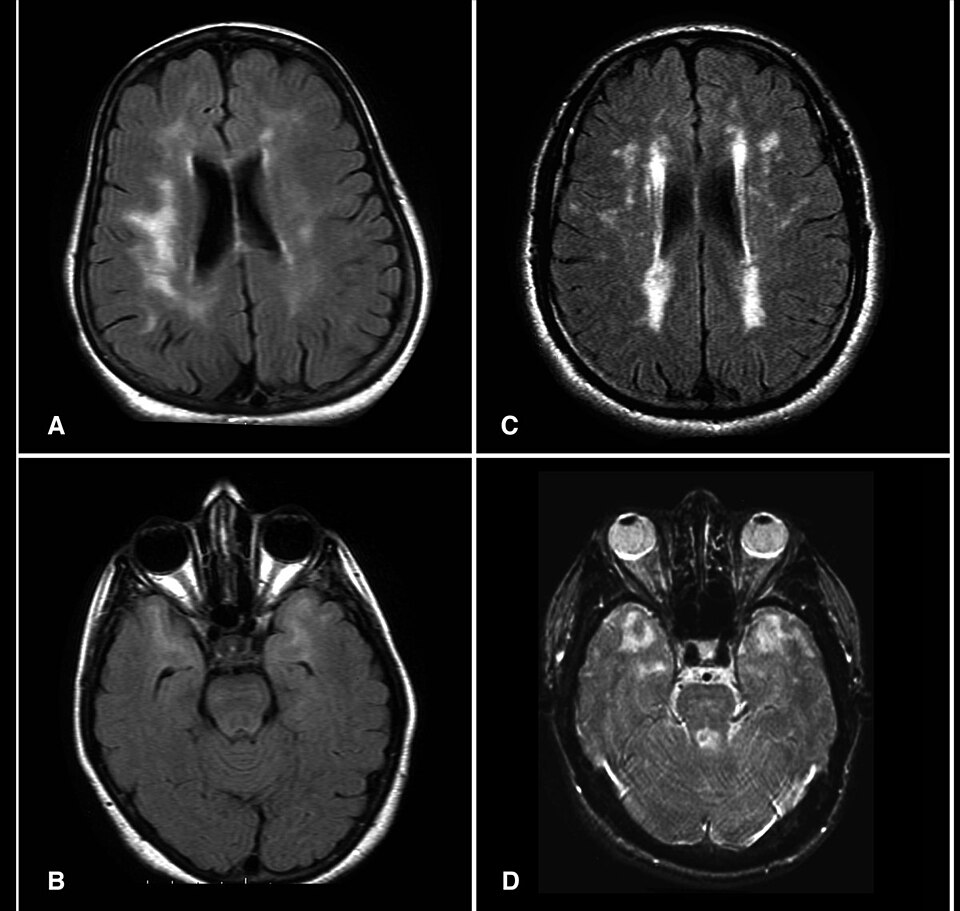
Cerebral Autosomal Dominant Arteriopathy with Subcortical Infarcts and Leukoencephalopathy is the most common heritable cause of stroke and vascular cognitive impairment in adults, and the archetypal monogenic cerebral small-vessel disease.
Genetics/etiology
CADASIL is caused by mutations in NOTCH3 on chromosome 19p13.12, inherited in an autosomal dominant pattern with near-complete penetrance. The pathogenic signature is stereotyped: virtually all disease-causing variants are missense changes that add or remove a cysteine residue within one of the 34 epidermal growth factor-like repeats (EGFr) of the NOTCH3 extracellular domain, producing an odd number of cysteines in that repeat and disrupting disulphide bonding. Mutations cluster in exons 3 and 4 (encoding EGFr 1–5), so targeted sequencing typically begins there before extending across the mutational hotspot (exons 2–24). The location of the cysteine-altering variant carries prognostic weight: mutations in EGFr 1–6 produce the classic, earlier and more severe phenotype, whereas variants in EGFr 7–34 are associated with milder, later-onset disease and lower lifetime stroke burden.
Pathophysiology
The mutant NOTCH3 ectodomain misfolds and accumulates at the surface of vascular smooth-muscle cells and pericytes, aggregating into pathognomonic granular osmiophilic material (GOM) visible by electron microscopy within the arterial media. Progressive degeneration and loss of vascular smooth-muscle cells, basement-membrane thickening and luminal narrowing of penetrating arterioles produce chronic hypoperfusion, impaired autoregulation and blood-brain barrier dysfunction, culminating in subcortical lacunar infarction and diffuse white-matter injury.
Clinical features
- Migraine with aura — often the earliest manifestation (30s), frequently with atypical or prolonged/hemiplegic aura.
- Recurrent subcortical ischemic events — lacunar syndromes and TIAs beginning in the 40s–50s, typically without conventional vascular risk factors.
- Mood disturbance — depression and apathy (the latter partly independent of depression).
- Progressive cognitive decline — a subcortical vascular dementia with executive dysfunction and processing-speed impairment; gait disturbance and pseudobulbar affect in advanced disease. Median survival is into the mid-60s.
Diagnostic workup
MRI is highly suggestive: confluent, symmetric T2/FLAIR white-matter hyperintensities with characteristic involvement of the anterior temporal poles (roughly 90% specific and largely absent in sporadic small-vessel disease) and the external capsule, alongside lacunar infarcts in the basal ganglia and thalamus and cerebral microbleeds on susceptibility sequences. Confirmation is by NOTCH3 sequencing. When genetics is unavailable or a variant is of uncertain significance, skin biopsy with electron microscopy demonstrating GOM in dermal arterioles, or immunostaining for NOTCH3 ectodomain accumulation, supports the diagnosis.
Management
No disease-modifying therapy exists. Care is supportive: aggressive control of blood pressure and smoking cessation, antiplatelet therapy used cautiously given the microbleed and intracerebral haemorrhage burden (dual antiplatelet and anticoagulation generally avoided unless a compelling indication exists). Intravenous thrombolysis is not absolutely contraindicated but is weighed against the elevated haemorrhagic-microangiopathy risk. Donepezil did not improve the primary cognitive endpoint in a randomised trial. Triptans and ergots are traditionally avoided for migraine on theoretical vasoconstrictive grounds. Genetic counselling is essential.
Clinical pearls
- Consider CADASIL in any young adult with lacunar stroke, migraine with aura, mood change and a leukoencephalopathy — particularly with a suggestive family history of stroke or early dementia.
- Anterior temporal pole white-matter involvement is the single most discriminating imaging clue versus hypertensive small-vessel disease and multiple sclerosis.
- A cysteine-altering NOTCH3 variant is pathogenic; cysteine-sparing variants require caution in interpretation.
Key references: Chabriat et al. CADASIL (Lancet Neurol 2009); Joutel et al. NOTCH3 mutations (Nature 1996); Rutten et al. NOTCH3 EGFr position and phenotype (Genet Med 2019); Di Donato et al. CADASIL review (BMC Med 2017).
CARASIL (HTRA1 Arteriopathy)
Cerebral Autosomal Recessive Arteriopathy with Subcortical Infarcts and Leukoencephalopathy (Maeda syndrome) is a rare hereditary small-vessel disease distinguished by its extraneurological triad of alopecia and spondylosis.
Genetics/etiology
Classic CARASIL results from biallelic loss-of-function mutations in HTRA1 (HtrA serine peptidase 1) on chromosome 10q26, inherited autosomal recessively. It is reported predominantly in individuals of Japanese and Chinese ancestry. Importantly, heterozygous HTRA1 mutations are now recognised as a more prevalent autosomal dominant cerebral small-vessel disease (HTRA1-related dominant SVD), typically milder and later in onset than biallelic CARASIL and lacking the full extraneurological triad.
Pathophysiology
HTRA1 is a secreted serine protease that represses signalling through the transforming growth factor-β (TGF-β) family. Loss of protease activity dysregulates TGF-β signalling, producing arteriopathy with intimal thickening, loss of smooth-muscle cells and adventitial fibrosis of small penetrating arteries. Unlike CADASIL, there is no granular osmiophilic material.
Clinical features
- Premature alopecia — scalp hair loss beginning in adolescence.
- Spondylosis / degenerative disc disease — early, severe lumbar and cervical spondylosis with acute disc herniation and low back pain.
- Progressive subcortical stroke and dementia — gait disturbance, pyramidal and pseudobulbar signs, and cognitive decline typically from the 20s to 40s, characteristically in non-hypertensive young adults.
Diagnostic workup
MRI shows diffuse leukoencephalopathy, lacunar infarcts and microbleeds; an arc-shaped pontine hyperintensity ("arc sign") is described. Anterior temporal involvement is less consistent than in CADASIL. Diagnosis rests on HTRA1 sequencing, with biallelic variants in classic CARASIL and monoallelic variants in the dominant form.
Management
Supportive only — vascular risk-factor control, physiotherapy and orthopaedic management of spondylosis, and antiplatelet therapy weighed against microbleed burden. Genetic counselling should address the recessive inheritance and the dominant heterozygous phenotype in carrier relatives.
Clinical pearls
- The combination of early alopecia, spondylotic back pain and subcortical stroke in a young normotensive adult is essentially pathognomonic.
- Heterozygous HTRA1 disease is increasingly recognised and should be considered in otherwise "sporadic" early small-vessel disease.
Key references: Hara et al. HTRA1 mutations in CARASIL (N Engl J Med 2009); Verdura et al. Heterozygous HTRA1 dominant SVD (Brain 2015); Nozaki et al. CARASIL/HTRA1 review (Stroke 2016).
Fabry Disease (GLA / α-Galactosidase A Deficiency)
Fabry disease is an X-linked lysosomal storage disorder and a treatable cause of stroke in the young, disproportionately affecting the posterior circulation.
Genetics/etiology
Mutations in GLA at Xq22.1 cause deficient activity of the lysosomal enzyme α-galactosidase A. Hemizygous males are typically affected with the classic phenotype; heterozygous females can be symptomatic (often later and milder) owing to skewed X-inactivation, so a normal enzyme level never excludes disease in a woman. Later-onset variants restricted to cardiac or renal disease exist.
Pathophysiology
Enzyme deficiency causes progressive accumulation of globotriaosylceramide (Gb3/GL-3) and its deacylated form lyso-Gb3 in vascular endothelium and smooth muscle, cardiomyocytes, renal podocytes and neurons. The resulting vasculopathy affects both small and large vessels and produces endothelial dysfunction, a prothrombotic state and characteristic dolichoectasia, notably of the vertebrobasilar system.
Clinical features
- Early/classic — acroparesthesias (small-fibre neuropathic pain), angiokeratomas, hypohidrosis, cornea verticillata (whorl keratopathy on slit lamp), heat/exercise intolerance.
- Systemic organ involvement — proteinuria progressing to renal failure, hypertrophic cardiomyopathy and conduction disease, gastrointestinal symptoms.
- Cerebrovascular — ischemic stroke and TIA at young age, often posterior circulation, related to small-vessel disease, dolichoectasia and cardioembolism; white-matter lesions and the pulvinar sign (T1 hyperintensity in the posterior thalamus).
Diagnostic workup
In males, measure leukocyte or plasma α-galactosidase A activity (low/absent). In females, enzyme activity is unreliable and GLA gene sequencing is required. Plasma lyso-Gb3 is a useful biomarker and can help interpret variants of uncertain significance. Slit-lamp examination for cornea verticillata, echocardiography, renal function/proteinuria and MRI (dolichoectasia, pulvinar sign) complete the evaluation. Routine screening of all young cryptogenic strokes has a low yield, so target patients with supportive features.
Management
Disease-specific therapy comprises:
- Enzyme replacement therapy — agalsidase beta (Fabrazyme); agalsidase alfa (Replagal, not FDA-approved in the US); and pegunigalsidase alfa (Elfabrio), a PEGylated enzyme FDA-approved in 2023 with a longer circulating half-life.
- Oral pharmacological chaperone — migalastat (Galafold) for patients with amenable (chaperone-responsive) missense GLA variants.
- Adjunctive — antiplatelet therapy and vascular risk-factor control; cardiology/nephrology co-management; genetic counselling and family screening.
Clinical pearls
- Suspect Fabry in young stroke with left-ventricular hypertrophy, renal disease, neuropathic pain or a maternal family history; cornea verticillata is a quick bedside clue.
- Genetic testing, not enzyme assay, is required to diagnose affected women.
Key references: Sims et al. Fabry and stroke, Fabry Registry (Stroke 2009); Rolfs et al. Fabry in young stroke (Lancet 2005); Germain, Fabry disease (Orphanet J Rare Dis 2010); FDA pegunigalsidase alfa approval (2023).
MELAS (Mitochondrial Encephalomyopathy, Lactic Acidosis, Stroke-like Episodes)
MELAS is the prototypical mitochondrial cause of stroke-like episodes that characteristically defy vascular territories.
Genetics/etiology
MELAS is a maternally inherited mitochondrial DNA disorder. The m.3243A>G mutation in MT-TL1 (tRNA-Leucine) accounts for roughly 80% of cases; other variants include m.3271T>C and m.13513G>A (MT-ND5). Expression depends on heteroplasmy with a tissue-specific threshold effect, so clinical severity varies within a maternal lineage. The same m.3243A>G variant also causes maternally inherited diabetes and deafness (MIDD), illustrating a phenotypic continuum.
Pathophysiology
Defective oxidative phosphorylation, a mitochondrial angiopathy with proliferation of dysfunctional mitochondria in vascular smooth muscle and endothelium, neuronal hyperexcitability and a state of nitric oxide deficiency underlie the stroke-like episodes. The lesions represent a metabolic-energetic rather than purely ischemic failure, which is why they cross arterial boundaries.
Clinical features
- Stroke-like episodes — usually before age 40, often posterior (occipital/parietal/temporal), migrating, with cortical visual and encephalopathic features and frequently seizures.
- Recurrent migraine-like headache, seizures, encephalopathy, sensorineural hearing loss, short stature, diabetes, cardiomyopathy and exercise-intolerant myopathy.
Diagnostic workup
- Elevated lactate in serum and CSF; MR spectroscopy shows a lactate doublet (~1.3 ppm) even in normal-appearing tissue.
- MRI — cortical/subcortical lesions crossing vascular territories, posterior predominance, often with vasogenic-type (preserved or increased ADC) as well as cytotoxic components.
- Muscle biopsy — ragged-red fibres (Gomori trichrome) and COX-negative fibres.
- Genetic testing — blood may be falsely negative because of low leukocyte heteroplasmy; urinary sediment, buccal mucosa or muscle offer higher yield.
Management
There is no cure. Evidence-supported and guideline-endorsed (notably Japanese) measures include:
- L-arginine — intravenous during acute stroke-like episodes and oral prophylaxis, replenishing nitric oxide substrate and reducing episode frequency/severity.
- Taurine supplementation reduces stroke-like episode recurrence (approved in Japan for this indication); coenzyme Q10, riboflavin and other "mitochondrial cocktail" cofactors are commonly used.
- Aggressive seizure control and supportive multisystem care.
- Avoid valproate (mitochondrial hepatotoxicity and can precipitate crises), metformin (lactic acidosis) and aminoglycosides (ototoxicity).
Clinical pearls
- A young patient with a "stroke" that crosses vascular territories, elevated lactate and a lactate peak on MRS should prompt mitochondrial evaluation.
- Do not thrombolyse a mitochondrial stroke-like episode reflexively; and avoid valproate and metformin.
Key references: El-Hattab et al. MELAS review (Mol Genet Metab 2015); Koga et al. L-arginine in MELAS (Neurology 2005); Ohsawa et al. taurine trial in MELAS (J Neurol Neurosurg Psychiatry 2019).
Moyamoya Disease & Syndrome
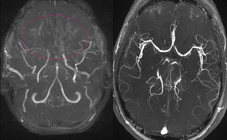
Moyamoya is a progressive steno-occlusive arteriopathy of the distal internal carotid arteries with a compensatory basal collateral network resembling a "puff of smoke" (Japanese moyamoya).
Genetics/etiology
Moyamoya disease denotes the idiopathic, typically bilateral entity; moyamoya syndrome refers to the same angiographic picture occurring with an associated condition (sickle-cell disease, neurofibromatosis type 1, Down syndrome, cranial irradiation, Graves disease, and others). The strongest genetic susceptibility factor in East Asians is the RNF213 variant p.R4810K (17q25.3); homozygotes have early-onset, severe disease. There is a bimodal age distribution (a childhood peak around 5–10 years and an adult peak in the 40s).
Pathophysiology
Progressive intimal hyperplasia (non-inflammatory, non-atherosclerotic) narrows the supraclinoid ICA and proximal anterior and middle cerebral arteries. Fragile dilated lenticulostriate and thalamoperforator collaterals develop and are prone to rupture. Children usually present with ischemia (often triggered by hyperventilation-induced hypocapnia — crying, exertion, hot food); adults present with either ischemia or haemorrhage (intraventricular/basal ganglia) from these fragile vessels or associated aneurysms.
Suzuki angiographic stages
| Stage | Angiographic finding |
|---|---|
| I | Narrowing of the ICA bifurcation |
| II | Initial appearance of basal moyamoya collaterals with ICA dilatation |
| III | Intensification of moyamoya vessels; progressive ICA/ACA/MCA narrowing |
| IV | Minimisation of moyamoya; external carotid collaterals develop |
| V | Reduction of moyamoya vessels; increasing ECA supply |
| VI | Disappearance of moyamoya; cerebral perfusion entirely via ECA collaterals |
Diagnostic workup
Catheter angiography (DSA) is the reference standard; MRA/CTA and MRI often suffice. The ivy sign (leptomeningeal FLAIR hyperintensity from slow pial collateral flow) is characteristic. Haemodynamic assessment with perfusion imaging and acetazolamide-challenge cerebrovascular reserve guides revascularisation. RNF213 testing is informative in Asian populations.
Management
- Surgical revascularisation is the mainstay: direct (STA-MCA bypass), indirect (encephaloduroarteriosynangiosis/EDAS, encephalomyosynangiosis, pial synangiosis — favoured in children), or combined procedures.
- Antiplatelet therapy for the ischemic phenotype; the JAM trial showed direct bypass reduces rebleeding in the haemorrhagic type.
- Perioperative care: maintain euvolemia and normocapnia; avoid hypotension, dehydration and hyperventilation, all of which provoke ischemia.
Clinical pearls
- Hyperventilation-triggered TIAs in a child (e.g., after crying or blowing) strongly suggest moyamoya.
- The ivy sign and reduced cerebrovascular reserve identify haemodynamically vulnerable territories that benefit most from bypass.
Key references: Suzuki & Takaku (Arch Neurol 1969); Kamada et al. RNF213 (J Hum Genet 2011); Miyamoto et al. JAM Trial (Stroke 2014); Scott & Smith moyamoya review (N Engl J Med 2009).
Marfan, Loeys-Dietz & Vascular Ehlers-Danlos Syndromes
These heritable connective-tissue disorders share a predisposition to arterial aneurysm and dissection and are important causes of stroke in the young, especially via cervical artery dissection and cardioembolism.
Genetics/etiology
| Disorder | Gene(s) | Protein/pathway | Inheritance |
|---|---|---|---|
| Marfan syndrome | FBN1 | Fibrillin-1 (dysregulated TGF-β) | Autosomal dominant |
| Loeys-Dietz syndrome | TGFBR1, TGFBR2, SMAD3, TGFB2, TGFB3 | TGF-β signalling | Autosomal dominant |
| Vascular EDS (type IV) | COL3A1 | Type III procollagen | Autosomal dominant |
Pathophysiology & features
- Marfan — aortic root aneurysm/dissection, ectopia lentis (usually superior lens dislocation), tall stature with arachnodactyly and dural ectasia. Stroke is typically cardioembolic (aortic/valvular disease) or from cervical artery dissection.
- Loeys-Dietz — the most aggressive: arterial tortuosity and aneurysm/dissection throughout the arterial tree (including cervical and intracranial vessels), hypertelorism, bifid uvula or cleft palate. High rates of intracranial aneurysm and dissection warrant whole-arterial surveillance.
- Vascular EDS — spontaneous rupture and dissection of medium arteries, characteristic thin translucent skin, easy bruising, and a distinctive facies; spontaneous carotid-cavernous fistula is a hallmark neurovascular event. Bowel and uterine rupture occur.
Diagnostic workup
Targeted or panel genetic testing; echocardiography and whole-body non-invasive arterial imaging (MRA/CTA); connective-tissue examination (skeletal, ocular, skin, palate). In vascular EDS, catheter angiography is relatively contraindicated because of arterial fragility — favour non-invasive imaging.
Management
- Marfan — β-blockers and/or angiotensin-receptor blockers (losartan) for aortic protection; prophylactic aortic root surgery at threshold diameters; avoid isometric/contact sport.
- Loeys-Dietz — earlier surgical thresholds; blood-pressure control; aneurysm surveillance across the arterial tree.
- Vascular EDS — celiprolol reduced arterial events in the BBEST trial and is first-line; strict blood-pressure control; conservative/endovascular management of complications with great caution.
Clinical pearls
- Suspect a connective-tissue disorder in young or recurrent arterial dissection and in spontaneous carotid-cavernous fistula (vascular EDS).
- Lens dislocation direction helps distinguish Marfan (upward) from homocystinuria (downward); a bifid uvula points to Loeys-Dietz.
Key references: Loeys et al. Ghent nosology (J Med Genet 2010); Loeys & Dietz LDS (GeneReviews); Ong et al. BBEST celiprolol trial in vascular EDS (Lancet 2010); Byers et al. vascular EDS management (Am J Med Genet 2017).
Homocystinuria & Hyperhomocysteinemia
Elevated homocysteine spans a spectrum from severe inborn errors of metabolism causing early thromboembolic stroke to mild, common elevations of uncertain therapeutic relevance.
Genetics/etiology
- Classic homocystinuria — autosomal recessive cystathionine β-synthase (CBS) deficiency, causing marked hyperhomocysteinemia and homocystinuria. Pyridoxine (B6)-responsive and non-responsive forms exist.
- Remethylation defects — severe MTHFR deficiency and cobalamin metabolism defects (e.g., MMACHC/cobalamin C).
- Mild hyperhomocysteinemia — the MTHFR C677T thermolabile polymorphism, nutritional deficiency of B12/folate/B6, renal impairment and older age.
Pathophysiology
Homocysteine is directly toxic to endothelium, promotes oxidative stress, impairs nitric-oxide bioavailability and creates a prothrombotic milieu, predisposing to both arterial and venous thromboembolism.
Clinical features
Classic homocystinuria presents in childhood/adolescence with a marfanoid habitus but downward ectopia lentis, intellectual disability, osteoporosis, livedo reticularis, and premature arterial and venous thromboembolism — the leading cause of death and a cause of early stroke and cerebral venous thrombosis. Mild hyperhomocysteinemia is at most a modest, independent risk marker.
Diagnostic workup
Plasma total homocysteine and methionine, urinary amino/organic acids, serum B12/folate and methylmalonic acid (elevated in cobalamin defects, normal in CBS deficiency), and CBS (or relevant) gene testing. In young stroke or CVT with a marfanoid phenotype, measure homocysteine.
Management
- Classic homocystinuria — high-dose pyridoxine (a trial to identify responders), methionine-restricted diet, folate and B12 repletion, and betaine to promote remethylation and lower homocysteine; antithrombotic prophylaxis around surgery.
- Mild hyperhomocysteinemia — correct nutritional deficiency, but note that homocysteine-lowering with B vitamins did not reduce recurrent stroke overall in the large VISP, HOPE-2 and VITATOPS trials (possible benefit only in subgroups such as low-B12/non-fortified populations). Routine supplementation for secondary stroke prevention is not supported.
Clinical pearls
- Downward lens dislocation plus early thromboembolism suggests homocystinuria, not Marfan.
- Do not over-rely on B vitamins for stroke prevention in folate-fortified populations; the target is the rare severe metabolic disorder.
Key references: Toole et al. VISP trial (JAMA 2004); Lonn et al. HOPE-2 (N Engl J Med 2006); VITATOPS Trial Group (Lancet Neurol 2010); Morris et al. CBS deficiency guidelines (J Inherit Metab Dis 2017).
Hereditary Hemorrhagic Telangiectasia (Osler-Weber-Rendu)
HHT is an autosomal dominant fibrovascular dysplasia in which arteriovenous malformations cause both paradoxical embolic stroke and brain abscess — largely preventable by pulmonary AVM screening.
Genetics/etiology
| Type | Gene | Protein/pathway | Notable associations |
|---|---|---|---|
| HHT1 | ENG | Endoglin (TGF-β/BMP9) | Higher pulmonary and cerebral AVM burden |
| HHT2 | ACVRL1 (ALK1) | Activin receptor-like kinase 1 | More hepatic AVMs, pulmonary hypertension |
| JP-HHT | SMAD4 | SMAD4 signalling | Overlap with juvenile polyposis |
Pathophysiology
Defective TGF-β/BMP9 endothelial signalling produces telangiectasias and arteriovenous malformations lacking an intervening capillary bed. A right-to-left shunt through a pulmonary AVM removes the capillary filter, allowing paradoxical passage of thrombi (ischemic stroke/TIA) and septic emboli (brain abscess). Cerebral AVMs additionally pose a haemorrhage risk.
Clinical features & Curaçao criteria
Recurrent spontaneous epistaxis is the hallmark; mucocutaneous telangiectasias (lips, tongue, fingertips) and visceral AVMs (pulmonary, hepatic, cerebral, GI) develop over time. Diagnosis uses the Curaçao criteria:
| Criterion | Detail |
|---|---|
| Epistaxis | Spontaneous, recurrent nosebleeds |
| Telangiectasias | Multiple at characteristic sites |
| Visceral lesions | Pulmonary, hepatic, cerebral or GI AVM |
| Family history | First-degree relative with HHT |
Three or more criteria = definite; two = possible/suspected.
Diagnostic workup
Screen for pulmonary AVM with agitated-saline (bubble) transthoracic echocardiography and, if positive, chest CT; brain MRI to detect cerebral AVMs; genetic testing (ENG, ACVRL1, SMAD4).
Management
- Transcatheter embolisation of pulmonary AVMs (feeding artery ≥2–3 mm) prevents paradoxical stroke and abscess, even when asymptomatic.
- Antibiotic prophylaxis before dental and other bacteraemia-prone procedures, and meticulous avoidance of intravenous air (air-filter use), reduce abscess and air-embolism risk.
- Iron replacement/transfusion for anaemia; bevacizumab (anti-VEGF) for severe epistaxis, GI bleeding or high-output hepatic AVMs; cerebral AVMs managed on standard principles.
Clinical pearls
- Young cryptogenic stroke or brain abscess with epistaxis and telangiectasias should trigger bubble-echo screening for pulmonary AVM.
- Counsel every HHT patient about IV air-embolism precautions and endocarditis/abscess antibiotic prophylaxis.
Key references: Faughnan et al. Second International HHT Guidelines (Ann Intern Med 2020); Shovlin et al. Curaçao criteria (Am J Med Genet 2000); Kjeldsen et al. cerebrovascular complications of HHT.
ACTA2 Arteriopathy (Smooth-Muscle α-Actin)
Mutations in ACTA2 cause a distinctive cerebrovascular arteriopathy that mimics — but is mechanistically and angiographically distinct from — moyamoya.
Genetics/etiology
ACTA2 encodes smooth-muscle α-actin, essential for vascular smooth-muscle contraction; mutations are autosomal dominant. The recurrent p.R179H (Arg179His) variant produces the severe multisystemic smooth-muscle dysfunction syndrome, while other variants cause thoracic aortic aneurysm and dissection (TAAD) with variable cerebrovascular disease.
Pathophysiology
Impaired smooth-muscle contractility yields a characteristic arteriopathy: abnormally straight (non-tortuous), broad-based dilatation of the proximal arteries with occlusive disease of the terminal internal carotid arteries — but crucially without the basal moyamoya collateral network. This "moyamoya-like" but collateral-poor pattern distinguishes it from true moyamoya. Periventricular white-matter lesions and a persistently patent, dilated arterial phenotype are typical.
Clinical features
The R179 multisystemic syndrome combines congenital fixed dilated pupils (congenital mydriasis), patent ductus arteriosus, malrotation, hypotonic bladder and gut, pulmonary hypertension, and early cerebrovascular disease with childhood ischemic stroke. Other ACTA2 variants present with TAAD, livedo reticularis, iris flocculi and premature stroke.
Diagnostic workup
Genetic testing (ACTA2), cerebral and whole-arterial imaging (demonstrating straightened vessels and terminal ICA occlusion without moyamoya collaterals), and echocardiography for aortic disease.
Management
No specific therapy; vascular risk-factor control and antiplatelet therapy for ischemia. Aortic surveillance and prophylactic surgery follow TAAD principles. Standard moyamoya revascularisation is of uncertain benefit because the pathology and collateral biology differ.
Clinical pearls
- Fixed dilated pupils in a child with stroke and aortic disease is highly suggestive of ACTA2 p.R179.
- Terminal ICA occlusion with abnormally straight arteries and an absent basal collateral network argues for ACTA2 arteriopathy rather than classic moyamoya.
Key references: Guo et al. ACTA2 R179H multisystemic smooth muscle dysfunction (Am J Hum Genet 2009); Milewicz et al. ACTA2 cerebrovascular disease (Am J Med Genet 2010); Munot et al. distinctive arteriopathy (Brain 2012).
Sturge-Weber Syndrome
Sturge-Weber syndrome is a sporadic neurocutaneous disorder in which a leptomeningeal capillary-venous malformation drives seizures, stroke-like episodes and progressive hemispheric injury.
Genetics/etiology
Sturge-Weber is caused by a somatic mosaic activating mutation in GNAQ (p.R183Q) (occasionally GNA11), arising post-zygotically — it is therefore not inherited. The distribution of affected tissue reflects the developmental timing of the mutation.
Pathophysiology
A leptomeningeal capillary-venous malformation (pial angioma), usually parieto-occipital and ipsilateral to a facial capillary malformation, impairs cortical venous drainage. Chronic venous stasis and hypoperfusion produce progressive cortical ischemia, laminar necrosis, gyriform ("tram-track") cortical calcification and hemispheric atrophy.
Clinical features
- Facial port-wine birthmark (capillary malformation), classically involving the forehead/upper eyelid (embryonic vascular/V1 distribution).
- Seizures — frequently early-onset and drug-resistant.
- Stroke-like episodes — often provoked by seizures, migraine or minor trauma, with transient or fixed hemiparesis and hemianopia, and cognitive impairment over time.
- Glaucoma in the ipsilateral eye.
Diagnostic workup
Contrast MRI shows leptomeningeal enhancement and the pial angioma with an enlarged ipsilateral choroid plexus; SWI depicts abnormal deep venous drainage; CT shows gyriform calcification; EEG characterises the epilepsy. Ophthalmological assessment screens for glaucoma.
Management
- Aggressive seizure control, with epilepsy surgery (including hemispherectomy) for refractory cases.
- Low-dose aspirin is widely used and observationally associated with fewer stroke-like episodes and seizures.
- Avoid dehydration and other provocateurs of venous stasis; treat migraine; manage glaucoma; laser therapy for the port-wine stain.
Clinical pearls
- A V1-distribution port-wine stain with seizures and gyriform "tram-track" calcification is the classic triad.
- Aspirin and prevention of dehydration reduce the stroke-like episode burden.
Key references: Shirley et al. GNAQ mutation in Sturge-Weber (N Engl J Med 2013); Comi, Sturge-Weber pathophysiology and management (Neurologist 2011); Bay et al. aspirin in SWS (J Child Neurol 2011).
RVCL-S (Retinal Vasculopathy with Cerebral Leukoencephalopathy; TREX1)
RVCL-S is a rare autosomal dominant systemic microvascular endotheliopathy that classically masquerades as a brain tumour or demyelinating disease.
Genetics/etiology
RVCL-S is caused by C-terminal frameshift/truncating mutations in TREX1 (three-prime repair exonuclease 1), autosomal dominant. These are mechanistically distinct from the N-terminal loss-of-function TREX1 variants that cause Aicardi-Goutières syndrome and systemic lupus: the C-terminal mutations preserve exonuclease activity but mislocalise the protein, producing a gain-of-function endotheliopathy. RVCL-S unifies previously separate eponymous entities (cerebroretinal vasculopathy, hereditary vascular retinopathy, and hereditary endotheliopathy with retinopathy, nephropathy and stroke/HERNS).
Pathophysiology
A systemic small-vessel endotheliopathy affects the retina, brain, kidney, liver and other organs, producing progressive capillary dropout and tissue infarction.
Clinical features
Onset is in the fourth to sixth decades:
- Retinal vasculopathy — progressive visual loss from capillary dropout, telangiectasia and macular oedema.
- Cerebral leukoencephalopathy — subacute focal deficits, cognitive decline, seizures, and characteristically contrast-enhancing pseudotumoral mass lesions with surrounding oedema and calcification that mimic glioma or tumefactive demyelination.
- Systemic — Raynaud phenomenon, nephropathy, hepatopathy with elevated liver enzymes, anaemia, hypertension, GI bleeding and subclinical hypothyroidism. The course is progressive and often fatal within 5–10 years of neurological onset.
Diagnostic workup
TREX1 sequencing focused on the C-terminus is diagnostic. Fluorescein angiography demonstrates the retinal vasculopathy; MRI shows enhancing white-matter lesions with oedema and calcification. Brain biopsy shows a non-inflammatory vasculopathy and should generally be avoided, as pseudotumoral lesions are frequently mistaken for neoplasm and resected unnecessarily.
Management
No proven disease-modifying therapy; immunosuppression is generally ineffective (helping to distinguish it from an inflammatory vasculitis). Anti-VEGF therapy is used for retinal complications. The anti-P-selectin antibody crizanlizumab showed possible benefit in a phase II study. Management is otherwise supportive, and unnecessary biopsy/resection of pseudotumoral lesions should be avoided.
Clinical pearls
- Enhancing "mass" lesions plus retinopathy and systemic organ involvement in a patient with an autosomal dominant family history should raise RVCL-S before tumour is diagnosed.
- Lack of response to steroids and a C-terminal TREX1 variant confirm the diagnosis.
Key references: Richards et al. TREX1 mutations cause RVCL (Nat Genet 2007); Stam et al. RVCL-S clinical spectrum (Brain 2016); Hedderich et al./JCI crizanlizumab phase II in RVCL-S (J Clin Invest 2024).
Genetic Cerebral Small-Vessel Diseases (COL4A1/2 & Overview)
Beyond CADASIL, a growing catalogue of monogenic small-vessel diseases causes stroke, haemorrhage and leukoencephalopathy; COL4A1/COL4A2 disease is the prototype of the haemorrhage-prone group.
Genetics/etiology — COL4A1/2
COL4A1 and COL4A2 encode the α1 and α2 chains of type IV collagen, a core basement-membrane component; mutations are autosomal dominant (many de novo). Impaired collagen IV assembly weakens the vascular basement membrane, producing a fragile, haemorrhage-prone small-vessel arteriopathy.
Clinical spectrum
- Perinatal/infantile — porencephaly and fetal/neonatal intracerebral haemorrhage (often provoked by minor trauma or delivery), infantile hemiparesis.
- Adult — spontaneous (frequently deep) intracerebral haemorrhage, small-vessel ischemic disease and leukoencephalopathy, intracranial aneurysm, and the HANAC syndrome (hereditary angiopathy with nephropathy, aneurysms and muscle cramps).
- Systemic clues — retinal arteriolar tortuosity, cataracts and nephropathy/haematuria.
Overview of monogenic cerebral small-vessel diseases
| Disease | Gene | Inheritance | Distinguishing features |
|---|---|---|---|
| CADASIL | NOTCH3 | AD | Anterior temporal WMH; GOM; migraine with aura |
| CARASIL / HTRA1-SVD | HTRA1 | AR (dominant if heterozygous) | Alopecia, spondylosis; arc sign |
| COL4A1/2 angiopathy | COL4A1/COL4A2 | AD | Porencephaly, ICH, retinal tortuosity, nephropathy |
| Fabry disease | GLA | X-linked | Enzyme deficiency; dolichoectasia; pulvinar sign |
| RVCL-S | TREX1 | AD | Retinopathy; pseudotumoral enhancing lesions |
| CARASAL | CTSA | AD | Late-onset SVD, hypertension, pain |
| PADMAL | COL4A1 3'UTR (miR-29) | AD | Pontine autosomal dominant microangiopathy |
| Cerebral cavernous malformations | KRIT1/CCM1, CCM2, PDCD10/CCM3 | AD | Multiple cavernomas; haemorrhage, seizures |
Diagnostic workup
MRI (haemorrhage burden on susceptibility imaging, leukoencephalopathy pattern, porencephaly), multigene small-vessel-disease panels, and screening for extracerebral clues (dilated fundus examination for retinal tortuosity, urinalysis/renal function, ophthalmology).
Management
- In haemorrhage-prone genotypes (especially COL4A1/2), avoid or minimise antithrombotics and anticoagulation, control blood pressure strictly, and counsel against contact sports and head trauma.
- Antiseizure therapy as needed; genetic counselling for autosomal dominant transmission and de novo risk.
Clinical pearls
- COL4A1 unites porencephaly, spontaneous ICH, retinal arteriolar tortuosity and nephropathy — a fundus examination can point to the diagnosis.
- Classify a monogenic small-vessel disease as ischemic- or haemorrhage-predominant before choosing antithrombotic therapy.
Key references: Gould et al. COL4A1 mutations and cerebral haemorrhage (Science 2005; N Engl J Med 2006); Meuwissen et al. COL4A1/2 spectrum (Genet Med 2015); Mancuso et al. monogenic cerebral SVD review (J Neurol 2020).
Approach to CNS Vasculitis
CNS vasculitis is an inflammatory arteriopathy that produces multifocal, multi-territory ischemia and haemorrhage; a structured classification-by-vessel-size and rigorous exclusion of mimics guide the workup.
Classification
The first division is primary (angiitis confined to the CNS — PACNS) versus secondary (systemic vasculitides, infection, connective-tissue disease, drugs, malignancy). Systemic vasculitides are then stratified by predominant vessel size (Chapel Hill Consensus Conference 2012):
| Vessel size | Representative diseases | Neurovascular relevance |
|---|---|---|
| Large vessel | Giant cell arteritis, Takayasu arteritis | Aorta/great-vessel and cranial-artery stenosis; hemodynamic/embolic stroke, AION |
| Medium vessel | Polyarteritis nodosa, Kawasaki disease | Microaneurysms, small deep infarcts, ICH; mononeuritis multiplex |
| Small vessel (ANCA-associated) | GPA, EGPA, MPA | Pachymeningitis, cranial/peripheral neuropathy, rare cerebral vasculitis |
| Small vessel (immune-complex) | Cryoglobulinemic, IgA, anti-GBM, hypocomplementemic urticarial | Small-vessel CNS involvement |
| Variable vessel | Behçet disease, Cogan syndrome | Venous thrombosis and parenchymal (Behçet); large-vessel/aortitis (Cogan) |
Systemic & CNS features suggesting vasculitis
Suspect CNS vasculitis with subacute headache, encephalopathy, cognitive decline, seizures, and strokes of differing ages across multiple arterial territories, especially alongside systemic inflammation (constitutional symptoms, elevated inflammatory markers, multiorgan disease, mononeuritis multiplex, sinopulmonary or renal disease).
Diagnosis
Laboratory: ESR/CRP, ANCA, ANA/dsDNA, complement, cryoglobulins, hepatitis B/C and HIV serologies, syphilis testing, blood cultures. CSF typically shows lymphocytic pleocytosis and elevated protein and helps exclude infection and RCVS. Imaging: MRI/MRA, and vessel-wall MRI (concentric, smooth mural enhancement favours vasculitis; eccentric enhancement suggests atheroma; uniform without much enhancement suggests RCVS). Catheter angiography may show alternating stenosis and dilation ("beading") but is not specific (RCVS, atherosclerosis and vasospasm mimic it) and can be normal in small-vessel vasculitis. Brain-and-leptomeningeal biopsy is the reference standard for PACNS.
Treatment
Treatment is cause-specific. Before immunosuppression, exclude infection and RCVS, since immunosuppression is harmful in RCVS and infection. Primary and idiopathic secondary vasculitides are treated with glucocorticoids plus a steroid-sparing/cytotoxic agent (see specific topics).
Clinical pearls
- Angiographic "beading" is a pattern, not a diagnosis — correlate with CSF, vessel-wall imaging and clinical context.
- Always consider mimics: RCVS, intravascular lymphoma, moyamoya, CADASIL, Susac, infective (VZV, TB, syphilis, fungal) arteritis, and cardioembolism.
Key references: Jennette et al. Revised Chapel Hill Consensus (Arthritis Rheum 2013); Salvarani et al. adult CNS vasculitis (Lancet Neurol 2012); Mandell et al. vessel-wall imaging expert consensus (AJNR 2017).
Giant Cell (Temporal) Arteritis
Giant cell arteritis is the commonest primary systemic vasculitis of older adults and a neuro-ophthalmic emergency because of its threat to vision.
Vessel size
A granulomatous large- and medium-vessel vasculitis of the aorta and its major branches and of the extracranial cranial arteries (superficial temporal, occipital, ophthalmic, posterior ciliary and vertebral arteries).
Systemic features
Age over 50 (usually over 70), new-onset headache, jaw claudication (high specificity), scalp tenderness, an abnormal temporal artery, constitutional symptoms and fever. Polymyalgia rheumatica coexists in roughly 40–50%.
CNS & neuro-ophthalmic features
- Arteritic anterior ischemic optic neuropathy (AION) — sudden painless monocular visual loss with pallid ("chalky-white") disc oedema; may become bilateral if untreated.
- Amaurosis fugax, central retinal artery occlusion and diplopia.
- Stroke — predominantly in the vertebrobasilar territory from vertebral artery arteritis; carotid-territory stroke is less common.
Diagnosis
Elevated ESR (often >50 mm/h) and CRP (more sensitive), frequently with thrombocytosis. Temporal artery biopsy (a 1–2 cm segment; granulomatous transmural inflammation with giant cells and intimal hyperplasia; skip lesions occur, so a contralateral biopsy may follow a negative result) remains the reference standard and stays informative for up to ~2 weeks after starting steroids. Temporal and axillary artery ultrasound (non-compressible hypoechoic "halo" sign) enables rapid diagnosis in fast-track pathways; MRI and PET assess large-vessel involvement. The 2022 ACR/EULAR classification criteria formalise these features.
Treatment
- Start high-dose glucocorticoids immediately — do not wait for biopsy. With visual symptoms/AION, use IV methylprednisolone 500–1000 mg daily for 3 days followed by oral prednisone ~1 mg/kg/day; without visual symptoms, oral prednisone 40–60 mg/day.
- Tocilizumab (IL-6 receptor antagonist) is an established steroid-sparing agent that improved sustained glucocorticoid-free remission in the GiACTA trial and is FDA-approved for GCA.
- Upadacitinib (oral JAK inhibitor, 15 mg) achieved sustained remission in the phase 3 SELECT-GCA trial and received FDA approval for GCA in April 2025 — the first JAK inhibitor for this indication.
- Methotrexate offers modest steroid-sparing benefit; low-dose aspirin is used selectively; provide bone protection and PJP prophylaxis; taper glucocorticoids over roughly 1–2 years.
Clinical pearls
- The immediate priority is protecting the fellow eye — treat on suspicion, then confirm.
- A normal ESR does not exclude GCA; check CRP and platelets, and pursue ultrasound/biopsy.
Key references: Stone et al. GiACTA tocilizumab trial (N Engl J Med 2017); Blockmans et al. SELECT-GCA upadacitinib (N Engl J Med 2025); Ponte et al. 2022 ACR/EULAR GCA classification criteria (Ann Rheum Dis 2022); Dejaco et al. EULAR large-vessel vasculitis recommendations (Ann Rheum Dis 2020).
Takayasu Arteritis
Takayasu arteritis ("pulseless disease") is a large-vessel granulomatous vasculitis of young women that causes stenosis, occlusion and aneurysm of the aorta and its main branches.
Vessel size
Large-vessel vasculitis of the aorta and its major branches (subclavian, common carotid, vertebral, renal and pulmonary arteries).
Systemic features
Onset typically before age 40, with a female and Asian predominance. An early inflammatory phase (constitutional symptoms, arthralgia, carotidynia) precedes an occlusive "pulseless" phase with limb claudication, absent or asymmetric pulses, inter-arm blood-pressure discrepancy (>10 mm Hg), vascular bruits and renovascular hypertension.
CNS features
Stenosis of the carotid, vertebral and subclavian arteries produces TIA and stroke (usually hemodynamic or artery-to-artery embolic), syncope, dizziness, visual symptoms and subclavian steal. Malignant/renovascular hypertension adds haemorrhagic and hypertensive risk.
Diagnosis
Imaging is central: MRA/CTA show mural thickening, long-segment stenosis, occlusion and aneurysm; PET-CT demonstrates arterial-wall FDG uptake indicating active inflammation; ultrasound shows the homogeneous circumferential "macaroni" carotid-wall thickening. ESR/CRP correlate imperfectly with disease activity. Classification uses the 1990 ACR and 2022 ACR/EULAR criteria; the Numano/Hata angiographic types (I–V) describe the distribution. The 2023 EULAR imaging update endorses MRI/PET/ultrasound for diagnosis and monitoring.
Treatment
- Glucocorticoids are first-line, with early addition of a steroid-sparing agent (methotrexate, azathioprine, mycophenolate or leflunomide).
- Biologics for relapsing/refractory disease — TNF inhibitors (infliximab) and tocilizumab are both effective; comparative data suggest broadly similar efficacy.
- Revascularisation (angioplasty or bypass) is best performed during disease quiescence, as intervention during active inflammation carries high restenosis rates. Antiplatelet therapy and careful blood-pressure management complete care.
Clinical pearls
- A young woman with inter-arm BP discrepancy, bruits and raised inflammatory markers should prompt large-vessel imaging.
- Measure blood pressure in the least-affected limb (or centrally) to avoid under-treating hypertension masked by subclavian stenosis.
Key references: Maz et al. 2021 ACR/VF GCA and Takayasu management guideline (Arthritis Rheumatol 2021); Grayson et al. 2022 ACR/EULAR Takayasu criteria (Ann Rheum Dis 2022); Dejaco et al. 2023 EULAR imaging in large-vessel vasculitis (Ann Rheum Dis 2024).
Polyarteritis Nodosa
Polyarteritis nodosa is a necrotizing medium-vessel vasculitis that spares glomeruli and lungs, is ANCA-negative, and reaches the CNS relatively late.
Vessel size
A necrotizing vasculitis of medium (and small) muscular arteries without glomerulonephritis or alveolar haemorrhage, characteristically ANCA-negative — features that separate it from the ANCA-associated small-vessel vasculitides.
Systemic features
Constitutional symptoms with mononeuritis multiplex (a hallmark peripheral neuropathy), cutaneous disease (livedo reticularis, nodules, ulcers, digital ischemia), renovascular hypertension with renal microaneurysms (not glomerulonephritis), mesenteric ischemia, orchitis, and myalgia/arthralgia. A subset is hepatitis B-associated.
CNS features
CNS involvement typically occurs months to years into the disease: encephalopathy, small deep ischemic infarcts, intracerebral haemorrhage from ruptured microaneurysms, seizures and cranial neuropathies.
Diagnosis
Biopsy of an affected nerve, muscle or skin shows necrotizing arteritis; visceral (renal/mesenteric) catheter angiography reveals microaneurysms and beading. HBsAg identifies HBV-associated disease. ANCA is negative. Apply the 2022 ACR/EULAR criteria and carefully distinguish from microscopic polyangiitis (small-vessel, ANCA-positive, with glomerulonephritis).
Treatment
- Idiopathic PAN — glucocorticoids plus cyclophosphamide for severe/organ-threatening disease.
- HBV-associated PAN — a distinct strategy of antiviral therapy, plasma exchange and a short glucocorticoid course, avoiding prolonged immunosuppression that would perpetuate viral replication.
Clinical pearls
- Mononeuritis multiplex plus renal/mesenteric microaneurysms in an ANCA-negative patient suggests PAN — and mandates hepatitis B testing.
- CNS disease is usually a late feature; early neurological involvement should prompt reconsideration of the diagnosis.
Key references: Forbess & Bannykh PAN review (Rheum Dis Clin North Am 2015); Guillevin et al. HBV-associated PAN treatment (Medicine 2005); 2022 ACR/EULAR PAN classification criteria (Ann Rheum Dis 2022).
Primary Angiitis of the CNS (PACNS)
PACNS is a rare vasculitis confined to the brain, spinal cord and leptomeninges; its central diagnostic challenge is separating it from reversible cerebral vasoconstriction syndrome (RCVS) and other mimics, because the treatments are opposite.
Diagnosis (Calabrese & Mallek criteria)
Diagnosis requires: (1) an acquired, otherwise unexplained neurological deficit; (2) angiographic or histological evidence of CNS angiitis; and (3) no systemic vasculitis or other condition that could account for the findings.
CNS features
Insidious headache, encephalopathy, cognitive decline, multifocal strokes, seizures and, in some, tumefactive mass lesions. Histological subtypes include granulomatous (sometimes with vascular amyloid — amyloid-β-related angiitis/ABRA in the elderly), lymphocytic and necrotizing patterns; small-vessel-predominant disease may have normal angiography.
Diagnostic workup
- MRI is abnormal in over 90% — multi-territory infarcts of different ages, T2/FLAIR white-matter lesions, leptomeningeal or parenchymal gadolinium enhancement, microbleeds and occasionally mass lesions.
- CSF is abnormal in ~80–90% (mild lymphocytic pleocytosis, elevated protein); a normal CSF strongly favours RCVS.
- Catheter angiography may show segmental stenosis and dilation but has limited specificity and can be normal in small-vessel PACNS.
- Vessel-wall MRI may reveal concentric wall enhancement.
- Brain-and-leptomeningeal biopsy is the reference standard but has a false-negative rate up to ~25% from patchy involvement; target an enhancing, accessible lesion and sample cortex plus leptomeninges.
Mimics — especially RCVS
| Feature | PACNS | RCVS |
|---|---|---|
| Headache | Insidious, progressive | Recurrent thunderclap |
| CSF | Usually inflammatory | Usually normal/near-normal |
| Angiographic vasoconstriction | Fixed/progressive | Reversible within ~12 weeks |
| Triggers | None specific | Postpartum, vasoactive drugs, exertion |
| Vessel-wall enhancement | Often concentric/present | Usually minimal |
Other mimics: intracranial atherosclerosis, infection (VZV, TB, syphilis, fungal, HIV), moyamoya, intravascular lymphoma, Susac syndrome, CADASIL, cardioembolism/myxoma, and drug (cocaine/sympathomimetic) vasculopathy.
Treatment
Induction with glucocorticoids (IV methylprednisolone then oral prednisone), adding cyclophosphamide for severe or progressive disease; rituximab is an alternative. Maintenance uses azathioprine, mycophenolate or methotrexate for 12–18 months. Angiographically defined large-vessel PACNS may respond to steroids alone. Critically, RCVS must not be immunosuppressed — it is managed with calcium-channel blockade (nimodipine), removal of precipitants and supportive care.
Clinical pearls
- CSF analysis and the headache pattern are the most useful bedside discriminators between PACNS and RCVS.
- A negative biopsy does not exclude PACNS; conversely, always exclude infection before committing to immunosuppression.
Key references: Calabrese & Mallek diagnostic criteria (Medicine 1988); Salvarani et al. Mayo PACNS cohort (Ann Neurol 2007; Lancet Neurol 2012); Beuker et al. PACNS diagnosis and treatment (Ther Adv Neurol Disord 2018).
Eosinophilic Granulomatosis with Polyangiitis (Churg-Strauss)
EGPA is an eosinophil-rich, granulomatous ANCA-associated vasculitis arising on a background of asthma; its neurological signature is mononeuritis multiplex, and cardiac involvement drives both mortality and cardioembolic stroke.
Vessel size
Small-to-medium-vessel necrotizing vasculitis with eosinophilic tissue infiltration and extravascular granulomas; ANCA (usually anti-MPO/p-ANCA) is positive in only ~30–40%.
Systemic features
Adult-onset asthma, blood and tissue eosinophilia, allergic rhinosinusitis and nasal polyps, migratory pulmonary infiltrates, and eosinophilic myocarditis/cardiomyopathy (a major cause of death). The disease evolves through prodromal (atopic), eosinophilic-infiltrative and vasculitic phases.
CNS features
Mononeuritis multiplex is very common (up to ~75%). CNS involvement is less frequent but includes ischemic and haemorrhagic stroke, cranial neuropathy and, importantly, cardioembolic stroke from eosinophilic cardiac disease and intracavitary thrombus.
Diagnosis
Peripheral eosinophilia (>1500/µL or >10%), ANCA (MPO), and biopsy showing eosinophilic vasculitis or granuloma; apply the 2022 ACR/EULAR criteria. ANCA-positive disease tends toward vasculitic/neuropathic/renal features, while ANCA-negative disease is more cardiac and pulmonary.
Treatment
- Glucocorticoids for all; add cyclophosphamide or rituximab for severe disease (cardiac, neurological, or high Five-Factor Score).
- Mepolizumab (anti-IL-5) is an approved steroid-sparing agent; benralizumab (anti-IL-5 receptor) is an emerging option.
Clinical pearls
- Asthma plus eosinophilia plus mononeuritis multiplex is the classic triad.
- Screen the heart (echocardiography, cardiac MRI) — eosinophilic cardiomyopathy is both a mortality driver and a source of cardioembolic stroke.
Key references: Wechsler et al. mepolizumab in EGPA/MIRRA (N Engl J Med 2017); Grayson et al. 2022 ACR/EULAR EGPA classification criteria (Ann Rheum Dis 2022); Comarmond et al. EGPA cohort and outcomes (Arthritis Rheum 2013).
Granulomatosis with Polyangiitis (GPA)
GPA (formerly Wegener granulomatosis) is a PR3-ANCA-associated necrotizing granulomatous vasculitis with a characteristic sinopulmonary-renal triad and a distinctive neurological predilection for hypertrophic pachymeningitis.
Vessel size
Small-vessel necrotizing granulomatous vasculitis, classically associated with c-ANCA/PR3 antibodies.
Systemic features
Upper-airway disease (chronic sinusitis, saddle-nose deformity, subglottic stenosis, otitis media), lower-airway disease (pulmonary nodules, cavities, alveolar haemorrhage) and rapidly progressive pauci-immune glomerulonephritis.
CNS features
- Hypertrophic pachymeningitis — thickened, enhancing dura causing headache and cranial neuropathies.
- Cranial and peripheral neuropathies, including from contiguous granulomatous extension from the sinuses/orbit.
- Orbital granuloma (proptosis), pituitary involvement, and — less commonly — cerebral vasculitis with stroke.
Diagnosis
c-ANCA/PR3 positivity, biopsy showing necrotizing granulomatous vasculitis, and the 2022 ACR/EULAR classification criteria; MRI demonstrates dural thickening and enhancement in pachymeningitis.
Treatment
- Induction — glucocorticoids plus either rituximab (non-inferior and superior for relapsing disease in the RAVE trial) or cyclophosphamide.
- Plasma exchange has a limited, selective role (severe renal disease or diffuse alveolar haemorrhage) following PEXIVAS.
- Avacopan (oral C5a-receptor antagonist) reduces glucocorticoid exposure and improved sustained remission in the ADVOCATE trial; maintenance is typically rituximab.
Clinical pearls
- New pachymeningitis with sinonasal disease and PR3-ANCA is highly suggestive of GPA.
- Rituximab has become the preferred induction and maintenance backbone, and avacopan meaningfully spares steroids.
Key references: Stone et al. RAVE trial (N Engl J Med 2010); Jayne et al. ADVOCATE avacopan trial (N Engl J Med 2021); Walsh et al. PEXIVAS (N Engl J Med 2020); 2022 ACR/EULAR GPA criteria (Ann Rheum Dis 2022).
Systemic Lupus Erythematosus & Antiphospholipid Syndrome
SLE and antiphospholipid syndrome (APS) cause stroke through multiple, mechanistically distinct routes; the pivotal clinical task is separating thrombotic disease (anticoagulate) from inflammatory disease (immunosuppress).
Systemic features
Neuropsychiatric SLE (NPSLE) encompasses 19 syndromes in the 1999 ACR nomenclature, including stroke, cognitive dysfunction, seizures, psychosis, myelitis, chorea, aseptic meningitis, headache and neuropathy. APS is defined by persistent antiphospholipid antibodies — lupus anticoagulant, anticardiolipin and anti-β2-glycoprotein I — with vascular thrombosis (arterial stroke, venous thrombosis, cerebral venous thrombosis) and/or pregnancy morbidity; it may be primary or secondary to SLE.
CNS features / stroke mechanisms
- Antiphospholipid-mediated thrombosis — the leading cause of arterial stroke in SLE/APS.
- Libman-Sacks endocarditis — non-bacterial verrucous valvular vegetations causing cardioembolism.
- Accelerated atherosclerosis from chronic inflammation and corticosteroid exposure.
- True CNS vasculitis (rare), thrombotic microangiopathy, and PRES.
Diagnosis
Confirm antiphospholipid antibodies on two occasions ≥12 weeks apart (revised Sapporo/Sydney and the 2023 ACR/EULAR APS criteria); triple positivity denotes the highest thrombotic risk. Characterise SLE (ANA, dsDNA, complement) and image the brain and heart (echocardiography for Libman-Sacks). Recognise catastrophic APS (CAPS) — rapid multiorgan microthrombosis.
Treatment
- APS-related stroke — long-term anticoagulation with warfarin (target INR 2–3). Direct oral anticoagulants are inferior and should be avoided, particularly in arterial events and triple-positive patients (the TRAPS trial showed excess events with rivaroxaban).
- Hydroxychloroquine reduces thrombosis in SLE and is recommended broadly.
- Inflammatory NPSLE (true vasculitis, myelitis, psychosis) — glucocorticoids plus cyclophosphamide or rituximab.
- CAPS — combination anticoagulation, glucocorticoids and plasma exchange/IVIG, with rituximab or eculizumab in refractory cases.
Clinical pearls
- Decide thrombotic versus inflammatory mechanism at the outset — the therapies diverge sharply.
- Check antiphospholipid antibodies in every young or cryptogenic stroke; use warfarin, not DOACs, for established APS.
Key references: Pengo et al. TRAPS trial (Blood 2018); Barbhaiya et al. 2023 ACR/EULAR APS classification criteria (Ann Rheum Dis 2023); Tektonidou et al. EULAR APS management recommendations (Ann Rheum Dis 2019); ACR NPSLE nomenclature (Arthritis Rheum 1999).
Behçet Disease
Behçet disease is a variable-vessel vasculitis with a unique venous predilection; its neurological involvement (neuro-Behçet) splits cleanly into parenchymal and vascular (chiefly venous) forms.
Vessel size
A variable-vessel vasculitis affecting arteries and veins of all sizes, distinctive for its strong venous tropism.
Systemic features
Recurrent oral and genital aphthous ulcers, uveitis (sometimes with hypopyon), skin lesions (erythema nodosum, papulopustular), a positive pathergy reaction, arthritis, gastrointestinal ulceration and vascular disease (venous thrombosis, pulmonary and systemic arterial aneurysm). It follows a "Silk Road" geographic distribution and is associated with HLA-B51.
CNS features
- Parenchymal neuro-Behçet (~80%) — a subacute meningoencephalitis favouring the brainstem and mesodiencephalic junction, with cranial neuropathies, pyramidal signs, behavioural change and headache.
- Non-parenchymal/vascular neuro-Behçet — cerebral venous sinus thrombosis with headache, raised intracranial pressure and occasionally haemorrhagic infarction; arterial aneurysm and arterial stroke are less common. Parenchymal and vascular forms rarely coexist.
Diagnosis
Clinical diagnosis using the International Criteria for Behçet's Disease (and older ISG criteria), supported by the pathergy test and HLA-B51. MRI shows the characteristic brainstem/mesodiencephalic lesion with enhancement; CSF is inflammatory (neutrophilic or lymphocytic) with elevated IL-6 in parenchymal disease; MR venography demonstrates sinus thrombosis.
Treatment
- Parenchymal — high-dose glucocorticoids plus immunosuppression (azathioprine first, escalating to TNF inhibitors, infliximab or adalimumab, or interferon-α for severe/refractory disease). Avoid cyclosporine, which is neurotoxic and can itself mimic neuro-Behçet.
- Cerebral venous thrombosis — glucocorticoids plus anticoagulation, after screening for a pulmonary artery aneurysm (in which anticoagulation risks fatal haemorrhage).
Clinical pearls
- Oral and genital ulcers with a brainstem lesion or cerebral venous thrombosis should raise neuro-Behçet.
- Exclude a pulmonary artery aneurysm before anticoagulating CVT, and avoid cyclosporine.
Key references: Kalra et al. neuro-Behçet consensus recommendations (J Neurol 2014); Hatemi et al. EULAR Behçet management update (Ann Rheum Dis 2018); Al-Araji & Kidd neuro-Behçet review (Lancet Neurol 2009).
Cogan Syndrome
Cogan syndrome is a rare autoimmune disorder defined by the combination of inflammatory eye disease and audiovestibular dysfunction, sometimes with a systemic large-vessel vasculitis.
Vessel size
A variable-vessel vasculitis; in the systemic form it behaves as a large-vessel vasculitis with aortitis.
Systemic features
The defining combination is interstitial keratitis (ocular pain, redness, photophobia) and Ménière-like audiovestibular dysfunction (sensorineural hearing loss, vertigo, tinnitus). Systemic vasculitis develops in roughly 10–15%, including aortitis with aortic regurgitation and large-vessel arteritis.
CNS features
Cerebrovascular events (TIA/stroke) from large-vessel vasculitis and aortitis-related complications, cranial neuropathy, and — untreated — progression of hearing loss to deafness.
Diagnosis
Clinical (typical versus atypical forms), supported by slit-lamp examination (interstitial keratitis), audiometry, and MRI/MRA/PET for vasculitis and aortitis. Exclude syphilis (a classic mimic of interstitial keratitis) and infective causes.
Treatment
- Urgent high-dose glucocorticoids to preserve hearing, which is a time-critical target.
- Steroid-sparing immunosuppression (methotrexate, azathioprine, cyclophosphamide) and biologics (TNF inhibitors/infliximab, tocilizumab) for systemic vasculitis or refractory disease.
- Cochlear implantation for established deafness.
Clinical pearls
- Interstitial keratitis plus sensorineural hearing loss is the diagnostic dyad; treat hearing loss emergently.
- Screen for aortitis, and distinguish from Susac syndrome (which features branch retinal artery occlusions rather than keratitis).
Key references: Grasland et al. Cogan syndrome cohort (Rheumatology 2004); Gluth et al. Mayo Cogan syndrome series (Mayo Clin Proc 2006); Espinoza et al. Cogan syndrome review (Curr Rheumatol Rep 2020).
Neurosarcoidosis
Neurosarcoidosis is granulomatous inflammation of the nervous system that can cause stroke through perivascular and mural granulomatous infiltration of cerebral vessels.
Vessel size / mechanism
Stroke in sarcoidosis usually reflects a small-to-medium-vessel granulomatous vasculitis from perivascular cuffing and vessel-wall infiltration (lacunar and territorial infarcts), with occasional large-vessel or venous involvement.
Systemic features
Systemic sarcoidosis features noncaseating granulomas with pulmonary/hilar lymphadenopathy, and ocular and cutaneous disease; the nervous system is involved in ~5–15% of patients.
CNS features
- Cranial neuropathy — the facial nerve (VII) most commonly, then the optic nerve (II).
- Basal leptomeningitis with a predilection for the hypothalamic-pituitary axis (diabetes insipidus, hypopituitarism) and for hydrocephalus.
- Parenchymal masses, myelopathy, aseptic meningitis, seizures, and ischemic stroke from granulomatous vasculitis.
Diagnosis
Apply the 2018 Neurosarcoidosis Consortium (Stern) criteria — definite (compatible clinical/imaging picture, exclusion of alternatives, and nervous-system biopsy showing noncaseating granuloma), probable (as above with extraneural biopsy evidence of systemic sarcoidosis but no CNS biopsy), and possible. MRI shows leptomeningeal/dural enhancement (basal predominance), parenchymal and spinal lesions; CSF shows lymphocytic pleocytosis, elevated protein, sometimes low glucose, and oligoclonal bands. Serum and CSF ACE are unreliable; chest CT, whole-body PET/gallium (lambda/panda sign) and biopsy of an accessible extraneural site (lymph node, lung, skin) support the diagnosis.
Treatment
- Glucocorticoids first-line.
- Steroid-sparing agents — methotrexate, azathioprine or mycophenolate.
- Infliximab (TNF inhibitor) is increasingly favoured and effective for refractory or severe neurosarcoidosis; rituximab is an alternative. Treatment courses are prolonged.
Clinical pearls
- Basal meningitis with facial palsy and diabetes insipidus is a classic neurosarcoid pattern.
- Biopsy an accessible systemic site rather than the CNS where possible; do not rely on ACE levels.
Key references: Stern et al. Neurosarcoidosis Consortium diagnostic criteria (JAMA Neurol 2018); Fritz et al. infliximab in neurosarcoidosis (systematic review, Ann Clin Transl Neurol 2024); Gelfand et al. infliximab in pathology-confirmed neurosarcoidosis (Neurol Neuroimmunol Neuroinflamm 2017).
Varicella Zoster Virus Vasculopathy
VZV vasculopathy is a productive viral arteritis and a treatable, frequently under-recognised cause of stroke — including in patients with no rash.
Vessel size / pathophysiology
Following reactivation, VZV spreads transaxonally from cranial or dorsal-root ganglia to the adventitia of cerebral arteries, then transmurally, producing inflammation, disrupted internal elastic lamina, intimal thickening and thrombosis. It affects both large and small vessels: unifocal large-artery disease (classically after trigeminal/ophthalmic zoster) or, especially in immunocompromised hosts, a mixed large- and small-vessel multifocal vasculopathy. It can occur weeks to months after zoster, or without any rash (zoster sine herpete).
CNS features
- Ischemic or haemorrhagic stroke and TIA, often delayed weeks after herpes zoster ophthalmicus with contralateral hemiparesis.
- Encephalitis, myelopathy and retinal necrosis; in children, post-varicella focal cerebral arteriopathy with basal-ganglia infarction.
Diagnosis
The most useful CSF test is the anti-VZV IgG antibody index — demonstrating intrathecal antibody synthesis (a reduced serum:CSF antibody ratio) is more sensitive than VZV DNA PCR, which is often negative later in the disease. CSF may show only mild pleocytosis (or none). Vascular imaging (MRA/DSA) shows stenosis, occlusion or beading, and vessel-wall MRI shows mural enhancement.
Treatment
- Intravenous acyclovir 10–15 mg/kg every 8 hours for 14 days.
- Adjunctive corticosteroids are commonly given to reduce the inflammatory arteritic component (evidence is limited).
- Antiplatelet therapy and standard stroke care.
Clinical pearls
- Send the CSF anti-VZV IgG antibody index, not PCR alone — antibody is the more sensitive marker in vasculopathy.
- Consider VZV even without a rash (sine herpete), particularly in stroke following recent zoster or in immunocompromised patients.
Key references: Nagel & Gilden VZV vasculopathy (Curr Neurol Neurosci Rep 2015; J Infect Dis 2018); Nagel et al. CSF anti-VZV IgG versus PCR diagnostic yield (Neurology 2007); Gilden et al. VZV and cerebrovascular disease review (Lancet Neurol 2009).
Stroke & HIV Infection
HIV raises stroke risk through a broad set of mechanisms, and the diagnostic priority is to identify treatable infective and metabolic contributors, which vary with immune status.
Mechanisms
- HIV-associated vasculopathy/vasculitis — direct viral endothelial and vessel-wall injury.
- Accelerated atherosclerosis — chronic immune activation plus antiretroviral metabolic effects (dyslipidaemia and insulin resistance, notably with some protease inhibitors and abacavir).
- Opportunistic infections — VZV, tuberculous meningitis (basal vasculitis), neurosyphilis, cryptococcus and CMV, particularly at low CD4 counts.
- Cardioembolism from HIV cardiomyopathy; coagulopathy (protein S deficiency, antiphospholipid antibodies); and, in children, an HIV-associated fusiform aneurysmal arteriopathy.
- CNS lymphoma and immune reconstitution phenomena.
Clinical & CNS features
Patients are younger than typical stroke populations and carry higher relative risk; both ischemic and haemorrhagic strokes occur. A low CD4 count shifts suspicion toward infective vasculopathy, whereas well-controlled HIV shifts it toward atherosclerotic mechanisms.
Diagnosis
CD4 count and viral load; CSF studies for opportunistic pathogens (VZV, TB, syphilis, cryptococcus); vascular imaging; echocardiography; thrombophilia and antiphospholipid testing; syphilis serology; and a lipid/metabolic profile.
Treatment
- Optimise antiretroviral therapy and treat identified opportunistic infections.
- Standard secondary prevention (statin, antiplatelet, risk-factor control), with attention to antiretroviral drug interactions and modification of metabolically adverse regimens.
Clinical pearls
- In HIV-related stroke — especially with a low CD4 count — actively search for a treatable opportunistic vasculopathy (VZV, TB, syphilis) before defaulting to atherosclerosis.
- Antiretroviral therapy is both protective (viral suppression) and, through metabolic effects, a contributor to vascular risk.
Key references: Benjamin et al. HIV and stroke review (Lancet Neurol 2012); Ovbiagele & Nath, HIV stroke epidemiology (Neurology 2011); Chow et al. HIV, inflammation and cardiovascular/cerebrovascular risk (Circulation reviews).
Neuroborreliosis & Stroke
Lyme neuroborreliosis is a rare but antibiotic-responsive cause of cerebral vasculitis and stroke, mechanistically analogous to meningovascular syphilis.
Etiology / mechanism
Lyme disease is caused by Borrelia burgdorferi (North America) and B. garinii/B. afzelii (Europe, more neurotropic). Stroke arises from a meningovascular vasculitis of the basal and posterior-circulation arteries, typically months after infection, producing recurrent TIA/stroke.
Systemic / typical features
Classic neuroborreliosis presents with lymphocytic meningitis, cranial neuritis (facial palsy, often bilateral) and painful radiculitis (Bannwarth syndrome, prominent in Europe). Vasculitic stroke is an uncommon manifestation.
CNS features / diagnosis
Two-tier serology (ELISA then Western blot) on serum, plus CSF showing lymphocytic pleocytosis and elevated protein with a positive intrathecal Borrelia antibody index (CSF:serum), which is the key confirmatory test; CXCL13 is a supportive marker. Vascular imaging demonstrates stenosis/vasculitis, often in the posterior circulation. A history of endemic exposure supports the diagnosis.
Treatment
Intravenous ceftriaxone 2 g/day for 14–28 days for CNS/vasculitic disease (oral doxycycline is acceptable for some presentations); antiplatelet therapy as adjunct. Stroke and vasculitis typically improve with antibiotic treatment.
Clinical pearls
- Consider neuroborreliosis in unexplained posterior-circulation stroke with CSF pleocytosis in an endemic region.
- The intrathecal antibody index — not serum serology alone — establishes CNS involvement; treat with ceftriaxone.
Key references: Halperin, Lyme neuroborreliosis (Neurol Clin 2018); Mygland et al. EFNS neuroborreliosis guidelines (Eur J Neurol 2010); Garkowski et al. cerebral vasculitis in Lyme neuroborreliosis (systematic review, Front Neurol 2017).
Susac Syndrome
Susac syndrome is an autoimmune endotheliopathy of the microvasculature of the brain, retina and inner ear, presenting with a characteristic clinical triad and distinctive central callosal lesions.
Etiology / mechanism
An immune-mediated microangiopathy (anti-endothelial cell antibodies implicated) affecting the precapillary arterioles of three organs, predominantly in young women aged 20–40.
Clinical triad
| Component | Manifestation | Key test |
|---|---|---|
| Encephalopathy | Confusion, behavioural change, often migrainous headache | MRI |
| Branch retinal artery occlusions | Visual field defects/scotomata | Fluorescein angiography |
| Sensorineural hearing loss | Low-to-mid-frequency loss, vertigo, tinnitus | Audiometry |
CNS features
The hallmark MRI finding is central corpus callosum lesions — "snowball" or "hole-punch" lesions in the central callosal fibres (contrasting with the peripheral/undersurface callosal lesions of multiple sclerosis) — with "string of pearls" lesions in the internal capsule and leptomeningeal enhancement.
Retinal & cochlear features
Branch retinal artery occlusions cause visual loss; fluorescein angiography shows the occlusions and, characteristically, Gass plaques (arterial-wall hyperfluorescence remote from bifurcations). Audiometry confirms sensorineural hearing loss. The triad is frequently incomplete at onset and evolves over time.
Diagnosis
Combine MRI (central callosal snowballs), fluorescein angiography (branch retinal artery occlusions, arterial-wall hyperfluorescence), audiometry and OCT; European consensus diagnostic criteria (Kleffner 2016) formalise the approach. Screen the retina and hearing even when the presentation is monosymptomatic.
Treatment
Aggressive immunosuppression: high-dose glucocorticoids plus IVIG, with steroid-sparing agents (mycophenolate, rituximab, or cyclophosphamide for severe disease). Susac syndrome is an autoimmune, not primarily thrombotic, process, so antiplatelet therapy alone is inadequate.
Clinical pearls
- Central callosal "snowball" lesions distinguish Susac from multiple sclerosis (peripheral callosal lesions).
- An incomplete triad is the rule — actively screen the retina (fluorescein angiography) and hearing, and treat as autoimmune.
Key references: Kleffner et al. European Susac consensus diagnostic criteria (J Neurol Neurosurg Psychiatry 2016); Dörr et al. Susac syndrome review (Nat Rev Neurol 2013); Rennebohm et al. treatment algorithm (Int J Stroke/J Neurol Sci).
Stroke in Inflammatory Bowel Disease
Crohn disease and ulcerative colitis confer a prothrombotic state that increases both venous and arterial cerebrovascular events, most strikingly cerebral venous thrombosis during flares.
Mechanism
Active intestinal inflammation drives a hypercoagulable milieu — elevated fibrinogen and factors V and VIII, thrombocytosis, and reduced antithrombin — superimposed on hyperhomocysteinemia (from malabsorption of folate and B12), dehydration and immobility. The net effect is increased venous and arterial thromboembolism, with a notably raised risk of cerebral venous sinus thrombosis, particularly in younger patients during disease flares.
CNS features
- Cerebral venous sinus thrombosis — headache, raised intracranial pressure, seizures or haemorrhagic venous infarction in a young patient during an IBD flare.
- Arterial ischemic stroke — elevated risk, especially in younger patients and women and during periods of active disease.
Diagnosis
MR venography for suspected CVT; thrombophilia evaluation; homocysteine with B12/folate; and assessment of IBD disease activity, since active inflammation is the principal driver.
Treatment
- Anticoagulation for CVT and systemic venous thromboembolism — generally safe and beneficial despite concern over intestinal bleeding.
- Control of IBD activity (the inflammation itself is prothrombotic), pharmacological thromboprophylaxis in hospitalised IBD patients, correction of B12/folate deficiency, and standard secondary prevention for arterial events.
Clinical pearls
- New headache in a young IBD patient during a flare should prompt evaluation for cerebral venous thrombosis.
- Anticoagulate CVT even in the setting of active colitis, and correct malabsorption-related hyperhomocysteinemia.
Key references: Zöller et al. risk of venous and arterial thromboembolism in IBD (population studies, Lancet 2012); Nguyen & Sam, IBD hypercoagulability and thromboprophylaxis (Am J Gastroenterol 2008); Katsanos et al. cerebrovascular disease in IBD review (QJM 2013).
Stroke & Celiac Disease
Celiac disease is linked to cerebrovascular events through a combination of chronic inflammation, nutritional deficiency and autoimmune associations, with an overall modest and partly homocysteine-mediated effect.
Mechanism
Proposed contributors include chronic systemic inflammation, hyperhomocysteinemia from malabsorption of folate, B6 and B12, other nutritional deficiencies, associated autoimmune disorders and antiphospholipid antibodies, and rare celiac-associated CNS vasculitis. Epidemiological data suggest a modestly increased ischemic stroke risk.
Neurological features
The broader neurological spectrum of gluten-related disease includes gluten ataxia, peripheral neuropathy, encephalopathy and the CEC syndrome (celiac disease, epilepsy and bilateral occipital cerebral calcifications), alongside the cerebrovascular associations.
Diagnosis
Tissue transglutaminase (tTG) IgA with total IgA, endomysial antibodies, and confirmatory duodenal biopsy; homocysteine and B-vitamin levels; and consideration of celiac disease in young cryptogenic stroke with malabsorption, other autoimmune disease or characteristic occipital calcification.
Treatment
- A gluten-free diet and correction of nutritional deficiencies (folate, B12, B6), which lowers homocysteine.
- Standard secondary stroke prevention; direct evidence that dietary treatment reduces stroke is limited and observational.
Clinical pearls
- The celiac-stroke association is modest and partly mediated by homocysteine — correct the vitamin deficiencies.
- Bilateral occipital calcification with epilepsy (CEC syndrome) is a recognisable clue to underlying celiac disease.
Key references: Ludvigsson et al. celiac disease and vascular/thrombotic risk (population cohorts, JAMA/Circulation); Hadjivassiliou et al. neurological manifestations of gluten sensitivity (Lancet Neurol 2010); Gobbi et al. celiac disease, epilepsy and cerebral calcifications/CEC syndrome (Lancet 1992).
Inherited & Acquired Thrombophilia in Ischemic Stroke
Definition & mechanism
Thrombophilias are inherited or acquired states that shift the haemostatic balance toward thrombosis. Their causal relationship with arterial ischemic stroke is far weaker than with venous thromboembolism (VTE), a distinction that dominates rational testing. The common inherited defects — factor V Leiden (activated protein C resistance), the prothrombin G20210A variant, and deficiencies of the natural anticoagulants protein C, protein S and antithrombin (AT) — are predominantly venous risk factors. Their most credible route to arterial brain infarction is paradoxical embolism through a patent foramen ovale (PFO) or other right-to-left shunt, rather than primary arterial thrombogenesis. Acquired thrombophilias (antiphospholipid syndrome, malignancy, nephrotic syndrome, paroxysmal nocturnal haemoglobinuria, oestrogen exposure, hyperhomocysteinaemia) carry more direct arterial relevance and are, in general, the higher-yield targets of evaluation.
Clinical features & when to test
Indiscriminate "hypercoagulable panels" in unselected stroke patients are low-yield, expensive and frequently misleading. Testing should be reserved for clinical contexts in which a positive result would change management or counselling: young patients (commonly <50–55 years) with cryptogenic infarction; a documented right-to-left shunt with cryptogenic stroke (evaluating paradoxical embolism); personal or strong family history of unprovoked VTE or recurrent pregnancy loss; recurrent thrombosis despite therapy; infarction in a patient with cerebral venous sinus thrombosis; and features suggesting antiphospholipid syndrome. Antiphospholipid antibody testing has the broadest indication in arterial stroke because it can mandate anticoagulation.
Diagnostic workup
| Defect | Assay | Acute-phase / anticoagulant pitfalls | Arterial stroke relevance |
|---|---|---|---|
| Factor V Leiden | APC-resistance functional assay, confirm with F5 genotype | Genotype unaffected by acute event or drugs | Weak; mainly via paradoxical embolism |
| Prothrombin G20210A | Targeted genotype | Unaffected | Weak; paradoxical embolism |
| Protein C | Functional activity | Falls in acute thrombosis; reduced by warfarin, vitamin K deficiency, liver disease | Rare; neonatal purpura fulminans if severe |
| Protein S | Free antigen + activity | Falls acutely; reduced by warfarin, pregnancy, oestrogen | Rare |
| Antithrombin | Functional (heparin cofactor) activity | Reduced by acute thrombosis, heparin, L-asparaginase, nephrotic loss | Rare; heparin resistance clue |
| Antiphospholipid antibodies | Lupus anticoagulant, anticardiolipin, anti-β2-glycoprotein I | LA invalidated by anticoagulants; confirm at 12 weeks | Strong — directly actionable |
Timing matters: protein C, protein S and AT are consumed during acute thrombosis and altered by anticoagulants, so functional deficiencies detected in the acute setting must be re-tested (typically ≥2 weeks off vitamin K antagonist, or after switching to a washout period) before being accepted. Genetic tests (factor V Leiden, prothrombin) can be drawn at any time.
Management
For most inherited thrombophilias discovered after an arterial stroke, standard antiplatelet secondary prevention is appropriate; the mutation alone does not convert an arterial event into an anticoagulation indication. Anticoagulation is reserved for a concurrent venous mechanism (paradoxical embolism with confirmed DVT/PE, or cerebral venous thrombosis) or for antiphospholipid syndrome. In a young cryptogenic stroke with a high-risk PFO, a co-existing prothrombotic state strengthens the rationale for closure and informs decisions about long-term antithrombotics and future oestrogen exposure. Homozygous or compound states and AT deficiency warrant haematology input and counselling on pregnancy and hormonal contraception.
Clinical pearls
- Order thrombophilia tests to answer a specific question — not as a reflex panel. A "positive" protein S drawn on warfarin the day after a stroke is usually an artefact.
- A heritable thrombophilia found after arterial stroke rarely changes antithrombotic therapy but frequently changes counselling (oestrogen avoidance, VTE prophylaxis in high-risk settings, family testing).
- Heparin resistance (failure of aPTT to rise) is a clue to antithrombin deficiency.
- Always pair a suspected paradoxical mechanism with lower-limb/pelvic venous imaging — the thrombophilia is only relevant if a venous source exists.
Key references: AHA/ASA 2021 Secondary Prevention Guideline (Kleindorfer et al., Stroke 2021); ASH 2018/2023 guidelines on thrombophilia testing; Morris et al. thrombophilia and arterial stroke reviews.
Antiphospholipid Syndrome and Stroke
Definition & mechanism
Antiphospholipid syndrome (APS) is an acquired autoimmune thrombophilia defined by persistent antiphospholipid antibodies (aPL) plus thrombosis and/or defined pregnancy morbidity. It is one of the few thrombophilias with a robust, actionable link to arterial ischemic stroke, particularly in younger patients. Mechanisms extend beyond simple hypercoagulability to endothelial and monocyte activation, complement activation, platelet activation, interference with natural anticoagulant pathways, and a distinctive valvulopathy (Libman–Sacks endocarditis) that can be a direct embolic source. APS may be primary or secondary to systemic lupus erythematosus.
Clinical features & classification
Stroke, TIA and multifocal or recurrent infarction in a young patient — especially with livedo reticularis/racemosa, thrombocytopenia, prior VTE, recurrent miscarriage, cardiac valve thickening/vegetations, or cognitive decline with white-matter disease — should prompt aPL testing. Sneddon syndrome (livedo racemosa plus stroke) overlaps with APS. Catastrophic APS (CAPS) is a fulminant multi-organ microthrombotic crisis with high mortality. The three laboratory criteria are the lupus anticoagulant (functional clotting assay, the strongest predictor of thrombosis), anticardiolipin and anti-β2-glycoprotein I antibodies (IgG/IgM). Persistence must be confirmed on two occasions ≥12 weeks apart at medium-to-high titre. The 2023 ACR/EULAR weighted classification criteria refined thresholds and improved specificity over the older Sydney criteria. Triple positivity (all three assays positive) confers the highest recurrence risk and is decisive for treatment choice.
Diagnostic workup
- Full aPL panel (LA, aCL IgG/IgM, anti-β2GPI IgG/IgM); repeat at ≥12 weeks to confirm persistence.
- Note that the lupus anticoagulant assay is unreliable while the patient is on heparin, warfarin or DOACs — interpret with the coagulation laboratory.
- Transthoracic/transoesophageal echocardiography for Libman–Sacks valvulopathy and vegetations.
- Screen for SLE (ANA, dsDNA, complement, renal indices) and for thrombocytopenia and haemolysis.
Management — anticoagulant choice
Confirmed thrombotic APS with arterial stroke is treated with long-term vitamin K antagonist (warfarin), generally targeting INR 2.0–3.0. The pivotal evidence against DOACs is the TRAPS trial (rivaroxaban vs warfarin in triple-positive APS), stopped early after an excess of arterial thrombotic events — including strokes and myocardial infarction — in the rivaroxaban arm; the RAPS and ASTRO-APS data reinforce that DOACs are inferior for arterial and high-risk APS. DOACs should therefore be avoided in triple-positive and in arterial-event APS. Some clinicians add low-dose aspirin to warfarin for arterial APS or target a higher INR (3.0–4.0) after breakthrough events, though the higher-intensity strategy increases bleeding without proven added efficacy in randomised data. Hydroxychloroquine (immunomodulatory, antithrombotic) is advised in SLE-associated APS. CAPS requires combination anticoagulation, high-dose corticosteroids, plasma exchange and/or IVIG, with complement inhibition (eculizumab) or rituximab in refractory cases.
| Scenario | Preferred therapy | Avoid |
|---|---|---|
| Arterial stroke + definite APS | Warfarin INR 2.0–3.0 (± low-dose aspirin) | DOACs |
| Triple-positive APS | Warfarin INR 2.0–3.0 | DOACs (TRAPS) |
| Recurrence on therapeutic warfarin | Higher-intensity VKA or add antiplatelet; reassess adherence/INR | Switching to DOAC |
| Single low-titre aPL, otherwise typical stroke | Standard antiplatelet; re-test for persistence | Over-diagnosis |
| Catastrophic APS | Anticoagulation + steroids + PLEX/IVIG ± rituximab/eculizumab | Monotherapy delay |
Clinical pearls
- A young stroke with unexplained aPTT prolongation, thrombocytopenia and livedo is APS until proven otherwise — the prolonged aPTT is the lupus anticoagulant, not a bleeding risk.
- Do not diagnose APS on a single positive test; transient aPL follow infection and thrombosis. Confirm persistence at 12 weeks.
- Triple positivity is the pivotal phenotype: it drives recurrence and forbids DOACs.
- Isolated low-titre IgM antibodies are weak; the lupus anticoagulant carries the greatest thrombotic weight.
Key references: Pengo et al. TRAPS (Blood 2018; 2-year outcomes JTH 2022); Barbhaiya et al. 2023 ACR/EULAR APS classification criteria; 15th International Congress on aPL treatment guidance; EULAR 2019 APS management recommendations.
Thrombotic Thrombocytopenic Purpura (TTP)
Definition & mechanism
TTP is a thrombotic microangiopathy (TMA) caused by a severe deficiency of ADAMTS13, the metalloprotease that cleaves ultra-large von Willebrand factor (VWF) multimers. Without cleavage, these multimers anchor platelets into VWF-rich microthrombi within the arterioles and capillaries of the brain, heart, kidneys and other organs, producing microvascular occlusion and mechanical (microangiopathic) haemolysis. Most adult cases are immune-mediated (autoantibody inhibitors of ADAMTS13); a minority are congenital (Upshaw–Schulman syndrome, biallelic ADAMTS13 mutations). Neurological injury is common and results from diffuse microvascular thrombosis rather than large-artery embolism.
Clinical features
The classic pentad (microangiopathic haemolytic anaemia, thrombocytopenia, neurological signs, renal impairment, fever) is present in a minority; waiting for all five delays treatment dangerously. Neurological manifestations are protean and fluctuating: headache, confusion, focal deficits mimicking stroke/TIA, seizures, and coma. Because deficits often wax and wane over hours, TTP can masquerade as recurrent TIAs or a stroke chameleon. The two indispensable findings are otherwise-unexplained thrombocytopenia and microangiopathic haemolysis (schistocytes on the smear, elevated LDH, low haptoglobin, negative direct antiglobulin test).
Diagnostic workup
- Blood film for schistocytes; CBC (thrombocytopenia, anaemia); LDH (high), haptoglobin (low), indirect bilirubin (high), reticulocytes (high); negative DAT.
- Normal or near-normal coagulation studies (PT/aPTT/fibrinogen) — this distinguishes TTP from DIC.
- ADAMTS13 activity <10% confirms TTP and, with an inhibitor/anti-ADAMTS13 IgG, defines immune TTP — but draw the sample before plasma exchange and do not wait for the result to treat.
- The PLASMIC score (platelets, haemolysis, absence of active cancer, no transplant, MCV, INR, creatinine) risk-stratifies the pre-test probability of severe ADAMTS13 deficiency and guides urgent empiric therapy.
Management
TTP is a haematologic emergency. The cornerstone is urgent therapeutic plasma exchange (TPE), which both removes autoantibody and replenishes ADAMTS13; it transformed mortality from ~90% to <20%. Add corticosteroids and, in immune TTP, caplacizumab — an anti-VWF nanobody that blocks platelet–VWF interaction (HERCULES/TITAN trials showed faster platelet normalisation, fewer exacerbations and less refractoriness). Rituximab is used for immune TTP to deplete the antibody, both up-front in severe/refractory cases and to reduce relapse. Critically, platelet transfusion is generally contraindicated unless there is life-threatening haemorrhage, because it can fuel microthrombosis. Congenital TTP is treated with plasma infusion or recombinant ADAMTS13. Neurologically, avoid attributing the deficits to primary stroke and withholding TPE; conversely, systemic thrombolysis and antithrombotics are inappropriate for the TMA itself.
Clinical pearls
- Thrombocytopenia + schistocytes + normal coagulation = TTP until proven otherwise; start plasma exchange the same day.
- Fluctuating focal neurology with a falling platelet count is a hallmark; do not call it "crescendo TIA" without a smear.
- Do NOT transfuse platelets except for major bleeding — it can precipitate catastrophic microthrombosis.
- Normal PT/aPTT/fibrinogen separates TTP from DIC; both can show schistocytes.
Key references: Scully et al. HERCULES caplacizumab (NEJM 2019); ISTH 2020 guidelines for the diagnosis and management of TTP; Bendapudi et al. PLASMIC score (Lancet Haematol 2017); Zheng et al. ADAMTS13 biology.
Heparin-Induced Thrombocytopenia (HIT)
Definition & mechanism
HIT (type II) is an immune, prothrombotic drug reaction in which IgG antibodies recognise complexes of platelet factor 4 (PF4) and heparin. Immune complexes cross-link platelet FcγRIIa receptors, causing platelet activation, thrombin generation and a paradoxical thrombotic tendency despite falling platelet counts. Arterial events, including ischemic stroke and limb ischaemia, occur alongside the more common venous thrombosis. HIT must be distinguished from the benign, non-immune type I dip in platelets. A closely related entity — vaccine-induced immune thrombotic thrombocytopenia (VITT), with anti-PF4 antibodies independent of heparin — can cause cerebral venous sinus thrombosis and is managed on similar principles.
Clinical features & the 4T score
Suspect HIT when the platelet count falls by ≥50% (or below ~150 ×10&sup9;/L) typically 5–10 days after heparin exposure (or within hours on re-exposure), especially with new thrombosis. Bleeding is uncommon; thrombosis dominates. The pre-test probability is estimated with the 4T score.
| Category (0–2 points each) | 2 points | 1 point | 0 points |
|---|---|---|---|
| Thrombocytopenia | Fall >50% and nadir ≥20 | Fall 30–50% or nadir 10–19 | Fall <30% or nadir <10 |
| Timing of fall | Day 5–10, or ≤1 day if prior heparin <30 days | Consistent but unclear, or after day 10, or ≤1 day if heparin 30–100 days ago | Fall ≤4 days without recent exposure |
| Thrombosis | New confirmed thrombosis, skin necrosis, or acute systemic reaction post-bolus | Progressive/recurrent or suspected thrombosis | None |
| oTher cause | None apparent | Possible other cause | Definite other cause |
Interpretation: 0–3 low probability (high negative predictive value — HIT effectively excluded); 4–5 intermediate; 6–8 high. Confirm with an anti-PF4/heparin immunoassay (ELISA, high sensitivity, quantify optical density) and a functional assay (serotonin release assay or heparin-induced platelet activation), which provides specificity.
Management
If HIT is clinically probable, act before confirmatory assays return: stop all heparin (including flushes and heparin-bonded catheters) and start a non-heparin anticoagulant at therapeutic dose — even without overt thrombosis, because thrombotic risk remains high for weeks. Options include argatroban (direct thrombin inhibitor, hepatic clearance, preferred in renal failure), bivalirudin, danaparoid or fondaparinux; DOACs are increasingly used once the patient is stable. Do not start warfarin until the platelet count recovers (typically ≥150 ×10&sup9;/L) because early warfarin depletes protein C and can precipitate venous limb gangrene and skin necrosis; overlap and bridge appropriately. Avoid prophylactic platelet transfusions. Document a durable heparin allergy label. In the stroke patient, HIT reframes antithrombotic choices entirely — heparin bridging for AF or DVT must be replaced by a non-heparin agent.
Clinical pearls
- HIT is a clotting disorder that presents as a low platelet count — thrombosis, not bleeding, is the danger.
- A low 4T score reliably excludes HIT and avoids unnecessary expensive alternative anticoagulants.
- Never give warfarin alone in acute HIT; wait for platelet recovery and overlap with a parenteral non-heparin agent.
- A positive ELISA with a low optical density and a negative functional assay often reflects clinically irrelevant antibodies — correlate with the 4T score.
Key references: Cuker et al. ASH 2018 guidelines on HIT; Lo et al. 4T score derivation/validation (JTH 2006); Warkentin, HIT reviews; Greinacher, NEJM.
Hyperviscosity Syndromes
Definition & mechanism
Hyperviscosity syndrome (HVS) results when abnormally elevated blood viscosity impairs microcirculatory flow, producing sludging, hypoperfusion and, at extremes, thrombosis. Three mechanisms dominate: (1) paraproteinaemic viscosity from large monoclonal proteins — classically IgM in Waldenström macroglobulinaemia (pentameric, highly viscous), and less often IgA or IgG3 in multiple myeloma; (2) cellular hyperviscosity from markedly elevated cell mass — polycythaemia (high haematocrit) or hyperleukocytosis/leukostasis in acute leukaemia; and (3) rare hereditary or acquired red-cell rigidity states. Cerebral consequences range from diffuse encephalopathy to focal ischemic stroke.
Clinical features
The classic triad is mucosal bleeding, visual disturbance, and neurological symptoms. Neurological features include headache, dizziness, tinnitus, hearing loss, somnolence, confusion, seizures, focal deficits and stroke. Fundoscopy is a bedside diagnostic tool: dilated, tortuous, "sausage-link" retinal veins, flame haemorrhages and papilloedema ("fundus paraproteinaemicus"). Symptoms of paraprotein HVS typically emerge at serum viscosity >4–5 centipoise (normal ~1.4–1.8), though the threshold is patient-specific.
Diagnostic workup
- Serum viscosity measurement; CBC with haematocrit and white-cell count; blood film.
- Serum protein electrophoresis with immunofixation, free light chains, quantitative immunoglobulins to identify a paraprotein.
- Bone-marrow biopsy and cross-sectional imaging as directed toward Waldenström, myeloma or leukaemia.
- Fundoscopy for retinopathy; neuroimaging to characterise ischemic lesions and exclude haemorrhage.
Management
HVS is a medical emergency directed at rapidly lowering viscosity and treating the underlying disease. For paraprotein-driven HVS, urgent plasmapheresis removes the offending monoclonal protein (especially efficient for intravascular IgM) and gives prompt symptomatic relief, bridging to definitive therapy (e.g., rituximab-based or BTK-inhibitor regimens for Waldenström; anti-myeloma therapy). For polycythaemia, phlebotomy lowers haematocrit; hyperleukocytosis with leukostasis may require leukapheresis plus induction chemotherapy and cytoreduction. Caution with red-cell transfusion in paraprotein or polycythaemic states — it can abruptly raise viscosity and precipitate deterioration. Hydration supports flow. Antithrombotic decisions must weigh the concurrent bleeding diathesis that hyperviscosity itself produces (acquired von Willebrand and platelet dysfunction).
Clinical pearls
- Look in the fundus: sausage-shaped retinal veins and haemorrhages provide an immediate clue to hyperviscosity.
- IgM (Waldenström) is the archetypal cause because pentameric IgM is intravascular and disproportionately raises viscosity.
- Avoid transfusing red cells before reducing viscosity — it can tip a compensated patient into crisis.
- Plasmapheresis relieves symptoms fast but is only a bridge; the paraprotein returns unless the clone is treated.
Key references: Stone & Bogen, hyperviscosity syndrome reviews (Blood); Gertz, Waldenström macroglobulinaemia management; NCCN/IWWM consensus on Waldenström.
Sickle Cell Disease & Stroke
Definition & mechanism
Sickle cell disease (SCD), especially HbSS and HbSβ⁰-thalassaemia, is among the most powerful risk factors for stroke in childhood. Chronic haemolysis, endothelial injury, adhesion of sickled cells, and a progressive vasculopathy produce a distinctive large-artery stenosis of the distal internal carotid and proximal middle/anterior cerebral arteries — frequently with moyamoya-type collateralisation. Overt ischemic stroke peaks in the first decade; silent cerebral infarcts are even more common and predict cognitive decline and future stroke. Haemorrhagic stroke becomes relatively more prominent in young adults. Acute chest syndrome, aplastic crises and hypoxaemia are triggers.
Clinical features & screening
Children may present with hemiparesis, aphasia, seizures or altered consciousness, or subtler cognitive/attentional decline from silent infarcts. The transformative advance is primary prevention by transcranial Doppler (TCD) screening. In the landmark STOP trial, children with HbSS aged 2–16 with an elevated time-averaged mean maximum velocity (≥200 cm/s in the terminal ICA or proximal MCA) had a markedly high stroke risk that chronic transfusion reduced by ~90%. Annual TCD screening from age 2 to 16 is standard of care; conditional velocities (170–199 cm/s) warrant closer surveillance.
| TCD velocity (TAMMV) | Category | Action |
|---|---|---|
| <170 cm/s | Normal | Continue annual screening (ages 2–16) |
| 170–199 cm/s | Conditional | Repeat in weeks–months; intensify monitoring |
| ≥200 cm/s (confirmed) | Abnormal / high-risk | Start chronic transfusion for primary prevention (STOP) |
Diagnostic workup
- Confirm SCD genotype; baseline haemoglobin and HbS fraction.
- MRI/MRA of brain and vessels for infarcts, silent infarction and vasculopathy/moyamoya; catheter angiography selectively.
- Annual TCD in children; MRI screening for silent infarct in selected patients.
Management
Acute overt stroke: prompt exchange transfusion to lower HbS to <30% and raise haemoglobin (avoid over-transfusing above ~10 g/dL/hyperviscosity), plus supportive care (oxygenation, hydration, treat precipitants). IV thrombolysis is not established in the SCD child; management centres on transfusion and cerebrovascular assessment. Primary prevention for abnormal TCD is chronic transfusion. In the TWiTCH trial, hydroxyurea was non-inferior to continued transfusion for maintaining TCD velocities in children with abnormal TCD but no severe vasculopathy who had already received ≥1 year of transfusion — establishing hydroxyurea as a transfusion-sparing option in that specific maintenance setting (not as replacement for initial transfusion after abnormal TCD, and not in those with prior stroke). For secondary prevention after overt stroke, indefinite chronic transfusion is standard; the earlier SWiTCH trial found hydroxyurea did not match transfusion for secondary prevention. Iron chelation manages transfusional overload. Haematopoietic stem-cell transplantation and emerging gene therapies offer curative potential. Hydroxyurea and, increasingly, disease-modifying agents reduce overall SCD morbidity. Adults with SCD warrant standard vascular risk-factor control and attention to haemorrhagic risk (aneurysms, moyamoya).
Clinical pearls
- TCD screening from age 2 is one of neurology's most effective primary stroke-prevention programmes — a confirmed velocity ≥200 cm/s mandates transfusion.
- Acute stroke in SCD is treated by exchange transfusion to HbS <30%, not by the standard adult acute-stroke pathway.
- TWiTCH allows switching stable, non-vasculopathic children from transfusion to hydroxyurea; it does not license hydroxyurea for secondary prevention after stroke (SWiTCH).
- Silent infarcts are common, cause cognitive decline, and justify surveillance MRI in selected children.
Key references: Adams et al. STOP (NEJM 1998); Ware et al. TWiTCH (Lancet 2016); Ware et al. SWiTCH (Blood 2012); DeBaun et al. ASH 2020 guidelines on cerebrovascular disease in SCD.
Hematologic Disorders Associated with Ischemic Stroke — Overview
Definition & mechanism
Beyond the classic thrombophilias, several primary haematologic diseases cause or contribute to ischemic stroke through hyperviscosity, cellular thrombosis, endothelial injury or associated cardioembolism. Recognising them matters because treatment is disease-specific and often diverges sharply from standard antiplatelet therapy.
Clinical features & specific entities
| Disorder | Mechanism of stroke | Diagnostic clue | Directed therapy |
|---|---|---|---|
| Polycythaemia vera | Hyperviscosity + arterial/venous thrombosis (incl. cerebral veins) | High haematocrit; JAK2 V617F | Phlebotomy to Hct <45%, low-dose aspirin, cytoreduction (hydroxyurea) |
| Essential thrombocythaemia | Platelet-mediated arterial thrombosis; microvascular events (erythromelalgia, TIA) | Persistent thrombocytosis; JAK2/CALR/MPL | Aspirin; cytoreduction if high-risk; treat acquired VWF disease if extreme counts |
| Paroxysmal nocturnal haemoglobinuria (PNH) | Complement-mediated haemolysis, platelet activation, venous > arterial thrombosis | Haemolysis, cytopenias, thrombosis at unusual sites; flow cytometry (GPI-anchor loss) | Complement inhibition (eculizumab/ravulizumab); anticoagulation for thrombosis |
| Leukaemia (esp. APL, hyperleukocytosis) | Leukostasis, DIC (haemorrhage and thrombosis), infection | Blasts, coagulopathy; APL = t(15;17) | Induction/ATRA for APL; leukapheresis for leukostasis; correct DIC |
| Myeloproliferative/JAK2 states generally | Thrombosis (arterial & splanchnic/cerebral venous) | JAK2 positivity even with normal counts | Cytoreduction, aspirin, risk-adapted anticoagulation |
| Intravascular lymphoma | Tumour occlusion of small vessels — stroke chameleon | Multifocal deficits, high LDH, B-symptoms; biopsy (skin/brain) | Chemotherapy (R-CHOP-based) |
Diagnostic workup
- CBC with differential and smear (thrombocytosis, erythrocytosis, blasts, schistocytes).
- JAK2 V617F (and CALR/MPL), flow cytometry for PNH clones when haemolysis/atypical thrombosis is present, LDH and haptoglobin.
- Coagulation profile including fibrinogen and D-dimer to detect DIC; bone-marrow evaluation as indicated.
Management
Treat the haematologic driver, not just the stroke. Cytoreduction and phlebotomy address myeloproliferative viscosity/thrombocytosis; complement inhibition plus anticoagulation addresses PNH; APL is a haematologic emergency needing ATRA and DIC management. Antithrombotic choices are individualised: aspirin for most myeloproliferative neoplasms, anticoagulation for PNH-associated or venous thrombosis, and caution in the presence of coagulopathy or acquired von Willebrand disease (which paradoxically raises bleeding risk at very high platelet counts). Early haematology collaboration is essential.
Clinical pearls
- Check a smear and CBC in every unexplained stroke — erythrocytosis, extreme thrombocytosis or blasts redirect the entire work-up.
- JAK2 can be positive despite normal counts in cerebral venous thrombosis of unknown cause.
- Consider PNH when stroke or unusual-site thrombosis coincides with Coombs-negative haemolysis and cytopenias.
- Intravascular lymphoma is a great imitator — multifocal ischaemia with sky-high LDH warrants tissue diagnosis.
Key references: Arber et al. WHO classification of myeloid neoplasms; Hill et al. PNH (Blood/NEJM reviews); Tefferi, MPN thrombosis management; AHA/ASA stroke evaluation guidance.
Patent Foramen Ovale (PFO)
Definition & mechanism
A PFO is a persistent flap-valve communication between the atrial septum primum and secundum, present in ~25% of adults. It permits transient right-to-left shunting during Valsalva or cough and is the anatomical substrate for paradoxical embolism — passage of venous thrombus into the systemic circulation. PFO is over-represented in young cryptogenic stroke, but its high background prevalence means that in any individual it may be an incidental bystander rather than the culprit. The clinical challenge is attribution.
Clinical features & risk stratification (RoPE, PASCAL)
PFO-related stroke is more likely in younger patients with few vascular risk factors and a cortical (embolic-appearing) infarct. The RoPE score estimates the probability that a PFO is causally related to the stroke: higher scores (younger, no hypertension/diabetes/smoking, no prior stroke/TIA, cortical infarct) indicate a higher attributable fraction.
| RoPE component | Points |
|---|---|
| No history of hypertension | 1 |
| No history of diabetes | 1 |
| No history of stroke/TIA | 1 |
| Non-smoker | 1 |
| Cortical infarct on imaging | 1 |
| Age 18–29 / 30–39 / 40–49 / 50–59 / 60–69 / ≥70 | 5 / 4 / 3 / 2 / 1 / 0 |
A high RoPE score (e.g., 7–10) corresponds to a PFO-attributable fraction of roughly 70–90%; a low score (0–3) suggests the PFO is likely incidental. The newer PASCAL framework (PFO-Associated Stroke Causal Likelihood) integrates the RoPE score with high-risk anatomical features — an atrial septal aneurysm (ASA, septal excursion ≥10 mm) and/or a large shunt (>20–30 microbubbles) — to classify causality as Unlikely, Possible or Probable, which best predicts benefit from closure.
| RoPE | High-risk feature (ASA / large shunt) | PASCAL category |
|---|---|---|
| Low | Absent | Unlikely |
| Low | Present | Possible |
| High | Absent | Possible |
| High | Present | Probable |
Diagnostic workup
- Confirm cryptogenic mechanism first: vessel imaging, prolonged rhythm monitoring to exclude AF, and evaluation for other sources — PFO closure is only justified once competing causes are excluded, particularly occult AF in older patients.
- Agitated-saline (bubble) study with transthoracic and especially transoesophageal echocardiography to confirm shunt, grade its size, and characterise septal anatomy (ASA, tunnel length); transcranial Doppler bubble study is highly sensitive for right-to-left shunt.
- Search for a venous source (lower-limb/pelvic venous imaging) and consider thrombophilia in selected patients.
Management — closure trials & selection
After three initially neutral trials (CLOSURE I, PC, and the primary RESPECT analysis), a second generation of trials with better patient selection established benefit. In patients ≤60 years with cryptogenic embolic stroke and a PFO — particularly with high-risk features — percutaneous closure plus antiplatelet therapy reduces recurrent stroke versus medical therapy alone, at the cost of a transient excess of (mostly self-limited) atrial fibrillation and rare device complications. Long-term follow-ups (RESPECT extended, and 5–6-year data from CLOSE and REDUCE) confirm durable benefit.
| Trial | Device / comparison | Key selection | Result |
|---|---|---|---|
| RESPECT (long-term) | Amplatzer PFO Occluder vs medical | Age 18–60, cryptogenic | ~45% relative reduction in recurrent stroke on extended follow-up |
| CLOSE | Closure vs antiplatelet vs anticoagulant | ASA and/or large shunt required | 0% vs ~6% recurrence; strongly favours closure in high-risk anatomy |
| REDUCE | Cardioform/Helex + antiplatelet vs antiplatelet | Moderate–large shunt | Significant reduction in recurrent stroke |
| DEFENSE-PFO | Closure vs medical | High-risk PFO (ASA, hypermobility, large shunt) | No recurrent stroke in closure arm |
Guideline synthesis (AHA/ASA, AAN practice advisory, ESO): in patients aged ~18–60 with a non-lacunar cryptogenic stroke and a PFO with high-risk features, closure is reasonable after shared decision-making, with the strongest benefit in PASCAL "Probable" cases. Decisions in patients >60, or with competing mechanisms, require individualisation. Antiplatelet therapy is standard after closure; anticoagulation is reserved for a concurrent indication (e.g., DVT/PE, hypercoagulable state).
Clinical pearls
- Closure benefits the well-selected young cryptogenic patient with high-risk anatomy — not the incidental PFO in an older patient with vascular risk factors, where occult AF must be excluded first.
- Post-closure atrial fibrillation is common, usually early and transient, but warrants monitoring and patient counselling.
- PASCAL "Probable" (high RoPE + ASA/large shunt) identifies those most likely to benefit; "Unlikely" cases should generally be managed medically.
- Always hunt for a venous source and exclude AF before crediting the PFO.
Key references: Saver et al. RESPECT long-term (NEJM 2017); Mas et al. CLOSE (NEJM 2017); Søndergaard et al. REDUCE (NEJM 2017); Kent et al. RoPE (Neurology 2013) and PASCAL (JAMA Neurol 2021); AAN 2020 practice advisory update; AHA/ASA 2021.
Paradoxical Embolism
Definition & mechanism
Paradoxical embolism is systemic arterial embolism arising from a venous thrombus that crosses a right-to-left shunt — most often a PFO, but also an atrial septal defect, pulmonary arteriovenous malformation (as in hereditary haemorrhagic telangiectasia), or a fenestrated/large ventricular septal defect. The shunt allows venous clot to bypass the pulmonary filter and lodge in the brain. Transient elevation of right-atrial pressure (Valsalva, coughing, straining at stool, or established pulmonary hypertension) promotes right-to-left flow. Documenting the full pathway — a venous source, a shunt, and a compatible embolic infarct — strengthens attribution.
Clinical features
Suspect paradoxical embolism in a young or middle-aged patient with an embolic-appearing (often cortical, multifocal) infarct, especially when temporally linked to a Valsalva event, recent immobilisation, long-haul travel, surgery, or a hypercoagulable state, and when standard arterial and cardiac sources are absent. Concurrent or antecedent deep-vein thrombosis or pulmonary embolism may be evident or clinically silent. Simultaneous pulmonary and systemic embolism can occur.
Diagnostic workup
- Establish the shunt: agitated-saline TTE/TEE and/or transcranial Doppler bubble study; grade shunt size and characterise septal anatomy.
- Establish the venous source: duplex ultrasound of the legs, and CT/MR venography of the pelvis (isolated pelvic DVT is a classic occult source missed by leg ultrasound alone).
- Exclude pulmonary AVM (contrast echo pattern, chest CT) when a shunt is present without an intracardiac defect, particularly with telangiectasia/epistaxis suggesting HHT.
- Thrombophilia evaluation in selected patients (young, recurrent, family history) — a hypercoagulable state raises the plausibility of the venous limb.
Management
Treatment addresses both the venous source and the shunt. Acute venous thromboembolism mandates anticoagulation; in the setting of a documented PFO and paradoxical stroke, closure may be considered to prevent recurrence, though closure decisions follow the same evidence base and selection criteria as for cryptogenic PFO stroke (favouring younger patients with high-risk anatomy). When a persistent prothrombotic state or recurrent VTE exists, long-term anticoagulation may supersede or accompany closure. Pulmonary AVMs are treated by transcatheter embolisation. Preventive measures for future venous thrombosis (mobilisation, prophylaxis during high-risk periods, avoidance of oestrogens in selected patients) are integral.
Clinical pearls
- Prove the pathway: a shunt without a venous source, or a source without a shunt, weakens the paradoxical diagnosis.
- Do not stop at negative leg ultrasound — image the pelvic veins, a frequent hidden source.
- A right-to-left shunt without an intracardiac defect should prompt a search for pulmonary AVM and HHT.
- Active VTE changes the calculus toward anticoagulation, sometimes alongside or instead of closure.
Key references: Windecker & Meier, paradoxical embolism reviews; PFO closure trials (RESPECT, CLOSE, REDUCE); Cramer et al. pelvic vein thrombosis in cryptogenic stroke; AHA/ASA 2021.
Left Atrial Appendage Occlusion (LAAO)
Definition & mechanism
In non-valvular atrial fibrillation, the great majority of thrombi form in the left atrial appendage (LAA). LAAO mechanically excludes the appendage — percutaneously with an endocardial device (Watchman/Watchman FLX, Amplatzer Amulet) or surgically/epicardially — to prevent cardioembolic stroke while sparing the patient long-term oral anticoagulation. It is chiefly a strategy for patients who cannot safely take, or repeatedly fail, anticoagulation.
Clinical features & indications
The principal candidate is a patient with non-valvular AF at meaningful stroke risk (elevated CHA₂DS₂-VASc) who has a contraindication or intolerance to long-term anticoagulation — recurrent or life-threatening bleeding, prior intracranial haemorrhage, cerebral amyloid angiopathy, or falls with bleeding — or who has had thromboembolism despite adequate anticoagulation. LAAO does not treat the arrhythmia and does not remove the need to manage other stroke risk factors.
Evidence base
| Trial | Comparison | Population | Result |
|---|---|---|---|
| PROTECT AF / PREVAIL | Watchman vs warfarin | AF eligible for warfarin | Non-inferior for stroke/embolism/CV death; fewer haemorrhagic strokes; long-term benefit in mortality/haemorrhage |
| PRAGUE-17 (long-term) | LAAO vs DOAC | High-risk AF, many with prior bleeding/stroke | Non-inferior for major cardiovascular/neurological events over extended (~4-year) follow-up |
| OPTION (2024) | Watchman FLX vs continued OAC after AF ablation | Post-ablation AF, CHA₂DS₂-VASc ~3.5 (95% DOAC in control) | Non-inferior composite death/stroke/systemic embolism (5.3% vs 5.8%); superior for non-procedural bleeding (8.5% vs 18.1%) |
| CATALYST | LAAO (Amulet/next-generation) vs DOAC | NVAF eligible for anticoagulation | Contemporary head-to-head vs DOAC extending evidence beyond warfarin-era trials |
The 2025 SCAI/HRS clinical practice guidelines formalise percutaneous LAAO as a reasonable alternative to oral anticoagulation in selected NVAF patients, with the strongest support in those with anticoagulation contraindications, and note the growing body of DOAC-comparator data (PRAGUE-17, OPTION, CATALYST) that moves the discussion beyond warfarin.
Management & procedural considerations
Devices are implanted under TEE or intracardiac echo guidance with careful sizing to avoid peri-device leak and device-related thrombus. Post-implant antithrombotic regimens vary — historically warfarin then dual antiplatelet, now often short-course DOAC or DAPT followed by single antiplatelet after imaging confirms seal at ~45 days. Complications include pericardial effusion/tamponade, device embolisation, peri-device leak, and device-related thrombus (a residual embolic risk requiring surveillance imaging). Neurologists are frequently the referring specialists after ICH or recurrent stroke on anticoagulation and should weigh the bleeding phenotype, the durability of the anticoagulation contraindication, and procedural risk.
Clinical pearls
- LAAO is primarily for the AF patient who cannot tolerate anticoagulation — not a routine substitute for a well-tolerated DOAC.
- The post-implant period still requires antithrombotic therapy until sealing is confirmed; "device instead of drugs" is not immediate.
- Device-related thrombus and peri-device leak are real residual embolic risks needing follow-up imaging.
- Contemporary trials (OPTION, CATALYST) increasingly benchmark LAAO against DOACs rather than warfarin, and highlight a consistent bleeding advantage.
Key references: Reddy et al. PROTECT AF/PREVAIL long-term (JACC); Osmancik et al. PRAGUE-17 (JACC 2020, long-term 2022); Wazni et al. OPTION (NEJM 2024/AHA 2024); 2025 SCAI/HRS LAAO guidelines; CATALYST (2025).
Cardioembolic Sources — Stratification
Definition & mechanism
Cardioembolism accounts for roughly 20–30% of ischemic strokes and typically produces larger, cortical, multi-territory infarcts with a propensity for early haemorrhagic transformation and abrupt maximal deficit. Rational management requires stratifying putative cardiac sources by their embolic potential, because "high-risk" sources justify anticoagulation or targeted intervention whereas "low- or uncertain-risk" sources rarely do and demand a search for alternative mechanisms.
Clinical features & risk stratification
| High embolic risk | Low / uncertain embolic risk |
|---|---|
| Atrial fibrillation/flutter (incl. paroxysmal) | PFO without high-risk features |
| Mechanical prosthetic valve | Small atrial septal aneurysm alone |
| Rheumatic mitral stenosis | Mitral annular calcification |
| Recent (<4 weeks) anterior MI; LV thrombus | Mitral valve prolapse |
| Dilated cardiomyopathy / LV systolic dysfunction with thrombus | Calcific aortic stenosis |
| Infective endocarditis (vegetation) | Isolated atrial high-rate episodes (context-dependent) |
| Non-bacterial thrombotic (marantic) endocarditis | Giant Lambl excrescences (uncertain) |
| Left atrial/ventricular myxoma or papillary fibroelastoma | Small patent foramen (incidental) |
| Mobile aortic arch / intracardiac thrombus |
Diagnostic workup
- ECG and prolonged rhythm monitoring (telemetry, ambulatory, and implantable loop recorders) to capture paroxysmal AF, the commonest occult high-risk source.
- Transthoracic echocardiography first-line; transoesophageal echocardiography for the LAA, prosthetic valves, vegetations, aortic arch atheroma, small mobile masses and interatrial septum.
- Cardiac MRI or contrast echocardiography to characterise apical thrombus, cardiomyopathy and masses; blood cultures when endocarditis is suspected.
- Multi-territory acute infarcts on DWI strongly suggest a proximal (cardiac or aortic) embolic source or a systemic prothrombotic state (e.g., malignancy).
Management
Therapy is source-specific. Non-valvular AF and most high-risk cardiac sources (LV thrombus, dilated cardiomyopathy with thrombus) warrant anticoagulation — DOACs for non-valvular AF, warfarin for mechanical valves and rheumatic mitral stenosis. Infective endocarditis is generally not anticoagulated for the stroke itself (haemorrhagic transformation and mycotic aneurysm risk) and is managed with antibiotics ± surgery. Marantic (non-bacterial thrombotic) endocarditis, usually cancer- or autoimmune-associated, is treated with heparin/LMWH and therapy of the underlying disease. Cardiac tumours (myxoma, papillary fibroelastoma) are treated surgically. Low-risk or uncertain sources should trigger continued evaluation rather than reflexive anticoagulation. The timing of anticoagulation after cardioembolic stroke balances recurrence against haemorrhagic transformation, individualised by infarct size and the specific source.
Clinical pearls
- Multi-territory simultaneous infarcts point to a proximal embolic source or systemic hypercoagulability — image the heart and aorta and consider cancer.
- Endocarditis-related stroke is a reason to withhold, not start, anticoagulation; hunt for mycotic aneurysms before any anticoagulant decision.
- A "cardioembolic-looking" stroke with a low-risk source still needs a rhythm-monitoring plan — occult AF is the usual true culprit.
- TEE outperforms TTE for the appendage, septum, prosthetic valves, small mobile masses and aortic atheroma.
Key references: Adams et al. TOAST cardioembolic definitions; Ay et al. embolic stroke phenotyping; AHA/ASA 2021 secondary prevention guideline; Pepi et al. EACVI echocardiography in cardiac source of embolism.
Mobile Aortic/Cardiac Thrombus as a Cause of Stroke
Definition & mechanism
Complex atherosclerotic plaque of the aortic arch — particularly plaque ≥4 mm thick, ulcerated, or bearing a mobile, pedunculated thrombus — is an under-recognised embolic source for cryptogenic stroke, especially when the culprit lies proximal to or at the origin of the great vessels. Similar mobile thrombi may form in the left atrium/appendage, on the interatrial septum, or on the ventricular wall. The mobile component embolises directly; the underlying plaque also seeds cholesterol emboli. Aortic arch atheroma correlates with widespread atherosclerosis and portends recurrent vascular events.
Clinical features
Presentation is that of an embolic infarct, often cortical and sometimes multi-territory when the arch is the source (simultaneous anterior and posterior or bilateral hemispheric infarcts implicate a proximal aortic origin). There may be coexisting peripheral or renal embolic phenomena and evidence of diffuse atherosclerosis. Mobile thrombi carry a notably high short-term re-embolisation risk.
Diagnostic workup
- Transoesophageal echocardiography is the reference standard for arch atheroma, plaque thickness, ulceration and mobile components; it also examines the atria and septum.
- CT angiography and cardiac/aortic MRI complement TEE, define plaque burden and can detect thrombus not well seen on TEE (e.g., the distal ascending aorta "blind spot").
- Multi-territory DWI lesions should prompt dedicated arch and cardiac imaging.
Management
Optimal therapy is not firmly established. Aggressive atherosclerosis risk-factor modification — high-intensity statin, blood-pressure control and antiplatelet therapy — is the foundation. The ARCH trial compared aspirin plus clopidogrel against warfarin for aortic arch plaque ≥4 mm and found no significant difference in vascular events (underpowered), leaving antithrombotic choice individualised. For a discrete mobile thrombus, many clinicians favour a period of therapeutic anticoagulation given the high re-embolic risk, with follow-up imaging to document resolution; surgical thromboendarterectomy is reserved for exceptional, recurrent, large mobile thrombi refractory to medical therapy because of substantial operative embolic risk. Statins reduce plaque-related embolic events and are strongly supported.
Clinical pearls
- Bilateral or multi-territory embolic infarcts without AF should trigger arch imaging — the aorta is a commonly missed embolic source.
- A mobile, pedunculated thrombus carries a high early re-embolic risk; short-term anticoagulation with follow-up imaging is a common (if evidence-limited) strategy.
- Statins are among the best-supported interventions for arch atheroma; they stabilise plaque and lower recurrence.
- Surgery is high-risk and reserved for refractory, recurrent large mobile thrombi.
Key references: Amarenco et al. aortic arch atheroma and stroke (NEJM); ARCH trial (Aortic Arch Related Cerebral Hazard); Di Tullio et al. aortic plaque and recurrent stroke; AHA/ASA 2021.
Fat & Air Embolism
Definition & mechanism
Fat embolism syndrome (FES) follows the release of marrow fat into the circulation, classically 24–72 hours after long-bone or pelvic fractures (or intramedullary instrumentation), with a mechanical component (fat globules occluding capillaries) and a biochemical component (free fatty acids injuring endothelium). Cerebral involvement produces a diffuse, often microembolic encephalopathy; paradoxical passage through a PFO or via the pulmonary capillary bed explains arterial brain involvement. Air (gas) embolism occurs when gas enters the vasculature — central venous catheter manipulation, neurosurgery in the sitting position, barotrauma/diving (arterial gas embolism from pulmonary overpressure), or cardiac/vascular procedures. Venous air can reach the brain paradoxically or by overwhelming the pulmonary filter; arterial gas embolism directly occludes cerebral arterioles.
Clinical features
FES presents with the classic triad of respiratory distress, neurological dysfunction, and a petechial rash (axillae, conjunctivae, oral mucosa), often with fever, tachycardia and thrombocytopenia, 1–3 days after trauma. Neurological features range from confusion and agitation to seizures, focal deficits and coma. Cerebral air embolism presents abruptly during or immediately after a precipitating procedure/dive with encephalopathy, seizures or focal deficits; sudden cardiovascular collapse may accompany a large venous air load.
Diagnostic workup
- FES is clinical (e.g., Gurd/Wilson criteria); MRI shows a characteristic "starfield" pattern of scattered punctate diffusion-restricting lesions.
- Fat-embolism supportive findings: fat in urine/sputum, unexplained anaemia and thrombocytopenia, hypoxaemia.
- Air embolism: CT may show intravascular/intracardiac gas; a high index of suspicion tied to the procedural context is essential because gas resorbs quickly. MRI can reveal ischemic lesions.
Management
Both are chiefly supportive. FES management is preventive and supportive: early fracture fixation reduces incidence; provide oxygenation/ventilation, haemodynamic support and thromboprophylaxis; corticosteroids have been used for prophylaxis in high-risk fractures but are not firmly established for treatment. For air embolism: stop further gas entry (occlude the catheter/wound, flood the surgical field), place the patient in the left lateral decubitus and Trendelenburg (Durant) position for venous air to trap gas in the right ventricular apex, administer 100% oxygen to accelerate nitrogen washout, provide haemodynamic support, and arrange urgent hyperbaric oxygen therapy for cerebral arterial gas embolism, which reduces bubble size and improves oxygen delivery. Closing a demonstrated PFO may be considered to prevent recurrent paradoxical events in selected patients.
Clinical pearls
- Confusion, hypoxaemia and petechiae 1–3 days after a femoral fracture is fat embolism syndrome; the MRI "starfield" pattern supports it.
- Sudden neurological deterioration during central-line placement, neurosurgery in the sitting position, or ascent from a dive is air embolism until proven otherwise.
- For cerebral arterial gas embolism, 100% oxygen and urgent hyperbaric therapy are the interventions that change outcome.
- Consider a right-to-left shunt when brain involvement occurs from a predominantly venous embolic source.
Key references: Gurd & Wilson fat embolism criteria; Kosova et al. air embolism review (Circulation 2015); Undersea & Hyperbaric Medical Society guidance on gas embolism; Muth & Shank, gas embolism (NEJM 2000).
Cholesterol Embolization Syndrome
Definition & mechanism
Cholesterol (atheroembolic) embolisation syndrome is a multisystem disorder caused by showers of cholesterol crystals released from ulcerated atherosclerotic plaques — typically of the aorta — that lodge in small arteries and arterioles, provoking mechanical occlusion and a foreign-body/inflammatory reaction with eosinophilia. It is usually iatrogenic, precipitated by arterial catheterisation/angiography, cardiac or vascular surgery, or anticoagulation/thrombolysis, though it can occur spontaneously in severe atherosclerosis. Cerebral involvement produces small scattered infarcts, retinal emboli and encephalopathy; the syndrome is distinguished from thromboembolism by its subacute, multi-organ, relapsing course.
Clinical features
The classic picture, days to weeks after an aortic instrumentation, is the combination of livedo reticularis, "blue/purple toes" with preserved pulses, and progressive renal insufficiency. Systemic features include fever, weight loss, myalgia, gastrointestinal bleeding and pancreatitis. Neurological manifestations include amaurosis fugax, Hollenhorst plaques (bright refractile cholesterol crystals at retinal artery bifurcations), TIA/stroke, confusion and, less commonly, focal deficits. The multi-organ, relapsing evolution and preceding procedure are key diagnostic clues.
Diagnostic workup
- Recognise the clinical constellation with a temporal link to arterial instrumentation.
- Laboratory clues: transient eosinophilia, elevated inflammatory markers, hypocomplementaemia, rising creatinine, eosinophiluria.
- Fundoscopy for Hollenhorst plaques; skin, renal or muscle biopsy showing needle-shaped cholesterol clefts within arterioles confirms the diagnosis when needed.
- Imaging of the aorta to define the embolic source.
Management
There is no specific proven therapy; care is supportive with aggressive atherosclerosis risk-factor control and high-intensity statin therapy (plaque stabilisation, and observationally associated with better renal and survival outcomes). Avoid further precipitants: minimise repeat arterial catheterisation, and note that anticoagulation and thrombolysis may precipitate or worsen cholesterol embolisation — their use should be reconsidered where they are not clearly indicated (although this is nuanced and not absolute). Manage organ-specific complications (renal support, blood-pressure control, wound care). Corticosteroids have been tried for the inflammatory component with inconsistent results. Surgical removal or exclusion of a discrete embolising aortic source is occasionally considered.
Clinical pearls
- Blue toes with intact pulses, livedo, and worsening renal function after a recent angiogram is cholesterol embolisation until proven otherwise.
- Transient eosinophilia and Hollenhorst plaques are helpful, inexpensive clues.
- Unlike thrombotic events, anticoagulation may aggravate rather than help — reassess its indication.
- Statins are the best-supported intervention; the syndrome is otherwise managed supportively.
Key references: Scolari & Ravani, cholesterol embolism (Lancet 2010); Fine et al. atheroembolic renal disease; Kronzon & Saric, cholesterol embolization (Circulation 2010).
Cancer-Related Stroke (Trousseau Syndrome)
Definition & mechanism
Active malignancy is an independent, increasingly recognised cause of ischemic stroke through a hypercoagulable state (Trousseau syndrome). Mechanisms include tumour-derived tissue factor and procoagulant mucins, cytokine-driven endothelial activation, platelet activation, NETs (neutrophil extracellular traps), non-bacterial thrombotic endocarditis (NBTE, sterile fibrin-platelet valvular vegetations), direct tumour compression/invasion of vessels, and treatment effects (chemotherapy, radiation vasculopathy, hormonal therapy). Adenocarcinomas — especially pancreatic, lung, gastric and ovarian — are classic. Cancer-associated stroke frequently manifests as an embolic-appearing, multi-territory pattern and may be the presenting event of an occult cancer.
Clinical features
Suspect cancer-related stroke when an embolic infarct is cryptogenic and multi-territory (simultaneous lesions in multiple vascular territories on DWI), particularly with a markedly elevated D-dimer, other thromboses (DVT/PE, migratory superficial thrombophlebitis), constitutional symptoms, or known/newly suspected malignancy. NBTE should be considered when embolic strokes recur despite therapy and echocardiography shows small, irregular, non-infected valvular vegetations. Disseminated intravascular coagulation may coexist.
Diagnostic workup
- MRI-DWI: multi-territory acute infarcts are the imaging signature of a proximal or hypercoagulable embolic mechanism.
- D-dimer is central — a high level in cryptogenic multi-territory stroke strongly suggests cancer-associated hypercoagulability and predicts recurrence.
- Transthoracic/transoesophageal echocardiography for NBTE; blood cultures to exclude infective endocarditis.
- Age-appropriate and symptom-directed cancer screening; consider CT chest/abdomen/pelvis and tumour markers when malignancy is suspected but occult.
Management
Treatment centres on the underlying cancer and on anticoagulation tailored to the hypercoagulable mechanism. Low-molecular-weight heparin (LMWH) has historically been preferred for cancer-associated thrombosis and Trousseau-type stroke, given its efficacy against tumour-driven hypercoagulability and the frequent failure of vitamin K antagonists in this setting; therapeutic enoxaparin (e.g., ~1 mg/kg twice daily) is typical. The role of DOACs is evolving: they are effective for cancer-associated VTE (with caution in luminal GI/genitourinary tumours because of bleeding), but robust evidence for arterial cancer-associated stroke is limited, and LMWH remains the common first choice, especially with high thrombotic burden or NBTE. Antiplatelet therapy alone is often inadequate for a strongly hypercoagulable state. NBTE is treated with anticoagulation (LMWH) plus cancer therapy; valve surgery is rarely needed. Prognosis is heavily driven by the malignancy, and goals-of-care discussions are integral.
Clinical pearls
- Multi-territory embolic infarcts plus a high D-dimer in a cryptogenic stroke should prompt a hunt for occult cancer.
- LMWH is generally favoured over VKA (and often over DOACs) for Trousseau-type arterial stroke and NBTE.
- NBTE valvular vegetations are sterile — blood cultures are negative and antibiotics do not help; anticoagulation and cancer therapy do.
- Cancer-associated stroke may be the sentinel event that unmasks the tumour.
Key references: Navi et al. cancer and ischemic stroke (JACC 2019; Stroke); Bang et al. D-dimer and cancer-related stroke mechanisms; ASCO/ITAC guidelines on cancer-associated thrombosis; el-Shami et al. NBTE reviews.
Cerebrovascular Events after Cardiac Interventions (TAVR, Cardiac Surgery, ECMO)
Definition & mechanism
Stroke is a feared complication of structural and surgical cardiac procedures. Mechanisms include embolisation of atheromatous or calcific debris during catheter/valve manipulation, thrombus from foreign surfaces and cannulae, air embolism, hypoperfusion during bypass or circulatory support, and new-onset atrial fibrillation postoperatively. Both overt strokes and abundant covert (silent) infarcts occur, the latter linked to later cognitive decline.
Clinical features by procedure
| Procedure | Typical stroke risk / timing | Dominant mechanisms |
|---|---|---|
| TAVR (transcatheter aortic valve replacement) | ~2–3% clinical stroke at 30 days; abundant covert infarcts on DWI | Calcific/atheromatous embolisation during valve crossing and deployment; new AF |
| Cardiac surgery (CABG/valve) | ~1–5%, higher with valve/combined and aortic atheroma | Aortic manipulation/cannulation emboli, hypoperfusion, postoperative AF |
| ECMO / mechanical circulatory support | High; both ischemic and haemorrhagic | Cannula thrombus, air, hypoperfusion, anticoagulation-related bleed, watershed injury |
| Left-heart catheterisation/ablation | Low but non-trivial | Thrombus, air, plaque disruption |
Peri-procedural stroke markedly increases early mortality (post-TAVR 30-day mortality several-fold higher with stroke). Onset is often intra- or peri-procedural, sometimes masked by sedation, demanding vigilance and low threshold for imaging.
Diagnostic workup
- Rapid neurological assessment and imaging (CT/CTA, MRI-DWI) for any new deficit peri-procedurally; recognise that anaesthesia can delay recognition.
- Evaluate for large-vessel occlusion amenable to thrombectomy — procedural strokes are frequently embolic and LVO-type.
- Assess for postoperative AF with telemetry.
Management & prevention
Acute embolic occlusions after cardiac procedures are candidates for mechanical thrombectomy; intravenous thrombolysis is often contraindicated by recent surgery/vascular access, so thrombectomy is the mainstay for LVO. For prevention during TAVR, cerebral embolic protection (Sentinel filter) was tested in PROTECTED TAVR, which did not significantly reduce all stroke (2.3% vs 2.9%; p=0.30) but showed a reduction in disabling stroke (0.5% vs 1.3%) in a secondary, non-powered analysis; routine use is therefore not established, and guidelines do not mandate it. Meticulous surgical technique (epiaortic scanning to avoid atheromatous aorta, careful de-airing), individualised anticoagulation on ECMO, and management of postoperative AF reduce risk. Antithrombotic strategy after TAVR has shifted toward single antiplatelet therapy (aspirin) rather than routine DAPT for patients without another anticoagulation indication (POPular TAVI/GALILEO informing practice). Neurology–cardiology co-management optimises timing of anticoagulation resumption after peri-procedural stroke.
Clinical pearls
- A new deficit on waking from a cardiac procedure is a stroke code — sedation masks onset, and LVO thrombectomy is often the only reperfusion option because lysis is contraindicated.
- Covert infarcts after TAVR are ubiquitous and contribute to cognitive decline even when no overt stroke occurs.
- PROTECTED TAVR did not meet its primary endpoint; embolic protection is optional, not standard.
- Post-TAVR antithrombotic therapy trends toward single antiplatelet unless AF or another indication mandates anticoagulation.
Key references: Kapadia et al. PROTECTED TAVR (NEJM 2022); Messe et al. stroke after cardiac surgery; Nazha et al. neurological complications of ECMO; POPular TAVI (NEJM 2020).
Pediatric Arterial Ischemic Stroke
Definition & mechanism
Childhood arterial ischemic stroke (AIS, beyond the neonatal period, ~29 days to 18 years) has an incidence of roughly 1–2 per 100,000 per year and a strikingly different aetiologic profile from adult stroke. Atherosclerosis is essentially absent; instead the leading causes are arteriopathies (the single most important predictor of recurrence), cardiac disease (congenital and acquired), and prothrombotic and systemic disorders. Focal cerebral arteriopathy (FCA) — unifocal, unilateral stenosis/irregularity of the distal ICA and proximal anterior-circulation arteries, frequently post-infectious (notably post-varicella, "FCA-inflammatory") — is a common and characteristic paediatric entity. Other arteriopathies include moyamoya, dissection, vasculitis, and sickle-cell vasculopathy.
Clinical features
Presentation is frequently delayed because stroke is under-suspected in children: acute hemiparesis, aphasia, hemisensory loss, visual field defect, ataxia or acute seizures. Seizures at onset are more common than in adults. FCA classically follows a recent (weeks-to-months) varicella or other viral illness and can progress over the first weeks before stabilising, a course that influences monitoring and therapy.
Diagnostic workup
- Urgent MRI with DWI and vascular imaging (MRA of head and neck, including vessel-wall imaging) — arteriopathy detection drives prognosis and recurrence risk; catheter angiography selectively.
- Cardiac evaluation (echocardiography, ECG) for congenital/acquired heart disease.
- Investigations for prothrombotic states, sickle cell disease, metabolic disorders, infection (including varicella serology/PCR) and inflammatory markers, guided by the clinical picture.
Management
Acute recanalisation: Evidence is extrapolated and observational. The TIPS (Thrombolysis in Pediatric Stroke) study closed early for poor accrual, leaving no randomised paediatric thrombolysis data; retrospective series (TIPSTER) suggest a symptomatic ICH rate (~2%) comparable to adults. IV thrombolysis is considered case-by-case in older children with confirmed AIS within adult time windows at experienced centres. Mechanical thrombectomy for large-vessel occlusion is supported by the observational SaveChildS study (73 children; low complication rate; ~87% achieving mRS 0–2 at 6 months) and is increasingly offered to older children with LVO. Antithrombotic therapy: most childhood AIS is treated with aspirin (commonly ~3–5 mg/kg/day) for secondary prevention; anticoagulation is favoured for cardioembolic sources, extracranial dissection, and defined hypercoagulable states. Sickle-cell stroke is treated with exchange transfusion. For FCA-inflammatory, corticosteroids added to antithrombotics are increasingly used and under active study, given the inflammatory, progressive nature of the arteriopathy. Comprehensive rehabilitation and management of post-stroke epilepsy are essential given the long life expectancy.
Clinical pearls
- Arteriopathy is the strongest predictor of recurrence — dedicated vessel and vessel-wall imaging is mandatory, not optional.
- Post-varicella focal cerebral arteriopathy can progress over the first weeks; corticosteroids plus antithrombotics are an evolving strategy.
- Seizures at onset are common in children and do not exclude stroke.
- Thrombectomy (SaveChildS) is reasonable for LVO in older children at experienced centres; randomised thrombolysis data are lacking (TIPS closed early).
Key references: Ferriero et al. AHA/ASA 2019 Management of Stroke in Neonates and Children; Sporns et al. SaveChildS (JAMA Neurol 2020); Rivkin et al. TIPS; Wintermark/VIPS focal cerebral arteriopathy studies.
Neonatal & Perinatal Stroke
Definition & mechanism
Perinatal stroke encompasses cerebrovascular events between 20 weeks' gestation and 28 postnatal days, subdivided into neonatal (acute, symptomatic) and presumed perinatal (retrospectively diagnosed in older infants) forms, and by pathology into arterial ischemic stroke, cerebral sinovenous thrombosis, and haemorrhagic/periventricular venous infarction. It is common — roughly 1 in 2,500–4,000 live births — and is a leading cause of hemiplegic cerebral palsy. The middle cerebral artery, particularly on the left, is most often involved. Contributing factors include a physiologically prothrombotic peripartum state, maternal and placental disorders (chorioamnionitis, pre-eclampsia, placental thrombosis), congenital heart disease, infection, hypoxia-ischaemia, thrombophilias, and difficult delivery; the placenta is a key and frequently overlooked embolic/thrombotic source.
Clinical features
The hallmark of acute neonatal AIS is focal seizures in the first days of life (often day 1–3), typically in an otherwise well-appearing term infant — the seizure may be the only sign, sometimes with apnoea or lethargy. In contrast, presumed perinatal stroke is diagnosed later, when an infant of 4–6 months or older presents with early hand preference (pathological before ~1 year) or emerging hemiparesis, developmental asymmetry, or later seizures. Sinovenous thrombosis may present with seizures, lethargy and a bulging fontanelle.
Diagnostic workup
- MRI with DWI is the imaging of choice (cranial ultrasound is insensitive for acute cortical infarction); MR/CT venography if sinovenous thrombosis is suspected.
- EEG for seizure characterisation and burden.
- Echocardiography for cardiac sources; targeted investigation for infection and, selectively, thrombophilia (yield and management impact are limited in isolated neonatal AIS).
- Placental pathology when available — a valuable and under-utilised source of mechanistic insight.
Management
Care is largely supportive: control seizures, maintain normal physiology (glucose, temperature, oxygenation, perfusion), and provide neuroprotective supportive care. Acute recanalisation therapies (thrombolysis, thrombectomy) are not used in neonatal AIS. Routine anticoagulation and antiplatelet therapy are generally not recommended for neonatal arterial ischemic stroke; anticoagulation is reserved for documented cardioembolic sources or, selectively, for cerebral sinovenous thrombosis (where anticoagulation may be considered, particularly with thrombus propagation, weighing haemorrhage risk). The cornerstone of long-term care is early, intensive rehabilitation — constraint-induced movement therapy and neurodevelopmental follow-up — alongside surveillance for and treatment of post-stroke epilepsy and cognitive/behavioural sequelae. Recurrence is uncommon in isolated perinatal stroke, and prognostic counselling should address the substantial likelihood of hemiplegic cerebral palsy with generally favourable survival.
Clinical pearls
- Focal seizures on day 1–3 in a well-looking term newborn are neonatal stroke until MRI proves otherwise.
- Early hand preference before ~1 year of age is pathological and suggests a presumed perinatal stroke.
- Neonatal AIS is generally not thrombolysed, thrombectomised or anticoagulated — management is supportive plus early rehabilitation.
- Send the placenta for pathology — it frequently reveals the mechanism.
Key references: Ferriero et al. AHA/ASA 2019 Management of Stroke in Neonates and Children; Dunbar & Kirton, perinatal stroke reviews (Lancet Neurol/Lancet Child Adolesc Health); International Pediatric Stroke Study data.
Acute Stroke Management During Pregnancy
Definition & mechanism
Stroke complicates pregnancy and the puerperium at an elevated rate relative to age-matched non-pregnant women, with risk concentrated in the third trimester and especially the early postpartum weeks. Distinct mechanisms predominate: pre-eclampsia/eclampsia (and its overlap with posterior reversible encephalopathy syndrome), the physiological hypercoagulable state, cerebral venous sinus thrombosis, reversible cerebral vasoconstriction syndrome (postpartum angiopathy), cardioembolism (peripartum cardiomyopathy), and paradoxical embolism. Both ischemic and haemorrhagic strokes are over-represented; hypertensive disorders of pregnancy drive much of the haemorrhagic and PRES-related burden.
Clinical features
Presentation follows standard stroke syndromes, but the differential is broadened by pregnancy-specific entities: thunderclap headache with vasoconstriction (RCVS), headache/seizure/visual disturbance with hypertension (eclampsia/PRES), and headache with focal deficits or seizures from cerebral venous thrombosis. A high index of suspicion and rapid, pregnancy-appropriate imaging are essential; pregnancy should not delay evaluation of a suspected stroke.
Diagnostic workup
- Non-contrast CT (with abdominal shielding) is acceptable and delivers negligible fetal dose; MRI without gadolinium is preferred when feasible.
- Vascular imaging as needed (CTA or MRA); MR/CT venography for suspected sinovenous thrombosis.
- Evaluate for pre-eclampsia (blood pressure, proteinuria, platelets, liver/renal indices) and for cardiac and hypercoagulable sources.
Management — reperfusion
Pregnancy was an exclusion criterion in the pivotal thrombolysis trials, so it is a relative (warning), not absolute, contraindication. Intravenous thrombolysis (alteplase; tenecteplase increasingly reported) can be considered when the anticipated benefit of treating a disabling stroke outweighs risks — the large tPA molecule does not appreciably cross the placenta, and reported outcomes in case series are broadly reassuring, though maternal and uterine/placental haemorrhage are the principal concerns. Mechanical thrombectomy for large-vessel occlusion is attractive in pregnancy because it avoids systemic lytic exposure and delivers only a small, shieldable radiation dose; it is increasingly used across trimesters at experienced centres. Decisions are individualised and multidisciplinary (neurology, maternal-fetal medicine, neurointervention, anaesthesia). For haemorrhagic stroke and eclampsia, prompt blood-pressure control (labetalol, hydralazine, nicardipine) and magnesium sulphate for seizure prophylaxis are central, with obstetric management including timing of delivery. The 2026 AHA/ASA acute stroke guideline reinforces individualised, multidisciplinary reperfusion decision-making in pregnancy rather than blanket exclusion.
Clinical pearls
- Pregnancy is a warning, not an absolute contraindication, to IV thrombolysis — treat a disabling stroke after individualised risk–benefit discussion.
- Thrombectomy is especially appealing in pregnancy: no systemic lytic, minimal shieldable radiation.
- Do not let pregnancy delay CT — fetal dose from head CT with shielding is negligible.
- Always consider cerebral venous thrombosis, RCVS and eclampsia/PRES in the peripartum stroke differential.
Key references: AHA/ASA acute ischemic stroke guidelines (2019/2026); Ladhani et al. Canadian pregnancy stroke consensus; Miller & Leffert, stroke in pregnancy reviews; case series of thrombolysis/thrombectomy in pregnancy.
Secondary Prevention & Antithrombotics in Pregnancy
Definition & mechanism
Antithrombotic secondary prevention in a pregnant or lactating woman must balance maternal stroke prevention against fetal teratogenicity, fetal/neonatal haemorrhage, and peripartum bleeding. Drug choice is governed by placental transfer (heparins do not cross; warfarin and DOACs do) and by trimester-specific risks. The physiological hypercoagulability of pregnancy heightens both the need for and the caution around anticoagulation.
Clinical features & agent selection
| Agent | Placental transfer | Pregnancy safety | Lactation | Role |
|---|---|---|---|---|
| Low-molecular-weight heparin | Does not cross | Preferred anticoagulant; weight-based dosing; monitor anti-Xa in selected cases | Compatible | First-line anticoagulation |
| Unfractionated heparin | Does not cross | Safe; used peri-delivery for reversibility/short half-life | Compatible | Peripartum window, renal failure |
| Low-dose aspirin (≤150 mg) | Minimal effect | Considered safe; also reduces pre-eclampsia risk in high-risk women | Compatible | Antiplatelet of choice |
| Warfarin | Crosses | Teratogenic (embryopathy ~6–12 weeks), fetal bleeding; sometimes used 2nd trimester–term for mechanical valves | Compatible (does not enter milk meaningfully) | Generally avoided; valve exception |
| DOACs (apixaban, rivaroxaban, dabigatran, edoxaban) | Cross | Avoid — insufficient safety data, potential fetal harm | Avoid | Not recommended |
| Clopidogrel | Limited data | Avoid unless essential; stop before neuraxial anaesthesia | Limited data; caution | Reserve |
| Statins | Cross | Traditionally stopped; FDA removed blanket contraindication (2021) but generally discontinued | Avoid | Usually paused |
Management
For most women needing antiplatelet secondary prevention, low-dose aspirin is the agent of choice and is compatible with pregnancy and breastfeeding; it additionally lowers pre-eclampsia risk in high-risk pregnancies. When anticoagulation is indicated (e.g., cardioembolic source, cerebral venous thrombosis, high-risk thrombophilia/APS), LMWH is first-line throughout pregnancy because it does not cross the placenta; doses require adjustment for the increasing volume of distribution, and anti-Xa monitoring is used in selected high-risk situations. Around delivery, LMWH is transitioned to unfractionated heparin or timed to permit neuraxial anaesthesia and to minimise peripartum haemorrhage. Mechanical heart valves pose a difficult trade-off: warfarin is most protective against valve thrombosis but is teratogenic in the first trimester, so regimens (dose-dependent warfarin vs LMWH strategies) are individualised with cardiology and maternal-fetal medicine. DOACs and, generally, statins are avoided. Postpartum, agents can be broadened (warfarin and LMWH are lactation-compatible; DOACs are avoided during breastfeeding).
Clinical pearls
- LMWH does not cross the placenta and is the anticoagulant of choice in pregnancy; DOACs and (usually) warfarin are avoided.
- Low-dose aspirin is safe and doubles as pre-eclampsia prophylaxis in high-risk women.
- Warfarin embryopathy risk is greatest at ~6–12 weeks; the mechanical-valve dilemma is a specialist, individualised decision.
- Plan the peripartum anticoagulation window in advance to allow neuraxial anaesthesia and limit haemorrhage.
Key references: AHA/ASA 2021 Secondary Prevention Guideline (pregnancy sections); ACOG guidance on thromboembolism in pregnancy; ESC 2018 cardiovascular disease in pregnancy; Bates et al. ASH 2018 VTE in pregnancy guidelines.
Peripartum Management, Contraception & HRT after Stroke
Definition & mechanism
Women with prior stroke require deliberate planning across three high-risk hormonal/haemostatic transitions: a subsequent pregnancy and delivery, contraception, and menopausal hormone therapy. Exogenous oestrogen and the peripartum period each independently amplify thrombotic risk; combined oestrogen-containing contraception roughly doubles-to-triples ischemic stroke risk in the general population and is compounded by additional risk factors (migraine with aura, smoking, hypertension, thrombophilia).
Clinical features & peripartum planning
A woman with prior stroke contemplating pregnancy benefits from pre-conception counselling: review of stroke mechanism, optimisation of blood pressure and risk factors, substitution of teratogenic or contraindicated drugs (warfarin, DOACs, statins, most ACE inhibitors/ARBs) with pregnancy-compatible alternatives, and a written peripartum antithrombotic plan. Low-dose aspirin is commonly continued (and recommended for pre-eclampsia prevention in high-risk women). Anticoagulation, when indicated, uses LMWH with a delivery-timing plan permitting neuraxial anaesthesia. Blood-pressure control and vigilance for pre-eclampsia, cerebral venous thrombosis and cardiomyopathy are heightened in the third trimester and postpartum.
Contraception
| Method | Suitability after stroke/TIA |
|---|---|
| Combined (oestrogen-containing) pills, patch, ring | Contraindicated (unacceptable risk) after ischemic stroke/TIA — MEC category 4 |
| Progestogen-only pill | Generally acceptable; continuation after a stroke on the method warrants review (broadly MEC 2–3) |
| Levonorgestrel intrauterine system | Preferred — highly effective, minimal thrombotic risk |
| Copper intrauterine device | Excellent — non-hormonal, no thrombotic risk |
| Depot medroxyprogesterone | Acceptable with caution; consider vascular risk profile |
| Barrier methods | Safe but less effective |
Management — menopausal hormone therapy
Systemic menopausal hormone therapy (oral oestrogen ± progestogen) increases ischemic stroke risk and should be avoided in women with prior stroke or TIA (WHI and subsequent data). When menopausal symptoms are severe, non-hormonal therapies are preferred; if hormone therapy is unavoidable, transdermal oestrogen at the lowest effective dose carries a lower thrombotic/stroke risk than oral preparations, though caution persists after stroke. For contraception, oestrogen-containing methods are contraindicated after ischemic stroke/TIA; progestogen-only or non-hormonal methods (especially the levonorgestrel IUS or copper IUD) are preferred. Migraine with aura is an additional reason to avoid combined oestrogen contraception. Individualised counselling documents the rationale.
Clinical pearls
- Combined oestrogen contraception is contraindicated after ischemic stroke/TIA; steer toward the levonorgestrel IUS or copper IUD.
- Systemic menopausal hormone therapy raises stroke risk and should be avoided after stroke; transdermal is lower-risk than oral if unavoidable.
- Pre-conception planning — swapping teratogenic drugs and writing a peripartum antithrombotic plan — prevents last-minute crises.
- Migraine with aura plus oestrogen plus smoking is a multiplicatively high-risk combination.
Key references: WHO Medical Eligibility Criteria for Contraceptive Use; AHA/ASA guideline on stroke prevention in women (Bushnell et al.); WHI stroke outcomes; AHA/ASA 2021 secondary prevention.
Neuroimaging in Pregnancy & Lactation
Definition & mechanism
Imaging a pregnant or lactating woman with suspected stroke requires balancing timely diagnosis against three concerns: ionising radiation to the fetus, iodinated and gadolinium contrast transfer, and drug/contrast entry into breast milk. In practice, necessary neuroimaging should not be withheld: the fetal radiation dose from cranial studies is negligible, and MRI without contrast is the workhorse.
Clinical features / modality considerations
| Modality / agent | Pregnancy | Lactation |
|---|---|---|
| MRI (non-contrast) | Preferred; no ionising radiation; 1.5–3 T considered safe (avoid unnecessary 1st-trimester scanning only by convention) | Fully compatible |
| Non-contrast head CT | Acceptable; fetal dose negligible with abdominal shielding (head CT far below the ~50–100 mGy threshold of concern) | Compatible |
| Gadolinium (MRI contrast) | Avoid unless essential — crosses placenta, associated with adverse fetal outcomes in observational data | Compatible — negligible transfer into milk/infant; no need to interrupt breastfeeding |
| Iodinated contrast (CT angiography/venography) | May be used when necessary; small theoretical risk of transient neonatal hypothyroidism (check neonatal thyroid function) | Compatible — minimal excretion; breastfeeding may continue |
| Catheter angiography (thrombectomy) | Low, shieldable fetal dose; justified for large-vessel occlusion | Compatible |
Diagnostic workup & principles
- Fetal deterministic effects (malformation, growth restriction) are not observed below roughly 50–100 mGy; diagnostic head/neck imaging is orders of magnitude lower, so radiation should not preclude indicated neuroimaging.
- Prefer non-contrast MRI; use non-contrast CT when speed or availability dictates, with abdominal shielding.
- Reserve gadolinium for cases where it materially changes management; use iodinated contrast when vascular imaging is needed, and consider neonatal thyroid monitoring after significant fetal exposure.
- Modern guidance (ACOG/ACR) confirms that both gadolinium and iodinated contrast are compatible with breastfeeding — "pump and dump" is unnecessary.
Management / communication
Counsel patients clearly that indicated neuroimaging is safe and should not be delayed; document the risk–benefit discussion. When contrast is required in pregnancy, use the minimum necessary; in lactation, reassure the mother that continued breastfeeding is appropriate after either contrast agent. The overriding principle is that a treatable maternal stroke jeopardises both mother and fetus, so diagnosis must not be compromised by unfounded imaging fears.
Clinical pearls
- Do not withhold indicated CT or MRI in pregnancy — cranial fetal dose is negligible and MRI uses none.
- Gadolinium is avoided in pregnancy but is fine during lactation; iodinated contrast is acceptable in both, with optional neonatal thyroid checks after fetal exposure.
- "Pump and dump" after contrast is unnecessary — breastfeeding continues.
- The 50–100 mGy fetal threshold reframes radiation anxiety: diagnostic neuroimaging is far below it.
Key references: ACOG Committee Opinion 723, Guidelines for Diagnostic Imaging During Pregnancy and Lactation; ACR Manual on Contrast Media; Tremblay et al. imaging pregnant/lactating patients (AJR).
Air Travel & Altitude Considerations after Stroke
Definition & mechanism
After a stroke, patients frequently ask when they may fly or travel to altitude. The relevant physiological stresses are mild cabin hypoxia (commercial cabins are pressurised to ~6,000–8,000 ft, giving an arterial oxygen tension equivalent to breathing ~15% oxygen at sea level), relative immobility promoting venous thromboembolism (and paradoxical embolism through a right-to-left shunt), dehydration, and the practical challenges of managing an acute neurological event in flight. True altitude exposure adds greater hypoxia and, at extremes, risks of high-altitude cerebral and pulmonary oedema.
Clinical features & risk considerations
There is no high-quality trial evidence dictating a fixed interval, so recommendations are pragmatic and individualised to clinical stability, residual deficit, stroke mechanism, and the risks of the specific journey. Concerns include the small hypoxic burden on recently injured brain, the fitness-to-fly of a patient with unstable deficits or seizures, aspiration risk with dysphagia, and the heightened VTE risk of long-haul flights — particularly relevant to patients with a PFO or prior venous thromboembolism.
Diagnostic workup / pre-travel assessment
- Confirm clinical stability and that acute investigations/treatment are complete before non-urgent travel.
- Assess residual disability, swallowing, seizure control, and mobility; arrange assistance and medications for the journey.
- Consider hypoxic challenge testing in patients with significant respiratory comorbidity; supplemental in-flight oxygen where indicated.
- Review the antithrombotic regimen and VTE-prevention plan for prolonged travel.
Management & practical guidance
Common, pragmatic advice (aligned with airline medical and stroke-association guidance) is to defer non-essential air travel until the patient is clinically stable — often a period on the order of about two weeks after an uncomplicated stroke or TIA, longer (several weeks) after severe or complicated stroke, intracranial haemorrhage, or if significant deficits, seizures or medical instability persist. For prolonged (>~4-hour) flights, mitigate VTE risk with hydration, regular mobilisation and calf exercises, and graduated compression stockings; pharmacologic prophylaxis is reserved for those at high thrombotic risk. Patients on anticoagulation should carry medication and documentation; those with a PFO and prior paradoxical embolism warrant particular VTE-prevention counselling. Continue all secondary-prevention medications on schedule across time zones. For genuine high-altitude travel, advise gradual ascent, awareness of hypoxia, and individualised assessment, especially in the early recovery period.
Clinical pearls
- No fixed rule exists; fitness to fly is judged on clinical stability, deficit and mechanism — roughly two weeks after an uncomplicated event is a common practical benchmark.
- Long-haul immobility is the main tangible hazard — hydrate, mobilise, and use compression stockings, especially with a PFO or prior VTE.
- Unstable deficits, uncontrolled seizures, significant dysphagia, or recent intracranial haemorrhage warrant longer deferral and formal fitness-to-fly assessment.
- Don't interrupt secondary-prevention drugs when crossing time zones; plan dosing around the schedule.
Key references: Aerospace Medical Association medical guidelines for air travel; UK/European stroke association fitness-to-fly guidance; airline in-flight medical recommendations; reviews of hypoxia and venous thromboembolism in air travel.
Overview of Imaging Modalities for Acute Stroke — A Decision Algorithm
Principle
Acute stroke imaging answers four sequential questions on which every downstream decision depends: (1) Is there haemorrhage? (2) Is there a large-vessel occlusion (LVO)? (3) How much brain is already irreversibly infarcted (core) versus salvageable (penumbra)? and (4) Is there a target for treatment within an acceptable time–tissue window? Modern hyperacute workflow is engineered to answer these in minutes, because reperfusion benefit decays steeply with time (“time is brain”: ~1.9 million neurons lost per minute of untreated proximal MCA occlusion).
Technique/protocol
Two dominant paradigms exist. The CT-first pathway — non-contrast CT (NCCT) → CT angiography (CTA) from arch to vertex → CT perfusion (CTP) — is fastest, most available, and MR-contraindication–agnostic; it is the default in most comprehensive stroke centres. The MR-first pathway (DWI, FLAIR, GRE/SWI, MRA, ± perfusion) offers superior core delineation and detects DWI-positive/FLAIR-negative “tissue clock” information for unknown-onset stroke, at the cost of speed, availability, and screening overhead.
| Question | First-line test | Key output | Decision |
|---|---|---|---|
| Haemorrhage? | NCCT (or MR GRE/SWI) | Blood vs no blood | Excludes thrombolysis; triages ICH/SAH pathway |
| LVO? | CTA head & neck | Occlusion site, tandem lesion, collaterals | Thrombectomy candidacy; access planning |
| Core vs penumbra? | CTP or MR-DWI/PWI | Core & mismatch volumes | Extended-window (6–24 h) selection |
| Tissue clock (unknown onset)? | MR DWI–FLAIR | FLAIR-negative → likely <4.5 h | IV thrombolysis in wake-up stroke |
Interpretation
Within 4.5 h of a known onset with a non-disabling NCCT, IV thrombolysis proceeds on NCCT alone — advanced imaging must never delay the bolus. For anterior LVO within 6 h, NCCT (ASPECTS) plus CTA is sufficient for thrombectomy (per HERMES-era trials). Between 6–24 h, perfusion- or clinical–core–mismatch selection (DEFUSE 3, DAWN) governs thrombectomy. The 2024–2025 large-core trials (SELECT2, ANGEL-ASPECT, TENSION, TESLA, LASTE) have pushed thrombectomy into ASPECTS 3–5 / large-core populations, shifting practice toward “treat unless clearly futile.”
Pitfalls & clinical pearls
- Perfusion mismatch is an adjunct, not a gatekeeper, within 6 h — do not deny thrombectomy to a <6 h LVO because CTP was equivocal or failed.
- A normal NCCT never excludes ischaemia; posterior-fossa and hyperacute infarcts are frequently occult.
- Always image the aortic arch and neck: tandem cervical ICA disease, dissection, and access anatomy change the interventional plan.
- “Scan-to-needle” and “scan-to-groin” metrics matter more than which fancy map you obtained.
Key references: Powers WJ, et al. 2019 AHA/ASA Guidelines for the Early Management of Acute Ischemic Stroke. Stroke. 2019;50:e344–e418. Albers GW, et al. DEFUSE 3. NEJM. 2018;378:708–18. Nogueira RG, et al. DAWN. NEJM. 2018;378:11–21. Sarraj A, et al. SELECT2. NEJM. 2023;388:1259–71.
Non-Contrast CT in Stroke: Early Ischemic Signs, Hyperdense Vessel & ASPECTS
Principle
NCCT remains the universal first study because it is fast, ubiquitous, and highly sensitive for acute haemorrhage. Ischaemic changes reflect the physics of tissue water: cytotoxic oedema increases parenchymal water content, and each ~1% rise in water lowers attenuation by roughly 1.8 Hounsfield units (HU). These few-HU shifts underlie every “early ischaemic sign” and demand narrow (stroke) window/level review (e.g. window width ~30–40 HU, level ~35 HU) to be seen.
Findings in stroke
- Hypoattenuation of grey matter with loss of grey–white differentiation — the fundamental sign of established infarct core.
- Insular ribbon sign — loss of the normal insular cortical ribbon; the insula is a watershed of both MCA and lenticulostriate supply and is highly vulnerable.
- Obscuration of the lentiform nucleus — early basal-ganglia hypodensity from lenticulostriate involvement.
- Cortical sulcal effacement from early swelling.
- Hyperdense vessel sign — intraluminal thrombus appears dense (typically ~43–77 HU). A hyperdense MCA sign, an MCA “dot” sign (M2/M3 branch in the Sylvian fissure), or a hyperdense basilar artery is specific but insensitive. An absolute thrombus attenuation >43–45 HU or a thrombus:contralateral ratio >1.2 supports true thrombus over a high-haematocrit or calcified pseudolesion.
ASPECTS scoring
The Alberta Stroke Program Early CT Score semi-quantifies early MCA-territory ischaemia on a 10-point scale across two standardized axial levels (ganglionic and supraganglionic). Start at 10 and subtract one point for each region showing hypoattenuation/oedema.
| Level | Regions (1 point each) |
|---|---|
| Ganglionic (basal ganglia/thalamus) | Caudate (C), Lentiform (L), Internal capsule (IC), Insular ribbon (I), M1 (anterior MCA cortex), M2 (MCA cortex lateral to insula), M3 (posterior MCA cortex) |
| Supraganglionic (~2 cm above) | M4, M5, M6 (anterior, lateral, posterior MCA cortex superiorly) |
ASPECTS 10 = normal; ≤7 correlates with larger core, worse outcome and higher symptomatic-haemorrhage risk after IV lysis; historically <6 was a thrombectomy exclusion, though large-core trials now treat selected ASPECTS 3–5 patients. A parallel pc-ASPECTS (10-point) grades the posterior circulation (thalami, cerebellar hemispheres, PCA territories, midbrain, pons).
Pitfalls
- Chronic microangiopathy, old infarcts and the “pseudo-hyperdense” artery of polycythaemia/high haematocrit mimic acute findings.
- ASPECTS suffers moderate inter-rater variability; automated software (e-ASPECTS, RAPID ASPECTS, Brainomix) improves consistency but is not infallible.
- Beam-hardening in the posterior fossa hides brainstem/cerebellar infarcts — a normal NCCT is worthless for excluding posterior-circulation stroke.
- Do not confuse the hyperdense artery of thrombus with mural calcification (much higher HU).
Key references: Barber PA, et al. ASPECTS. Lancet. 2000;355:1670–74. Puetz V, et al. pc-ASPECTS. Int J Stroke. 2008;3:230–36. AHA/ASA 2019 AIS Guidelines. Stroke. 2019;50:e344–e418.
CT Angiography: LVO Detection, Collaterals, Spot Sign & Clot Burden
Principle
CTA acquires a timed bolus (typically 60–90 mL of iodinated contrast, bolus-tracked on the aortic arch/ascending aorta) covering the arch to vertex. It defines the presence, site and length of occlusion, the collateral status, and the cervical access anatomy — the four pieces of information the neurointerventionalist needs before groin puncture.
Findings in stroke — LVO detection
“LVO” classically denotes occlusion of the intracranial ICA (including the T/L terminus), MCA M1, and often proximal M2, plus basilar and dominant vertebral occlusions in the posterior circulation. Findings include abrupt contrast cut-off, a filling defect, and non-opacification distal to the clot. Multiphase or delayed acquisitions reduce false-positive “occlusions” caused by slow antegrade or retrograde collateral filling.
Collateral assessment
Leptomeningeal collateral status is one of the strongest imaging predictors of infarct growth rate and outcome (“good collaterals buy time”). Several ordinal scales exist:
| Scale | Basis | Grading |
|---|---|---|
| Tan (single-phase) | % of occluded territory filled by collaterals | 0 (none), 1 (≤50%), 2 (>50–<100%), 3 (100%) |
| Maas | Collateral vessels vs contralateral | Reduced / equal / increased |
| Miteff | Reconstitution level of MCA | Good / moderate / poor |
| Menon multiphase (mCTA) | Extent & delay across 3 phases | 0–5 ordinal |
| ASITN/SIR | DSA collateral grade | 0–4 |
Multiphase CTA (Menon/Calgary) adds a temporal dimension, distinguishing truly absent from merely delayed collaterals, and correlates with CTP-defined mismatch without a separate perfusion acquisition.
Clot burden score
The Clot Burden Score (CBS) is a 10-point scale: 2 points are subtracted each for thrombus in the supraclinoid ICA, proximal M1 and distal M1, and 1 point each for infraclinoid ICA, A1, and each of two M2 branches. Lower scores (larger thrombus burden) predict worse recanalization and outcome. Thrombus length >8 mm on thin-section NCCT/CTA predicts poor response to IV thrombolysis alone.
Spot sign (in ICH)
In intracerebral haemorrhage, the spot sign is one or more foci of contrast extravasation within the haematoma on CTA source images — a marker of ongoing bleeding that predicts haematoma expansion, growth, and poor outcome. A delayed-phase or dual-phase acquisition increases sensitivity. (The PREDICT and SPOTLIGHT/STOP-IT work characterizes its predictive value; spot-sign–guided haemostatic therapy has not yet proven outcome benefit.)
Pitfalls & pearls
- Mistiming the bolus mimics occlusion (early phase) or washes out the spot sign.
- Venous contamination and calcified plaque degrade cervical stenosis grading — correlate with source images and MIP/curved reformats.
- Do not withhold CTA in acute LVO awaiting a creatinine in a patient without known renal disease — the AHA and ACR endorse proceeding.
Key references: Menon BK, et al. Multiphase CTA. Radiology. 2015;275:510–20. Puetz V, et al. Clot burden score. Int J Stroke. 2008;3:230–36. Demchuk AM, et al. PREDICT spot sign. Lancet Neurol. 2012;11:307–14.
CT Perfusion: Parameters, Core/Penumbra Thresholds, Automated Software & Pitfalls
Principle
CTP tracks a contrast bolus through the cerebral microvasculature over ~45–60 s of repeated whole-brain acquisitions. Voxel-wise time–attenuation curves are deconvolved against an arterial input function (AIF) to yield haemodynamic maps. The central volume theorem links them: MTT = CBV / CBF.
| Parameter | Meaning | Ischaemic core | Penumbra |
|---|---|---|---|
| CBF (mL/100 g/min) | Blood flow per tissue mass | Markedly reduced | Reduced but preserved |
| CBV (mL/100 g) | Blood volume per tissue mass | Reduced (autoregulation exhausted) | Normal/increased (autoregulatory vasodilation) |
| MTT / Tmax (s) | Transit / delay of residue function | Prolonged | Prolonged |
Core & penumbra thresholds
The operational, trial-validated thresholds used by automated packages (RAPID being the reference standard from DEFUSE 3/DAWN) are:
| Tissue compartment | Threshold |
|---|---|
| Ischaemic core | relative CBF (rCBF) < 30% of contralateral normal tissue |
| Critically hypoperfused / penumbra | Tmax > 6 s |
| Target mismatch ratio (penumbra volume ÷ core volume) | ≥ 1.8 |
| Absolute mismatch volume (penumbra − core) | ≥ 15 mL |
| Severe hypoperfusion (collateral surrogate) | Tmax > 10 s |
| Hypoperfusion Intensity Ratio (HIR) | (Tmax>10 s volume) ÷ (Tmax>6 s volume); >0.4–0.5 = poor collaterals, faster growth |
These thresholds anchor extended-window trials:
| Trial | Window | Imaging selection |
|---|---|---|
| DEFUSE 3 | 6–16 h | Core < 70 mL, mismatch ratio ≥ 1.8, mismatch volume ≥ 15 mL |
| DAWN (clinical–core mismatch) | 6–24 h | ≥80 y: core <21 mL & NIHSS ≥10; <80 y: core <31 mL & NIHSS ≥10, or core 31–<51 mL & NIHSS ≥20 |
Automated software
RAPID (iSchemaView), Viz.ai, Brainomix e-CTP and syngo.via produce colour core/penumbra overlays and volumes and drive workflow alerts. Different deconvolution algorithms and vendor thresholds are not interchangeable — a core volume from one package should not be applied to another’s cut-offs.
Pitfalls
- Ghost core / core overestimation in early (<3 h) fast presenters: benign oligaemia crosses the rCBF<30% threshold, overcalling irreversible tissue.
- Poor cardiac output, atrial fibrillation, severe proximal stenosis and a poorly chosen AIF distort deconvolution and inflate Tmax globally.
- Motion, truncation of the time–density curve (short acquisition), and delay/dispersion artefacts degrade maps.
- Deep white-matter and posterior-fossa perfusion is less reliable; CTP is insensitive to small/lacunar and many posterior-circulation strokes.
- Within 6 h, treat the patient (LVO + salvageable clinical picture), not the perfusion map alone.
Key references: Albers GW, et al. DEFUSE 3. NEJM. 2018;378:708–18. Nogueira RG, et al. DAWN. NEJM. 2018;378:11–21. Demeestere J, et al. Review of perfusion imaging in acute ischemic stroke. Stroke. 2020;51:1017–24.
MRI in Stroke — Sequences Overview (DWI/ADC, FLAIR, GRE/SWI, T2, MRA)
Principle
A hyperacute stroke MRI protocol interrogates several tissue properties in parallel: water diffusion (DWI/ADC) for the infarct core, water content/gliosis (FLAIR/T2) for lesion age, blood-breakdown susceptibility (GRE/SWI) for haemorrhage and thrombus, and flow (TOF/contrast MRA, ± perfusion) for the vasculature.
Sequence-by-sequence
| Sequence | What it shows | Stroke role |
|---|---|---|
| DWI (b~1000) + ADC map | Restricted diffusion of water | Detects core within minutes; most sensitive/specific for acute infarct |
| FLAIR | T2 signal with CSF suppression | Lesion becomes visible ~3–6 h; DWI–FLAIR mismatch = tissue clock; shows old infarcts, WMH, FLAIR vessel/hyperintense-vessel sign (slow collateral flow) |
| GRE T2* / SWI | Susceptibility (deoxy-Hb, haemosiderin, calcium) | Haemorrhage detection = CT; microbleeds; susceptibility vessel sign (blooming thrombus); cerebral amyloid/CAA burden |
| T2 TSE | Water content | Subacute/chronic infarct, mimics, oedema |
| TOF-MRA (non-contrast) | Flow-related enhancement | Circle of Willis, LVO, stenosis (tends to overestimate) |
| Contrast-enhanced MRA | Luminal contrast | Neck vessels, dissection, large-territory vascular map |
| DSC-PWI (perfusion) | Bolus tracking (Tmax, CBF, CBV) | Penumbra; same thresholds as CTP in automated pipelines |
Interpretation & pearls
- MR GRE/SWI is at least as sensitive as CT for acute parenchymal haemorrhage and far superior for chronic microbleeds — MRI-based lytic pathways are legitimate.
- The susceptibility vessel sign (blooming intraluminal hypointensity on GRE/SWI) marks red-cell–rich thrombus and can predict clot composition/recanalization.
- The FLAIR hyperintense vessel sign reflects slow retrograde collateral flow distal to an occlusion.
- Fat-saturated axial T1 of the neck reveals the crescentic mural haematoma of arterial dissection.
Key references: Chalela JA, et al. MRI vs CT in acute stroke. Lancet. 2007;369:293–98. Schellinger PD, et al. AAN evidence report on diffusion/perfusion MRI. Neurology. 2010;75:177–85.
Diffusion-Weighted Imaging & DWI–FLAIR Mismatch (Evolution, DWI-Negative Stroke)
Principle
Ischaemia causes energy failure, Na⁺/K⁺-ATPase shutdown, and a shift of water from the extracellular to the intracellular compartment (cytotoxic oedema). Restricted water motion raises the DWI signal and lowers the apparent diffusion coefficient (ADC) within minutes — the earliest reliable structural marker of infarct core. True restriction requires bright DWI plus dark ADC; DWI brightness alone can be T2 “shine-through.”
Temporal evolution
| Phase | DWI | ADC | FLAIR/T2 |
|---|---|---|---|
| Hyperacute (min–hours) | Bright | Dark (low) | Usually normal |
| Acute (~3–6 h onward) | Bright | Low | Becomes hyperintense |
| Subacute (days) | Bright | Rising; pseudonormalizes ~7–10 days | Bright |
| Chronic (weeks+) | Iso/low (T2 shine-through possible) | High (encephalomalacia) | Bright/gliosis |
DWI–FLAIR mismatch (the tissue clock)
Because DWI turns positive within minutes but FLAIR lags by hours, a lesion that is DWI-positive but FLAIR-negative implies onset within roughly the preceding 4.5 h. The PRE-FLAIR study established this pattern’s value, and the WAKE-UP trial used DWI–FLAIR mismatch to select wake-up/unknown-onset patients for IV alteplase, showing functional benefit. This lets MRI substitute a “tissue clock” for an unknown “wall clock.”
DWI-negative stroke
Roughly 5–7% of clinically definite acute ischaemic strokes are initially DWI-negative. Predisposing factors: brainstem/posterior-fossa location, small lacunar lesions, very early or very mild strokes, and low field strength/thick slices. A negative DWI never excludes stroke — repeat DWI at 24–72 h if the syndrome is convincing.
Pitfalls
- T2 shine-through (bright DWI with high ADC) mimics restriction — always read the ADC map.
- Non-ischaemic restriction: pyogenic abscess, epidermoid, hypercellular tumour (lymphoma), status epilepticus, hypoglycaemia, encephalitis, MELAS, transient peri-ictal changes, and CJD.
- DWI lesions can be partially or fully reversible after very early reperfusion — “core” is probabilistic, not absolute.
- Susceptibility/geometric distortion from air, metal, and haemorrhage degrades echo-planar DWI.
Key references: Thomalla G, et al. WAKE-UP. NEJM. 2018;379:611–22. Thomalla G, et al. PRE-FLAIR. Lancet Neurol. 2011;10:978–86. Edlow BL, et al. DWI-negative stroke. Neurohospitalist. 2017.
MRI Contraindications & Safety
Principle
MRI hazards derive from three fields: the static field (B₀, projectile/“missile” effect and device force/torque), the switching gradients (peripheral nerve stimulation, acoustic noise), and radiofrequency (tissue heating/SAR, lead-tip burns). Safety is organized by the ACR zoning system (Zones I–IV) and MR-personnel screening.
Device & implant labelling
| Label | Meaning |
|---|---|
| MR Safe | No known hazards in any MR environment (non-conducting, non-metallic) |
| MR Conditional | Safe only within specified conditions (field strength, spatial gradient, SAR, coil, positioning) |
| MR Unsafe | Known hazard in all MR environments |
Contraindications relevant to stroke care
- Non-conditional cardiac implantable electronic devices (older pacemakers/ICDs), abandoned or epicardial leads — modern MR-conditional devices are scannable under protocol.
- Certain cochlear implants, some neurostimulators/DBS, programmable shunt valves (require re-programming), and drug-infusion pumps.
- Ferromagnetic intracranial aneurysm clips of unknown/old type; intra-ocular or intracranial metallic foreign bodies (orbital metal warrants radiographic screening).
- Retained epicardial pacing wires, Swan–Ganz catheters, and other conductive lines (RF-heating risk).
- First-trimester pregnancy — MRI itself is not contraindicated when indicated, but gadolinium is generally avoided in pregnancy.
Practical safety in the acute setting
- Screen every patient and accompanying staff/equipment; ferromagnetic infusion pumps, oxygen cylinders and monitors are classic projectile disasters — use MR-conditional gear.
- Unresponsive/aphasic stroke patients cannot self-report implants; obtain radiographs, records or a reliable proxy before scanning.
- Retained transdermal medication patches with metallic backing can cause skin burns — remove them.
- Acoustic protection is mandatory; SAR limits are stricter at 3 T.
Key references: ACR Committee on MR Safety. ACR Manual on MR Safety, 2024. Kanal E, et al. ACR guidance document on MR safe practices. J Magn Reson Imaging. 2013;37:501–30.
Digital Subtraction Angiography: Indications & Complications
Principle
DSA remains the gold standard for cerebrovascular luminal imaging and the platform for all endovascular therapy. Mask subtraction removes bone/soft tissue, leaving a high-resolution, temporally resolved map of arterial, capillary and venous phases that no cross-sectional technique yet matches.
Indications
- Mechanical thrombectomy for LVO (the dominant acute indication) and intra-arterial thrombolysis.
- Detection/characterization and treatment of aneurysms, AVMs, and dural AV fistulae; SAH work-up when CTA is negative or equivocal.
- Problem-solving in vasculitis and reversible cerebral vasoconstriction (beading, “sausage-on-a-string”), suspected dissection, and cervical/intracranial stenosis quantification.
- Pre-operative mapping, balloon test occlusion, WADA/functional testing, and provocative testing.
Complications
| Category | Examples | Notes |
|---|---|---|
| Neurological | Thromboembolic stroke, TIA, dissection, air embolism | Permanent neurologic deficit is low (roughly <0.5–1% in experienced hands for diagnostic DSA) |
| Access-site | Groin haematoma, pseudoaneurysm, AV fistula, retroperitoneal bleed; radial-access spasm/occlusion | Most common complication class |
| Contrast | Allergic-like reaction, contrast-associated AKI | See iodinated contrast topic |
| Other | Radiation skin injury, cortical blindness (occipital contrast effect, usually transient), infection | Cumulative dose awareness in long neurointervention |
Pitfalls & pearls
- Diagnostic-only DSA is increasingly reserved for questions CTA/MRA cannot answer — weigh its small but real stroke risk.
- Meticulous catheter flushing and heparinized saline, limited contrast/fluoroscopy time, and closure-device technique reduce events.
- Transient cortical blindness/encephalopathy after high contrast loads is typically self-limited but alarming; recognize to avoid unnecessary intervention.
Key references: Kaufmann TJ, et al. Complications of diagnostic cerebral angiography. Radiology. 2007;243:812–19. AHA/ASA 2019 AIS Guidelines. Stroke. 2019;50:e344–e418.
Balloon Test Occlusion
Principle
Balloon test occlusion (BTO) predicts whether a patient can tolerate permanent sacrifice of the internal carotid artery (or, less commonly, another large vessel) — needed when tumour encasement, giant/complex aneurysm, blister lesion, uncontrollable epistaxis, or trauma may force deconstruction. It probes the adequacy of collateral cerebral perfusion (circle of Willis and leptomeningeal supply) under temporary occlusion.
Technique/protocol
- Under systemic heparinization, a compliant balloon is inflated in the target ICA; occlusion is maintained typically 15–30 min with serial neurological examination in the awake patient.
- Clinical monitoring is primary: any new deficit = failure and immediate deflation.
- Hypotensive (augmented) challenge: pharmacologically lower mean arterial pressure ~20–30% to stress marginal collaterals and unmask borderline tolerance.
- Adjuncts increase sensitivity: contralateral injection to assess cross-filling and venous-phase symmetry (a capillary/venous delay >~0.5–1 s on the occluded side predicts intolerance), stump-pressure measurement, quantitative perfusion (SPECT, xenon-CT, CTP), TCD, and EEG monitoring.
Interpretation
| Result | Implication |
|---|---|
| Passes clinical + hypotensive + adjunct testing | Low risk; permanent occlusion often tolerated (residual delayed-ischaemia risk remains) |
| Passes clinical only, fails adjunct/venous-phase | Intermediate; consider revascularization/bypass or staged approach |
| Fails clinical exam | High risk; requires bypass before sacrifice or vessel preservation |
Pitfalls & pearls
- A “pass” is not a guarantee — delayed haemodynamic and thromboembolic strokes still occur after permanent occlusion; add quantitative/hypotensive testing to reduce false-negatives.
- Thromboembolic complications of the test itself mandate anticoagulation and careful technique.
- Sedation blunts the clinical exam — keep the patient examinable.
Key references: American Society of Neuroradiology / ASITN practice standards for BTO. Mathis JM, et al. Temporary balloon test occlusion of the ICA. AJNR. 1995;16:749–54.
Iodinated Contrast Agents: Reactions, Premedication & Contrast-Associated Nephropathy
Principle
Modern iodinated agents are low-osmolar or iso-osmolar non-ionic monomers/dimers, far safer than the legacy high-osmolar ionic media. Adverse events divide into physiologic (dose/osmolality-related: warmth, nausea, vasovagal, arrhythmia) and allergic-like/hypersensitivity (not truly dose-dependent, not IgE-verified in most cases).
Acute reactions & management
| Severity | Features | Management |
|---|---|---|
| Mild | Limited urticaria, flushing, nausea, mild itching | Observation; antihistamine if needed |
| Moderate | Diffuse urticaria, bronchospasm, mild hypotension, laryngeal oedema | Oxygen, bronchodilator, IV fluids, H₁ antihistamine, consider epinephrine |
| Severe / anaphylactoid | Profound hypotension, severe bronchospasm, laryngeal oedema, arrest | IM epinephrine (0.3 mg 1:1000) — first line; airway, IV fluids, call code team |
Premedication (elective, prior reactor)
For patients with a prior moderate/severe allergic-like reaction, ACR-endorsed elective regimens:
- Prednisone 50 mg PO at 13, 7, and 1 h before contrast, plus diphenhydramine 50 mg IV/IM/PO 1 h before; or
- Methylprednisolone 32 mg PO 12 and 2 h before (± diphenhydramine).
- Accelerated IV regimens (e.g. hydrocortisone 200 mg IV q4h + diphenhydramine) exist for urgent scans but are of unproven efficacy when the interval is <4–5 h — in a code-stroke, do not delay a life-saving CTA to premedicate.
- Premedication reduces but does not eliminate breakthrough reactions; switching agents and observation are complementary.
Contrast-associated AKI (CA-AKI / CIN)
The causal contribution of modern IV contrast to AKI has been substantially revised downward; much historical “CIN” was confounded. Current ACR/NKF guidance:
- eGFR ≥ 30 mL/min/1.73 m²: risk of contrast-associated AKI is very low; prophylaxis generally unnecessary.
- eGFR < 30 (or AKI): individualize; IV isotonic volume expansion (normal saline or bicarbonate) is the only intervention with reasonable support; N-acetylcysteine is not recommended.
- Metformin: does not cause AKI, but continued in renal impairment during an AKI it risks lactic acidosis — hold and reassess in eGFR <30 or AKI per ACR.
- Dialysis patients: no need for urgent post-contrast dialysis solely for gadolinium/iodine clearance in anuric ESRD (individualize for residual function/fluid).
Key references: ACR Committee on Drugs and Contrast Media. ACR Manual on Contrast Media, 2024. Davenport MS, et al. ACR–NKF consensus on contrast-associated AKI. Radiology. 2020;294:660–68.
Gadolinium-Based Contrast Agents: NSF, Gadolinium Retention & Class Grouping
Principle
Free Gd³⁺ is toxic; clinical GBCAs chelate it in a linear or macrocyclic ligand, and as ionic or non-ionic complexes. Macrocyclic agents cage the ion in a rigid ring, conferring greater thermodynamic and kinetic stability and less dechelation than open-chain linear agents — the property that drives both NSF risk and tissue retention.
ACR class grouping (NSF risk)
| Group | Agents | Structure | NSF risk |
|---|---|---|---|
| I (greatest historical NSF risk) | Gadodiamide (Omniscan), Gadopentetate dimeglumine (Magnevist), Gadoversetamide (OptiMARK) | Linear (non-ionic or ionic) | Highest; associated with the majority of unconfounded NSF cases |
| II (lowest risk) | Gadobenate dimeglumine (MultiHance, ionic linear), Gadoterate meglumine (Dotarem/Clariscan, ionic macrocyclic), Gadobutrol (Gadavist, non-ionic macrocyclic), Gadoteridol (ProHance, non-ionic macrocyclic) | Macrocyclic (plus gadobenate) | Few, if any, unconfounded NSF cases; risk extremely low |
| III (limited data) | Gadoxetate disodium (Eovist/Primovist, non-ionic linear hepatobiliary) | Linear | Insufficient data; few reported cases |
With Group II agents, routine laboratory screening of renal function before contrast is generally unnecessary, and these agents may be given — when clinically necessary — even in advanced CKD or dialysis, as the incidence of NSF approaches nil.
Nephrogenic systemic fibrosis (NSF)
NSF is a rare, potentially fatal fibrosing disorder of skin, joints and viscera occurring almost exclusively in patients with severe renal impairment (eGFR <30, dialysis, or AKI) exposed to (predominantly Group I) GBCAs. With avoidance of high-risk agents and dose limitation in renal failure, incidence has fallen to near zero — a genuine success of pharmacovigilance.
Gadolinium retention
Gadolinium deposits measurably in the dentate nucleus, globus pallidus, bone and skin after repeated exposure — more with linear than macrocyclic agents (dentate T1 hyperintensity on unenhanced scans is the imaging signature). No definite clinical disease has been causally linked to retention in patients with normal renal function, but the FDA/EMA advise using the lowest effective dose and preferring macrocyclic agents, especially for repeated imaging, children and pregnancy. The EMA suspended/restricted several linear agents on this basis.
Pearls
- In stroke practice GBCA is rarely needed acutely (DWI/GRE do the work); reserve for vessel-wall imaging, vasculitis, tumour/mimic evaluation and CE-MRA.
- Avoid GBCA in pregnancy unless essential; it crosses the placenta.
- Document agent and cumulative dose in patients undergoing serial MRI.
Key references: ACR Manual on Contrast Media, 2024. Weinreb JC, et al. ACR–NKF consensus on GBCA use in CKD. Radiology. 2021;298:28–35. FDA Drug Safety Communication on gadolinium retention, 2017/2018.
Dual-Energy CT: Contrast Staining vs Haemorrhage Post-Thrombectomy
Principle
Dual-energy CT (DECT) acquires data at two photon energies (e.g. ~80 and 140 kVp, or with a rapid kV-switching/dual-source/dual-layer detector). Because iodine and haemorrhage have different energy-dependent attenuation (iodine’s attenuation rises sharply toward its k-edge at low energy; blood does not), material decomposition can separate them — producing a virtual non-contrast (VNC) image and an iodine (overlay) map from a single post-procedure acquisition.
The clinical problem
After mechanical thrombectomy, hyperdensity on immediate NCCT is common and ambiguous: it may be contrast staining/extravasation (benign, from blood–brain barrier disruption and reperfusion) or true haemorrhagic transformation — a distinction that dictates antithrombotic timing, blood-pressure targets and prognosis.
Interpretation
| Feature | Contrast staining | Haemorrhage |
|---|---|---|
| Iodine (overlay) map | Iodine present (bright) | No iodine |
| Virtual non-contrast image | Hyperdensity resolves | Hyperdensity persists |
| Typical attenuation | Often very high (>~90–100 HU) on conventional images | Usually ~40–70 HU |
| Follow-up NCCT (~24 h) | Washes out | Persists / evolves |
| Location | Often follows infarcted grey matter / basal ganglia | Any; may show mass effect, fluid level |
Pitfalls & pearls
- Mixed lesions (contrast and blood coexisting) are common — iodine on the overlay does not exclude concurrent haemorrhage; correlate with VNC density and follow-up.
- Persisting hyperdensity on VNC or on delayed NCCT, or interval increase, indicates haemorrhage.
- Dense contrast staining itself predicts higher subsequent haemorrhage risk and is not entirely “benign.”
- DECT also aids gout/urate work-ups, calculus characterization, and metal-artefact reduction, but its neuro-emergent value is chiefly this post-thrombectomy question.
- Beam-hardening near dense staining and small-volume blood can still deceive — a short-interval follow-up NCCT resolves doubt.
Key references: Gupta R, et al. DECT differentiation of intracerebral haemorrhage from iodinated contrast after thrombectomy. Stroke. 2010;41:2635–40. Bonatti M, et al. Dual-energy CT of the brain after endovascular stroke treatment. AJNR. 2018;39:1636–42.
Physical Principles of Ultrasound & Doppler
Principle
Diagnostic ultrasound uses pulsed high-frequency sound (~1–15 MHz) generated by piezoelectric crystals. Image formation depends on reflection at acoustic-impedance boundaries, and resolution/penetration trade off with frequency: higher frequency gives better axial/lateral resolution but shallower penetration (hence low-frequency 1–2 MHz phased-array probes for the thick temporal bone in TCD, and 5–12 MHz linear probes for the superficial carotid).
The Doppler effect
Moving red cells shift the frequency of reflected sound. The Doppler equation gives velocity:
v = (Δf × c) ÷ (2 × f₀ × cos θ)
where Δf is the frequency shift, c the speed of sound in tissue (~1540 m/s), f₀ the transmitted frequency, and θ the insonation angle between the beam and flow. Because velocity scales with 1/cos θ, angle correction is critical: at θ = 60° a small error causes large velocity error, and cos 90° = 0 means flow perpendicular to the beam is undetectable. Convention for carotid duplex is to keep the angle ≤60°.
Doppler modalities
| Mode | Feature | Use |
|---|---|---|
| Continuous-wave (CW) | No range gating; no aliasing; measures very high velocities | High-velocity jets |
| Pulsed-wave (PW) | Range-gated (depth-specific); limited by Nyquist | Spectral velocity at a chosen depth |
| Colour Doppler | Mean-velocity map overlaid on B-mode | Flow direction/turbulence, vessel ID |
| Power Doppler | Amplitude (energy) of Doppler signal | Angle-independent, sensitive to slow flow; no direction |
Pitfalls & pearls
- Aliasing occurs when the Doppler shift exceeds the Nyquist limit (½ the pulse-repetition frequency); raise the PRF/scale, lower the baseline, use a lower frequency, or switch to CW.
- Spectral broadening reflects turbulence but also excessive gain and a large sample volume.
- Attenuation, calcified plaque shadowing and a poor temporal bone window are the practical limits of neurosonology.
- Mechanical index and thermal index bound bioeffects (cavitation, heating); keep exposure ALARA, particularly for transcranial and ocular scanning.
Key references: Alexandrov AV, ed. Cerebrovascular Ultrasound in Stroke Prevention and Treatment. 2nd ed. Wiley-Blackwell; 2011. AIUM practice parameters for cerebrovascular/transcranial ultrasound.
Extracranial Carotid & Vertebral Duplex (Stenosis Velocity Criteria)
Principle
Carotid duplex combines B-mode plaque assessment with spectral Doppler velocities. A flow-limiting stenosis accelerates flow (continuity principle): the tighter the residual lumen, the higher the peak systolic velocity (PSV) — until near-occlusion, where velocity paradoxically falls. Grading follows the Society of Radiologists in Ultrasound (SRU) 2003 consensus, benchmarked to NASCET-method angiographic stenosis.
SRU velocity criteria
| Stenosis (NASCET) | ICA PSV (cm/s) | ICA/CCA PSV ratio | ICA EDV (cm/s) |
|---|---|---|---|
| Normal / <50% | <125 | <2.0 | <40 |
| 50–69% | 125–230 | 2.0–4.0 | 40–100 |
| ≥70% to near-occlusion | >230 | >4.0 | >100 |
| Near-occlusion | High, low, or undetectable | Variable | Variable |
| Total occlusion | Undetectable | N/A | No flow / “string” sign |
PSV and the presence of flow-limiting plaque are the primary parameters; the ICA/CCA ratio and EDV are supportive, especially useful when PSV is unreliable (e.g. cardiac, contralateral disease).
Vertebral duplex
- Interrogate origin, intertransverse (V2) and, when possible, atlas-loop (V3) segments. Origin stenosis is common and often missed.
- Subclavian steal: reversed (retrograde) or to-and-fro vertebral flow, provoked/augmented by ipsilateral arm hyperaemia (cuff test), from proximal subclavian occlusion — a spectrum from early-systolic deceleration (“bunny” waveform) to full reversal.
- A high-resistance, low-diastolic vertebral waveform suggests distal (intracranial) obstruction or hypoplasia; marked asymmetry (dominant vs hypoplastic vertebral) is normal variation.
Pitfalls & pearls
- Contralateral high-grade stenosis/occlusion raises ipsilateral velocities (compensatory flow) and overestimates stenosis — apply corrected thresholds or corroborate with CTA/MRA.
- Distinguish near-occlusion (“string” sign, distal ICA collapse, low/undetectable velocity) from total occlusion — management differs (near-occlusion is a distinct, lower-risk category in modern guidance).
- Heavy plaque calcification shadows the lumen; tortuosity, kinks and cardiac states (AF, low output, aortic regurgitation) distort velocities.
- Velocity criteria are laboratory-dependent — validate locally against angiography.
Key references: Grant EG, et al. Carotid artery stenosis: SRU consensus. Radiology. 2003;229:340–46. AbuRahma AF, et al. SVS clinical practice guidelines on extracranial carotid disease. J Vasc Surg. 2022;75:4S–22S.
Transcranial Doppler / Colour-Coded Duplex (Windows, Normal Velocities, MCA)
Principle
Transcranial Doppler (TCD, non-imaging) and transcranial colour-coded duplex (TCCD/TCCS, imaging) insonate the basal intracranial arteries through natural skull windows using low-frequency (~1–2.5 MHz) probes. Depth, flow direction relative to the probe, and window identify each vessel; TCCD adds a B-mode reference plane and colour, improving vessel identification and angle correction.
Acoustic windows & vessel identification
| Window | Vessels | Depth (mm) | Flow direction (to probe) |
|---|---|---|---|
| Transtemporal | MCA (M1) | ~45–65 | Toward |
| Transtemporal | ACA (A1) | ~60–75 | Away |
| Transtemporal | ICA terminus | ~60–65 | Toward |
| Transtemporal | PCA (P1/P2) | ~60–70 | P1 toward / P2 away |
| Transorbital | Ophthalmic a., ICA siphon | ~40–60 (½ power — ALARA) | Variable |
| Suboccipital (foramen magnum) | Vertebral, basilar | ~60–100+ | Away |
| Submandibular | Distal extracranial ICA | ~40–60 | Away |
Normal mean flow velocities (adult)
| Artery | Mean velocity (cm/s, approx.) |
|---|---|
| MCA | ~55±12 (upper normal ~80–90) |
| ACA | ~50±11 |
| PCA | ~40±10 |
| Vertebral / basilar | ~35–40 (basilar slightly higher) |
Velocities fall with age and rise with anaemia, fever, pregnancy, hyperthyroidism and hyperaemia; the pulsatility index (PI = [PSV−EDV]/MFV, normal ~0.5–1.1) rises with distal high resistance (raised ICP) and falls distal to a tight proximal stenosis (blunted, “tardus-parvus”-like).
Findings in stroke & pearls
- A focal >30% velocity step-up with turbulence and a downstream blunted waveform localizes intracranial stenosis; abrupt signal dropout with a stump upstream suggests occlusion.
- TCD provides continuous, bedside, real-time haemodynamics — ideal for emboli detection, vasospasm surveillance, reperfusion monitoring, VMR and shunt testing.
- ~10–20% of patients (older women especially) have an inadequate temporal window — contrast (agitated-saline or transpulmonary agents) can rescue insonation.
- Always confirm vessel identity by window + depth + direction + response to compression/tapping manoeuvres before interpreting velocities.
Key references: Aaslid R, et al. Noninvasive transcranial Doppler ultrasound. J Neurosurg. 1982;57:769–74. Alexandrov AV, et al. Practice standards for transcranial Doppler (TCD) ultrasound. J Neuroimaging. 2007/2012.
Cerebral Vasomotor Reactivity / Breath-Holding Index
Principle
Cerebral vasomotor reactivity (VMR), or cerebrovascular reserve, is the capacity of arterioles to vasodilate in response to a vasodilatory stimulus — chiefly a rise in PaCO₂. In a patient with a haemodynamically significant proximal stenosis/occlusion, distal arterioles are already maximally dilated to maintain flow; they cannot dilate further, so VMR is exhausted. TCD tracks MCA mean-velocity change as a surrogate for flow change during the stimulus.
Technique/protocol
- Breath-holding index (BHI): monitor MCA mean velocity, have the patient breath-hold ~30 s, and compute
BHI = [(MFVend − MFVbaseline) / MFVbaseline] × 100 ÷ (seconds of breath-hold). - CO₂ challenge: inhalation of 5–7% CO₂ (or carbogen) vs hyperventilation, expressing % velocity change per mmHg PaCO₂ (or per end-tidal CO₂).
- Acetazolamide (Diamox) challenge: 1 g IV induces cerebral vasodilation; percentage velocity increase indexes reserve.
Interpretation
| Measure | Normal | Impaired reserve |
|---|---|---|
| Breath-holding index | ≥ ~0.69 (commonly >1.0–1.2) | Low; <~0.69 marks exhausted reserve |
| CO₂/acetazolamide reactivity | ~3–5% MFV rise per mmHg CO₂ (robust augmentation) | Blunted, absent, or paradoxical (steal) |
Clinical significance & pearls
- Exhausted VMR distal to carotid occlusion/high-grade stenosis identifies a haemodynamically compromised hemisphere at elevated ipsilateral stroke risk — useful in selecting/counselling patients (e.g. symptomatic carotid occlusion, moyamoya).
- A paradoxical fall in velocity during vasodilation (intracerebral steal / “reversed Robin Hood”) is an ominous sign of severe reserve failure and has been linked to neurological worsening.
- Results depend on a stable window, cooperation with breath-hold, and controlled baseline CO₂; sedation, COPD and poor effort confound.
Key references: Markus HS, Harrison MJG. Breath-holding index. Stroke. 1992;23:668–73. Silvestrini M, et al. VMR and stroke risk in asymptomatic carotid stenosis. JAMA. 2000;283:2122–27.
Microembolic Signal Detection (HITS)
Principle
Solid or gaseous emboli traversing the Doppler sample volume produce a high-intensity transient signal (HITS) — a brief, high-amplitude, unidirectional signal within the flow spectrum, classically with a “chirp” or “whistle” audible signature. Because an embolus has different acoustic backscatter than blood, it stands out against the background spectrum.
Consensus detection criteria
The international consensus (1995) defines a true microembolic signal (MES) by:
- Random occurrence within the cardiac cycle;
- Short duration (typically <300 ms);
- Unidirectional within the velocity spectrum;
- High intensity (commonly ≥3–7 dB above background); and
- An accompanying audible “snap/chirp/moan.”
Multi-gate (multi-depth) and dual-frequency systems help distinguish true emboli (which appear at sequential depths as they travel, and have frequency-dependent behaviour) from artefact (probe tap, speech, movement — typically bidirectional, longer, or simultaneous across gates). Gaseous vs solid discrimination exploits the greater relative backscatter of bubbles.
Clinical applications
- Symptomatic carotid stenosis: MES presence (e.g. the ACES study) predicts higher ipsilateral stroke/TIA risk and identifies “active” plaques; MES may fall with antiplatelet intensification (CARESS/CLAIR showed dual therapy reduces emboli).
- Monitoring during carotid endarterectomy/stenting and cardiac/aortic surgery.
- Detecting a cardiac/aortic embolic source and right-to-left shunt (paradoxical gaseous emboli during bubble study).
- Prosthetic heart valves generate abundant (usually gaseous, benign) HITS — interpret with caution.
Pitfalls & pearls
- Prolonged fixed-probe monitoring (headframe) over 30–60 min is needed; counts are low and intermittent.
- Distinguishing artefact from true MES is the central skill — use standardized thresholds and multi-gate confirmation.
- Not yet a routine treatment-selection tool outside specialized centres/trials, but a powerful research and risk-stratification method.
Key references: Consensus Committee. Basic identification criteria of Doppler microembolic signals. Stroke. 1995;26:1123. Markus HS, et al. ACES. Lancet Neurol. 2010;9:663–71. Markus HS, et al. CARESS. Circulation. 2005.
TCD/TCCD Bubble Test for Right-to-Left Shunt
Principle
Agitated-saline contrast TCD (“bubble study”) detects a right-to-left shunt (RLS) — most often a patent foramen ovale (PFO), occasionally a pulmonary AV fistula — by monitoring an intracranial artery (usually the MCA) for microbubbles that reach the cerebral circulation, bypassing the pulmonary filter. It is highly sensitive and, unlike transoesophageal echo, requires no sedation and gives a functional, brain-side readout.
Technique/protocol
- Prepare contrast by agitating ~9 mL saline + ~1 mL air (± a drop of the patient’s blood) between two syringes via a three-way stopcock; inject into an antecubital vein.
- Insonate one or both MCAs with a fixed headframe; monitor for microbubbles (HITS) at rest and again with a calibrated Valsalva manoeuvre (which transiently raises right-atrial pressure and unmasks a shunt).
- Count MES appearing within the standard window (commonly ~10–25 s after injection; late arrival >~25 s suggests a pulmonary shunt).
Interpretation — grading
| Grade | Microbubbles (MES) in MCA | Interpretation |
|---|---|---|
| 0 | 0 | No shunt |
| I | 1–10 | Small shunt |
| II | >10, no curtain | Moderate |
| III / IV | Shower / “curtain” (uncountable, monitor saturated) | Large shunt |
(Spencer’s logarithmic scale grades 0–V similarly.) A large/“curtain” shunt and a shunt present at rest carry greater relevance for paradoxical embolism, informing PFO-closure decisions (with the RoPE score and TOE anatomy).
Pitfalls & pearls
- An inadequate temporal window yields a false-negative — confirm a good baseline MCA signal first; consider bilateral monitoring.
- An ineffective Valsalva is the commonest cause of a missed shunt — coach and standardize (e.g. release timed to injection arrival).
- TCD cannot localize the shunt (cardiac vs pulmonary) or define anatomy — pair with TOE when closure is contemplated; late/continuous bubbles favour pulmonary AVM.
- Sensitivity for PFO is excellent and often exceeds transthoracic echo; specificity for the cardiac level requires echo correlation.
Key references: Jauss M, Zanette E. Consensus on microbubble detection for RLS. Cerebrovasc Dis. 2000;10:490–96. Spencer MP, et al. Power M-mode TCD for diagnosis of RLS. J Neuroimaging. 2004;14:342–49.
Vasospasm Monitoring after SAH (Lindegaard Ratio, Thresholds)
Principle
After aneurysmal subarachnoid haemorrhage, TCD is the standard bedside tool for serial surveillance of large-artery vasospasm, the leading cause of delayed cerebral ischaemia. As a vessel narrows, flow velocity rises (continuity). The challenge is distinguishing true vasospasm from hyperaemia (globally raised flow from induced hypertension/hypervolaemia), which the Lindegaard ratio solves.
MCA velocity thresholds
| MCA mean flow velocity (cm/s) | Interpretation |
|---|---|
| <120 | Normal / no significant vasospasm |
| 120–149 | Mild vasospasm |
| 150–199 | Moderate vasospasm |
| ≥200 | Severe vasospasm (high risk of ischaemia) |
A rapid day-to-day rise (>~50 cm/s/24 h) is an early warning independent of the absolute value.
Lindegaard ratio (LR)
LR = MCA mean velocity ÷ ipsilateral extracranial ICA mean velocity (submandibular window). It normalizes for systemic hyperaemia:
| Lindegaard ratio | Interpretation |
|---|---|
| <3 | Hyperaemia (no vasospasm) |
| 3–6 | Mild–moderate vasospasm |
| >6 | Severe vasospasm |
For the posterior circulation, the analogous Sviri ratio (basilar mean velocity ÷ extracranial vertebral mean velocity) is used: >2 suggests basilar vasospasm and >3 with basilar velocity >85 cm/s indicates severe basilar spasm.
Pitfalls & pearls
- TCD is operator-dependent and highly specific but only moderately sensitive — a normal study does not exclude distal/branch vasospasm; a high velocity with high LR is fairly specific.
- Velocities are confounded by anaemia, fever, raised ICP, hyperdynamic states, and induced hypertension — interpret trends and the LR, not isolated numbers.
- Distal (A1/PCA/branch) and second-order vasospasm may be missed; correlate with clinical exam, CT/CTA/CT-perfusion and DSA when in doubt.
- Serial same-operator studies at the same depth/window maximize reliability; sudden velocity change should prompt clinical reassessment for DCI.
Key references: Lindegaard KF, et al. Cerebral vasospasm diagnosis by TCD velocity/ratio. Acta Neurochir. 1988;100:12–24. Sviri GE, et al. Basilar artery vasospasm ratio. Stroke. 2006;37:1738–42. Kumar G, et al. TCD for vasospasm diagnosis (meta-analysis). J Neurosurg. 2016.
TIBI Grading of Residual Flow
Principle
The Thrombolysis In Brain Ischemia (TIBI) flow-grading system is a 6-tier TCD scale (0–5) that describes residual flow at and distal to an intracranial occlusion, analogous to angiographic TIMI grades. It enables non-invasive, real-time assessment of recanalization during and after IV thrombolysis, and correlates with baseline stroke severity, early clinical recovery and mortality.
The TIBI grades
| Grade | Waveform | Description |
|---|---|---|
| 0 | Absent | No detectable flow signal |
| 1 | Minimal | Systolic spikes of variable velocity and duration; no diastolic flow |
| 2 | Blunted | Flattened, delayed systolic upstroke (slow acceleration); positive end-diastolic flow; reduced mean velocity |
| 3 | Dampened | Normal systolic upstroke but mean velocity reduced by ≥30% vs comparison (contralateral/upstream) segment |
| 4 | Stenotic | Mean velocity ≥80 cm/s and ≥30% higher than the comparison side, or turbulence/disturbed flow (recanalizing lumen) |
| 5 | Normal | <30% mean-velocity difference vs comparison side; comparable waveforms |
Interpretation & clinical use
- An increase of ≥1 grade during tPA infusion signals recanalization; a jump to TIBI 4–5 marks (partial-to-)complete recanalization and predicts better outcomes.
- Low grades (0–1) correspond to higher NIHSS, larger clot burden and worse prognosis.
- TIBI is the flow-grading substrate for sonothrombolysis monitoring (CLOTBUST) and for tracking spontaneous or drug-induced recanalization.
Pitfalls & pearls
- Requires accurate depth/vessel identification and a “comparison” segment; a poor window precludes grading.
- Reocclusion can occur after early recanalization — continuous or repeated monitoring captures it.
- Grade transitions, not a single snapshot, carry the prognostic information.
Key references: Demchuk AM, et al. TIBI flow grades predict severity, recovery, and mortality after IV tPA. Stroke. 2001;32:89–93. Alexandrov AV, et al. CLOTBUST. NEJM. 2004;351:2170–78.
Optic Nerve Sheath Diameter Ultrasound (Raised ICP)
Principle
The optic nerve sheath is contiguous with the dura and the subarachnoid space; a rise in intracranial pressure (ICP) transmits along it and distends the sheath, maximally in the retrobulbar segment. Bedside ocular ultrasound of the optic nerve sheath diameter (ONSD) is therefore a rapid, non-invasive surrogate for raised ICP — valuable when invasive monitoring is unavailable or contraindicated.
Technique/protocol
- High-frequency linear probe (7.5–10+ MHz) over the closed eyelid with copious gel; low mechanical/thermal index (ocular ALARA — keep exposure minimal).
- Identify the hypoechoic optic nerve posterior to the globe; measure the sheath diameter 3 mm posterior to the retina/optic disc, perpendicular to the nerve axis.
- Average measurements from both eyes and in two planes (axial and sagittal).
Interpretation
| ONSD (adult) | Interpretation |
|---|---|
| ≤ ~5.0 mm | Normal |
| ~5.0–5.7 mm | Borderline / suggestive of raised ICP (threshold varies by study/technique) |
| > ~5.7–6.0 mm | Strongly suggests ICP > 20 mmHg |
Meta-analyses report good sensitivity/specificity for ICP >20 mmHg, but the exact cut-off is technique- and population-dependent; a dynamic rise and the trend can be more informative than a single number. Optic-disc elevation >~0.6–1 mm on ultrasound corroborates papilloedema.
Pitfalls & pearls
- Operator-dependent with a real learning curve; measurement point (3 mm) and perpendicularity are critical — oblique planes overestimate.
- Confounders: optic nerve tortuosity, optic-nerve tumours/drusen, prior papilloedema (sheath may remain distended after ICP normalizes — hysteresis), and hypotony.
- Never apply pressure to a globe with suspected rupture; avoid in ocular trauma.
- Use as a screening/triage adjunct, not a substitute for definitive imaging or invasive ICP monitoring when management hinges on it.
Key references: Dubourg J, et al. ONSD for ICP: systematic review and meta-analysis. Intensive Care Med. 2011;37:1059–68. Robba C, et al. Non-invasive ICP assessment. Intensive Care Med. 2019.
Neurosonology in Brain Death Confirmation
Principle
Brain death is a clinical diagnosis; ancillary tests are used only when clinical/apnoea testing is impossible, incomplete, or confounded. TCD is an accepted ancillary that documents cerebral circulatory arrest: when ICP rises to and exceeds diastolic then systolic arterial pressure, net forward cerebral perfusion ceases, producing characteristic TCD waveforms in the basal arteries.
Confirmatory TCD patterns
Any one of the following, documented bilaterally in the anterior and posterior circulation, supports cerebral circulatory arrest:
| Pattern | Waveform |
|---|---|
| Reverberating (oscillating) flow | Forward systolic flow with equal reversed (retrograde) diastolic flow — net zero net flow |
| Systolic spikes | Small, sharp early-systolic spikes <~200 ms, <50 cm/s, with no diastolic flow |
| Disappearance of previously documented flow | Loss of a signal that was earlier clearly present at the same window/depth |
The key requirement: these patterns must be reproducible bilaterally and, ideally, the same vessels must have been insonable earlier — so that absence reflects arrest rather than a poor window. Typically two examinations ~30 min apart are recommended.
Pitfalls & pearls
- Absent signals in a patient never previously insonated cannot confirm brain death — a poor temporal window is indistinguishable from arrest; document a prior signal or use the transorbital/suboccipital windows and contrast.
- A decompressive craniectomy, open fontanelle, or large ventricular drain may prevent the ICP rise needed to produce arrest patterns — TCD may then be falsely reassuring (flow persists).
- TCD confirms the cerebral circulatory consequence, not whole-brain death per se; it must accompany, not replace, the clinical determination and follow local statutory/professional criteria.
- Requires an experienced operator; the 2010/updated AAN and 2023 consensus (adult & paediatric brain death/death-by-neurologic-criteria) list TCD among acceptable ancillary tests with these caveats.
Key references: Ducrocq X, et al. Consensus on TCD in brain death. J Neurol Sci. 1998;159:145–50. Greer DM, et al. Determination of brain death/death by neurologic criteria: consensus guideline. JAMA. 2023;329:470–79.
Sonothrombolysis (CLOTBUST, Current Status)
Principle
Ultrasound energy can mechanically potentiate enzymatic thrombolysis: acoustic pressure waves improve tPA delivery and penetration into the clot, expose fibrin binding sites, and induce microstreaming and cavitation at the thrombus surface — sonothrombolysis. Adding intravenous microbubbles (gaseous contrast) amplifies cavitation and, in some paradigms, may lyse clot even without tPA.
Evidence timeline
| Trial | Design | Result |
|---|---|---|
| CLOTBUST (2004) | Phase 2 RCT; 2-MHz diagnostic TCD + IV tPA vs tPA alone in MCA occlusion | Higher rates of complete recanalization/early recovery with TCD monitoring; no increase in symptomatic ICH — proof of concept |
| TUCSON (2009) | tPA + microbubbles + US, dose-escalation | Signal of enhanced recanalization; higher-dose bubble arm halted for ICH concern — safety caution |
| CLOTBUST-ER (2019, Lancet Neurol) | Phase 3, operator-independent 2-MHz US headframe + tPA | Neutral — no improvement in 90-day functional outcome; the pivotal trial that halted enthusiasm |
Current status
- Sonothrombolysis is not standard of care. The definitive phase 3 CLOTBUST-ER was neutral, and the field has been eclipsed by the dominance of mechanical thrombectomy for LVO.
- Possible reasons for failure: enrolment in the thrombectomy era, device-targeting/energy-delivery limitations of an operator-independent array, heterogeneous occlusion sites, and timing.
- Research continues on microbubble-enhanced and focused-ultrasound approaches, and on sonothrombolysis where thrombectomy is unavailable — but no regulatory-grade indication exists.
Pearls
- The enduring legacy of CLOTBUST is diagnostic: it validated TCD/TIBI monitoring of real-time recanalization, now used clinically and in research.
- Diagnostic-power TCD during tPA is safe; therapeutic-intent high-energy or high-dose-microbubble protocols carry a haemorrhage signal and remain investigational.
Key references: Alexandrov AV, et al. CLOTBUST. NEJM. 2004;351:2170–78. Molina CA, et al. TUCSON. Ann Neurol. 2009;66:28–38. Alexandrov AV, et al. CLOTBUST-ER. Lancet Neurol. 2019;18:338–47.
Anatomy of the Cerebral Arteries & the Circle of Willis
Anatomy — overview of the two arterial systems
The brain is perfused by two paired arterial trunks that anastomose at the base of the skull. The anterior (carotid) circulation arises from the internal carotid arteries (ICA) and supplies the anterior two-thirds of the cerebral hemispheres — most of the frontal, parietal and lateral temporal lobes, the basal ganglia and the anterior diencephalon. The posterior (vertebrobasilar) circulation arises from the vertebral arteries, which unite to form the basilar artery, and supplies the brainstem, cerebellum, occipital lobes, medial temporal lobes and much of the thalamus. The two systems are linked at the circle of Willis and, more distally, by leptomeningeal (pial) anastomoses. Roughly 80% of cerebral inflow is carotid and 20% vertebrobasilar.
Internal carotid artery — segmental anatomy (Bouthillier C1–C7)
The modern Bouthillier classification (1996) numbers the ICA in the direction of blood flow, from proximal (C1) to distal (C7); this has largely replaced Fischer's older reverse-numbered angiographic scheme. Each segment has a defining anatomic boundary and characteristic branches:
| Segment | Name | Course / boundaries | Principal branches |
|---|---|---|---|
| C1 | Cervical | Carotid bifurcation (≈C3–C5) to entry into the carotid canal of the petrous bone; contains the carotid sinus/bulb | None (normally) |
| C2 | Petrous | Within the carotid canal; vertical then horizontal (genu) as it turns anteromedially | Caroticotympanic; vidian (pterygoid/artery of the pterygoid canal) |
| C3 | Lacerum | Passes over (not through) the foramen lacerum; ascends to the petrolingual ligament | Occasional small clival/meningeal twigs |
| C4 | Cavernous | Within the cavernous sinus, forming the carotid siphon; medial to CN III, IV, V1, V2 and VI | Meningohypophyseal trunk; inferolateral trunk (of the cavernous sinus); McConnell capsular arteries |
| C5 | Clinoid | Short segment between the proximal (carotid-oculomotor membrane) and distal dural rings | None (usually) |
| C6 | Ophthalmic (supraclinoid) | From the distal dural ring — now intradural — to just proximal to the posterior communicating origin | Ophthalmic artery; superior hypophyseal arteries |
| C7 | Communicating (terminal) | From the posterior communicating origin to the carotid terminus, where it bifurcates into ACA and MCA | Posterior communicating artery; anterior choroidal artery |
The genu of the cavernous segment (C4) and the ophthalmic segment (C6) are the classic sites of intradural saccular aneurysms; the anterior choroidal and posterior communicating origins (C7) are important aneurysm sites and, because the ICA becomes intradural at the distal dural ring (mid-C5/C6), rupture proximal to this point does not produce subarachnoid haemorrhage.
External carotid artery — branches and dangerous anastomoses
Although the ECA does not directly perfuse brain, its branches provide critical collateral pathways to the intracranial circulation and are the source of "dangerous anastomoses" relevant to embolisation. From proximal to distal, the eight branches (mnemonic Some Anatomists Like F***ing Over Poor Medical Students) are: Superior thyroid, Ascending pharyngeal, Lingual, Facial, Occipital, Posterior auricular, Maxillary (internal maxillary) and Superficial temporal (a terminal branch). Clinically important ECA-to-ICA/vertebral anastomoses include: facial/angular artery to the ophthalmic artery (peri-orbital); internal maxillary to ICA via the middle meningeal, artery of the foramen rotundum and accessory meningeal; ascending pharyngeal to the ICA (via clival/caroticotympanic branches) and to the vertebral artery; and occipital artery to the vertebral (muscular branches at C1–C2). These become functional collaterals in chronic ICA occlusion.
Vertebrobasilar system
The vertebral artery (first branch of the subclavian) is divided into four segments: V1 (pre-foraminal/ostial) from origin to the transverse foramen of C6; V2 (foraminal) ascending through the transverse foramina C6–C2; V3 (extraspinal/atlantic) looping from C2 over the arch of the atlas (a common dissection site with neck rotation); and V4 (intradural/intracranial), which pierces the dura at the foramen magnum and joins its fellow at the pontomedullary junction to form the basilar. V4 gives the anterior spinal artery, the posterior inferior cerebellar artery (PICA), posterior spinal contributions and paramedian/lateral medullary perforators. The basilar artery runs in the prepontine cistern and gives paramedian and circumferential pontine perforators, the anterior inferior cerebellar artery (AICA), the labyrinthine (internal auditory) artery and the superior cerebellar artery (SCA), before terminating in the interpeduncular fossa as the paired posterior cerebral arteries (PCA).
The circle of Willis
This heptagonal anastomotic ring at the skull base connects the two carotids and the vertebrobasilar system. It comprises anteriorly the two A1 (precommunicating ACA) segments joined by the single anterior communicating artery (AComm), and posteriorly the two P1 (precommunicating PCA) segments joined to the ICA termini by the paired posterior communicating arteries (PComm). A textbook-complete, symmetric circle is present in only about 20–25% of individuals; the remainder harbour hypoplasia or absence of one or more components (most often a PComm or A1), which materially affects collateral capacity and stroke risk.
Clinical pearls
- The carotid siphon (C4–C6, the S-shaped cavernous-to-supraclinoid course) is a preferential site of atherosclerosis and a landmark on angiography.
- Because the ophthalmic artery arises from the intradural C6 segment and anastomoses with ECA branches, amaurosis fugax and retinal emboli localise disease to the ipsilateral ICA and orbit.
- A "non-bifurcating" or aberrant ICA and an aberrant course through the middle ear are rare but catastrophic pitfalls during ENT/myringotomy procedures.
- Junctional dilatation (infundibulum) at the PComm origin (<3 mm, funnel-shaped, artery arising from the apex) must be distinguished from a true aneurysm.
Key references: Bouthillier A, van Loveren HR, Keller JT. Segments of the internal carotid artery: a new classification (Neurosurgery 1996). Osborn AG. Diagnostic Cerebral Angiography, 2nd ed. Rhoton AL. The supratentorial arteries (Neurosurgery 2002). Standring S. Gray's Anatomy, 42nd ed.
Anterior Circulation — Anterior & Middle Cerebral Arteries and Their Stroke Syndromes
Anterior cerebral artery — segmental anatomy (A1–A5)
The ACA is the smaller terminal branch of the ICA. Its course is divided into five segments:
| Segment | Name | Course | Key branches |
|---|---|---|---|
| A1 | Precommunicating (horizontal) | ICA bifurcation to the AComm | Medial lenticulostriate perforators; recurrent artery of Heubner (often at A1/A2 junction) |
| A2 | Postcommunicating (infracallosal) | AComm to the genu of the corpus callosum, ascending in front of the lamina terminalis | Orbitofrontal (medial), frontopolar arteries; recurrent artery of Heubner (commonly) |
| A3 | Precallosal | Curves around the genu of the corpus callosum | Continues as the pericallosal artery; gives the callosomarginal artery |
| A4 / A5 | Supracallosal / postcallosal | Over the body and splenium of the corpus callosum | Paracentral, precuneal (superior internal parietal) and inferior parietal branches; splenial (posterior pericallosal) |
The pericallosal artery is the continuation of the ACA over the corpus callosum; the callosomarginal artery is its largest branch, running in the cingulate sulcus. Cortical branches (orbitofrontal, frontopolar, internal frontal, paracentral, parietal) supply the medial surface of the hemisphere from the frontal pole back to the parieto-occipital sulcus, and a variable strip over the superomedial convexity — critically, the leg area of the motor and sensory homunculus in the paracentral lobule.
Recurrent artery of Heubner & medial lenticulostriates
The recurrent artery of Heubner (medial striate artery), the largest of the medial lenticulostriate group, most often arises at the A1/A2 junction or proximal A2 and doubles back laterally along the A1 to perforate the anterior perforated substance. It supplies the anteroinferior head of the caudate, the anterior putamen and globus pallidus, the anterior limb of the internal capsule and part of the anterior hypothalamus/septal region. Isolated occlusion produces contralateral face and arm weakness (from anterior internal-capsule/caudate involvement), sometimes dysarthria and, on the dominant side, transient abulia or transcortical aphasia. Additional medial lenticulostriate perforators from A1 supply the anterior hypothalamus, septum pellucidum and anterior commissure.
ACA stroke syndromes
- Contralateral weakness and sensory loss, leg > arm, face spared — from paracentral lobule involvement (leg homunculus).
- Abulia, akinetic mutism, apathy — medial frontal / cingulate ischaemia; severe and prolonged with bilateral infarction (e.g., a single dominant A2 or AComm-region occlusion).
- Transcortical motor aphasia (dominant supplementary motor area) with preserved repetition.
- Alien hand / limb, callosal apraxia and left-hand agraphia — anterior corpus callosum (disconnection).
- Grasp reflex, gait apraxia, urinary incontinence — medial frontal.
- Bilateral ACA territory infarction (e.g., in AComm aneurysm vasospasm or an azygos ACA) produces a striking "man-in-the-barrel"-like paraparesis with profound abulia.
Middle cerebral artery — segmental anatomy (M1–M4)
The MCA is the larger terminal ICA branch and the vessel most commonly implicated in embolic stroke.
| Segment | Name | Course | Key branches |
|---|---|---|---|
| M1 | Sphenoidal (horizontal) | ICA bifurcation laterally to the limen insulae, where it bi-/tri-furcates (the "genu") | Lateral lenticulostriate perforators; anterior temporal artery; polar temporal artery |
| M2 | Insular | Six to eight stem trunks ascending over the insula within the Sylvian fissure | Superior and inferior divisions arise here |
| M3 | Opercular | Loops over the frontoparietal and temporal opercula, emerging from the Sylvian fissure | Transition to cortical branches |
| M4 | Cortical (terminal) | Over the lateral convexity | Orbitofrontal, prefrontal, precentral, central, parietal, angular, temporo-occipital, temporal branches |
The MCA usually divides into a superior (frontal-opercular) division supplying the inferior frontal and precentral cortex, and an inferior (temporoparietal) division supplying the superior temporal, supramarginal and angular gyri; a trifurcation (with a separate middle/temporopolar trunk) occurs in a substantial minority. The cortical territory covers almost the entire lateral convexity — including the face and arm homunculus, Broca and Wernicke language areas (dominant), and the parietal association cortex mediating spatial attention (non-dominant).
Lateral lenticulostriate arteries
These 5–17 perforators arise from the superior aspect of M1 (and occasionally the proximal M2) and ascend through the anterior perforated substance to supply the bulk of the putamen, the superior half of the internal capsule (posterior limb), the body of the caudate, the lateral globus pallidus and the adjacent corona radiata. They are functional end-arteries without collateral, so are the substrate for lacunar infarcts (pure motor hemiparesis from the posterior-limb capsule) and for the "striatocapsular" infarct seen when a proximal M1 occlusion is recanalised before the cortex infarcts but after the perforators have been lost.
MCA stroke syndromes
| Pattern | Territory | Deficits |
|---|---|---|
| Complete MCA (proximal M1) | Superficial + deep | Contralateral hemiplegia (face/arm > leg), hemisensory loss, homonymous hemianopia, ipsilateral gaze preference; dominant: global aphasia; non-dominant: hemineglect, anosognosia, apractagnosia |
| Superior division | Frontal-opercular | Face/arm weakness and sensory loss; dominant: Broca (expressive) aphasia; gaze preference; leg relatively spared |
| Inferior division | Temporoparietal | Little or no weakness; dominant: Wernicke (receptive) aphasia; non-dominant: neglect, dressing/constructional apraxia; contralateral superior quadrantanopia (Meyer loop) |
| Deep / lenticulostriate | Striatocapsular | Pure motor hemiparesis (face/arm/leg equal) ± cortical signs if large |
| Malignant MCA | Complete + oedema | Large hemispheric infarct with space-occupying oedema, declining consciousness and herniation — candidate for decompressive hemicraniectomy |
Clinical pearls
- An ACA-territory leg-predominant hemiparesis without face/arm involvement in an embolic setting should prompt a search for a cardiac or carotid source, since the ACA is a less common embolic destination than the MCA (in-line flow favours the MCA).
- A hyperdense MCA sign or "dot sign" on non-contrast CT indicates thrombus in M1 or an M2 branch.
- Lenticulostriate territory is spared by good leptomeningeal collaterals only at the cortex, never in the deep grey — hence early irreversible deep infarction despite penumbral cortex.
- Cortical hand knob weakness with sparing of the leg localises to the MCA precentral branch, not the whole territory.
Key references: Caplan LR. Caplan's Stroke: A Clinical Approach, 5th ed. Tatu L, et al. Arterial territories of the human brain: cerebral hemispheres (Neurology 1998). Rhoton AL. The supratentorial arteries (Neurosurgery 2002). Marinković S, et al. The perforating branches of the middle cerebral artery.
Posterior Circulation — PCA, Basilar, Vertebral and the Cerebellar Arteries
Posterior cerebral artery — segmental anatomy (P1–P4)
| Segment | Name | Course | Key branches |
|---|---|---|---|
| P1 | Precommunicating (mesencephalic/peduncular) | Basilar bifurcation to the PComm junction | Posterior thalamoperforating (paramedian) arteries; the artery of Percheron variant; short circumferential mesencephalic perforators |
| P2 | Postcommunicating (ambient) | Around the midbrain in the ambient cistern (P2A anterior, P2P posterior subsegments) | Thalamogeniculate arteries; peduncular perforators; medial and lateral posterior choroidal arteries; anterior/posterior temporal branches; hippocampal artery |
| P3 | Quadrigeminal | Within the quadrigeminal cistern, behind the midbrain | Collicular/quadrigeminal branches |
| P4 | Calcarine (cortical) | Within the calcarine and parieto-occipital fissures | Calcarine, parieto-occipital, splenial (posterior pericallosal) arteries |
The PCA cortical territory covers the occipital lobe (primary visual cortex on the calcarine banks), the inferomedial temporal lobe (fusiform and parahippocampal gyri, hippocampus), the splenium and the medial parietal precuneus. Its deep territory, via P1/P2 perforators, includes much of the thalamus and rostral midbrain (see the brainstem/thalamus topic).
PCA stroke syndromes
- Contralateral homonymous hemianopia with macular sparing — the hallmark; macular sparing reflects dual MCA/PCA supply to the occipital pole.
- Alexia without agraphia — dominant occipital cortex plus splenium (disconnecting intact right visual cortex from the left language area).
- Anton syndrome (visual anosognosia) and Balint syndrome (optic ataxia, oculomotor apraxia, simultanagnosia) with bilateral occipito-parietal infarction; cortical blindness from bilateral calcarine infarcts.
- Prosopagnosia, visual (associative) agnosia, achromatopsia — inferomedial occipitotemporal.
- Amnesia (bilateral or dominant hippocampal), peduncular hallucinosis, and thalamic pain syndromes when deep perforators are involved.
Basilar artery and its perforators
The basilar gives paramedian perforators (to the medial pons — corticospinal tracts, medial lemniscus, pontine nuclei, MLF, and abducens/facial nuclei), short and long circumferential branches (to the anterolateral and dorsolateral pons), and the three named cerebellar arteries. Occlusion produces a spectrum from bilateral pontine (locked-in syndrome) to the top-of-the-basilar syndrome (embolic occlusion of the rostral basilar → bilateral thalamic, midbrain and occipitotemporal ischaemia with visual, oculomotor and behavioural disturbance).
The three cerebellar arteries
| Artery | Origin | Territory | Signature syndrome |
|---|---|---|---|
| PICA (posterior inferior cerebellar) | Intracranial vertebral (V4) | Dorsolateral medulla; inferior vermis; posteroinferior cerebellar hemisphere; choroid plexus of the 4th ventricle | Lateral medullary (Wallenberg) syndrome; posteroinferior cerebellar infarct with vertigo, ataxia, headache |
| AICA (anterior inferior cerebellar) | Lower/mid basilar | Lateral caudal pons; middle cerebellar peduncle; flocculus; anteroinferior cerebellum; usually gives the labyrinthine artery | Lateral pontine syndrome with ipsilateral deafness and vertigo (inner-ear infarction), facial palsy, Horner, ataxia |
| SCA (superior cerebellar) | Rostral basilar, just below the PCA | Superior cerebellar hemisphere and vermis; dentate nucleus; superior and middle cerebellar peduncles; dorsolateral rostral pontine tegmentum | Ipsilateral limb ataxia and dysarthria, ipsilateral Horner, contralateral spinothalamic loss, contralateral IV-nerve palsy; prominent cerebellar swelling |
The labyrinthine (internal auditory) artery usually arises from AICA (occasionally directly from the basilar); it is an end-artery to the cochlea and vestibular labyrinth, so AICA occlusion characteristically produces sudden ipsilateral sensorineural hearing loss and vertigo — the only brainstem stroke that regularly causes deafness. Because the cerebellar arteries reciprocally supply overlapping territories, one dominant vessel may compensate for a hypoplastic neighbour.
Clinical pearls
- Cerebellar infarction (especially PICA and SCA) may present with isolated vertigo, gait ataxia and headache mimicking a peripheral vestibulopathy — the HINTS examination and vigilance for a "pseudo-vestibular" stroke are essential; malignant swelling can obstruct the 4th ventricle and cause fatal herniation.
- Top-of-the-basilar is usually embolic; look for bilateral, often asymmetric, thalamic and occipital signs, a fluctuating level of arousal and vertical gaze palsy.
- A fetal-type PCA (see variants topic) means an ICA embolus can cause an "occipital" stroke.
- Isolated fourth-nerve palsy from the SCA/quadrigeminal region is contralateral because the trochlear nerve decussates in the anterior medullary velum.
Key references: Caplan LR. Posterior Circulation Ischemia (Cerebrovasc Dis / Caplan's Stroke). Tatu L, et al. Arterial territories of the human brain: brainstem and cerebellum (Neurology 1996). Amarenco P. The spectrum of cerebellar infarctions (Neurology 1991). Savitz SI, Caplan LR. Vertebrobasilar disease (NEJM 2005).
Arterial Territories of the Brain — Cortical Maps, Watershed Zones and Deep vs Superficial Supply
Cortical (pial) territories
Each hemisphere's surface is partitioned among three arterial systems whose boundaries are the anastomotic leptomeningeal watersheds:
- ACA — the medial surface from the frontal pole to the parieto-occipital sulcus, plus a variable strip curving over the superomedial convexity (the parasagittal leg/foot cortex).
- MCA — almost the entire lateral convexity: lateral frontal, parietal and superior/lateral temporal lobes, the insula and operculum; the face and arm homunculus and the perisylvian language cortex.
- PCA — the occipital lobe, the inferomedial temporal lobe and the splenium; the medial parietal precuneus.
The anterior choroidal artery, though arising from the ICA, is best considered a deep-territory vessel (optic tract, posterior-limb internal capsule, medial temporal structures).
Watershed (borderzone) territories
Borderzones lie at the distal margins of adjacent arterial trees, where perfusion pressure is lowest and end-arteries are longest. Two categories are recognised:
| Type | Location | Between | Typical imaging pattern |
|---|---|---|---|
| External (cortical) borderzone — anterior | Superior frontal / parasagittal cortex | ACA and MCA | Wedge-shaped cortical/subcortical infarct near the vertex |
| External (cortical) borderzone — posterior | Parieto-occipital / temporo-occipital junction | MCA and PCA | Wedge-shaped infarct; may cause transcortical sensory aphasia or visual disturbance |
| Internal (subcortical/deep) borderzone | Corona radiata and centrum semiovale | Superficial MCA (medullary/pial penetrators) and deep lenticulostriate perforators | Linear or "rosary-like" chain of infarcts parallel to the lateral ventricle |
Borderzone infarcts arise by two overlapping mechanisms: haemodynamic hypoperfusion (systemic hypotension, severe ipsilateral carotid stenosis/occlusion) and micro-embolic clearance failure (emboli washed to the distal fields where flow is slowest). Bilateral anterior borderzone infarction produces the classic "man-in-the-barrel" proximal-arm-predominant weakness. Internal borderzone infarcts, in particular, are a marker of haemodynamic compromise and portend recurrent stroke without flow restoration.
Deep vs superficial supply — the perforator concept
The distinction between penetrating (perforating) end-arteries and the pial (leptomeningeal) network is the single most important organising principle of cerebral vascular territories:
- Perforators — the lenticulostriates (ACA/MCA), thalamoperforators and thalamogeniculates (PCA/PComm), anterior choroidal branches, recurrent artery of Heubner and brainstem paramedian/circumferential perforators — are functional end-arteries with negligible collateral. They supply the basal ganglia, internal capsule, thalamus and brainstem, and are the substrate of lacunar infarction and, when diseased by lipohyalinosis, of deep hypertensive haemorrhage and Charcot–Bouchard aneurysms.
- Pial arteries over the cortex form a rich anastomotic web across the watersheds, so cortical tissue at the borderzone can be rescued by collateral flow if perfusion pressure is maintained — the anatomic basis for penumbra salvage and for the therapeutic value of permissive hypertension and reperfusion.
Clinical pearls
- A chain of small infarcts in the centrum semiovale ipsilateral to a tight carotid stenosis is an internal-borderzone pattern and a red flag for haemodynamic failure — consider revascularisation and avoid over-treating blood pressure.
- Scattered cortical borderzone plus territorial infarcts across multiple territories suggest a proximal embolic source or cardiac arrest with hypoperfusion.
- Because perforators lack collateral, deep grey infarction on DWI is essentially always irreversible even when adjacent cortex remains penumbral.
Key references: Tatu L, et al. Arterial territories of the human brain (Neurology 1996/1998). Mangla R, et al. Border zone infarcts: pathophysiologic and imaging characteristics (RadioGraphics 2011). Momjian-Mayor I, Baron JC. The pathophysiology of watershed infarction (Stroke 2005). van der Zwan A, et al. Variability of the territories of the major cerebral arteries.
Anatomical Variants of the Cerebral Arteries
Why variants matter
The circle of Willis and its feeders are anatomically complete and symmetric in only a minority of people. Variants alter collateral reserve, redistribute embolic risk across territories, create characteristic infarct patterns, and predispose to aneurysm formation at points of haemodynamic stress. The neurologist should recognise the common ones on CTA/MRA.
Posterior communicating / PCA variants
- Fetal-type PCA (fetal PComm) — the PCA is supplied predominantly or entirely by the ICA through a large PComm, with a hypoplastic or absent P1. Present in ~20–30% (partial or complete, uni- or bilateral). Consequence: an anterior-circulation (carotid) embolus can produce a PCA-territory occipital infarct, and the occipital lobe is not protected by the basilar during vertebrobasilar disease.
- Artery of Percheron — a solitary dominant paramedian thalamoperforating trunk arising from one P1 that supplies both paramedian thalami (± the rostral midbrain). Occlusion causes a striking bilateral paramedian thalamic infarct: acute altered consciousness/hypersomnolence, vertical gaze palsy and memory impairment, often with a normal early CT.
Anterior circulation variants
- Azygos (unpaired) ACA — a single midline A2 supplying both hemispheres; associated with AComm aneurysm and, when occluded, bilateral ACA infarction; part of the holoprosencephaly spectrum.
- Bihemispheric ACA — one dominant A2 supplying territory of both hemispheres while the contralateral A2 is hypoplastic.
- A1 hypoplasia/aplasia — common; throws collateral load onto the AComm and is strongly associated with AComm aneurysm.
- Accessory ACA / median artery of the corpus callosum — a third A2 arising from the AComm.
- Duplicated or accessory MCA — a duplicated MCA arises from the distal ICA; an accessory MCA arises from the ACA (A1/A2) and parallels the main MCA.
- MCA fenestration / early bi- or trifurcation.
Persistent carotid–basilar (carotid–vertebrobasilar) anastomoses
These embryonic connections normally regress as the posterior communicating and vertebral arteries mature; persistence links the carotid to the posterior circulation. From cranial to caudal:
| Persistent artery | Connects | Notes |
|---|---|---|
| Trigeminal artery (most common) | Cavernous ICA → basilar (between SCA and AICA) | Saltzman types I–II; associated with aneurysms and, when compressing CN V/VI, trigeminal neuralgia or abducens palsy |
| Otic (acoustic) artery (rarest) | Petrous ICA → basilar, via the internal auditory canal | Very uncommon |
| Hypoglossal artery | Cervical ICA → basilar, through the hypoglossal canal | Second most common; often the sole posterior supply |
| Proatlantal intersegmental artery | Cervical ICA or ECA → vertebral artery, at C1–C2 | Enters through the foramen magnum rather than a transverse foramen |
Vertebrobasilar variants
- Vertebral artery hypoplasia/dominance — one VA (usually the left) is dominant; a hypoplastic VA may terminate in PICA ("PICA-ending vertebral") so that vessel supplies no basilar contribution.
- Basilar or vertebral fenestration/duplication — a segmental splitting of the lumen; the proximal fenestration limbs are aneurysm-prone.
- PICA/AICA reciprocal dominance — a large AICA-PICA complex compensating for a small counterpart.
Clinical pearls
- An acutely comatose patient with vertical gaze palsy and a normal CT — think artery of Percheron occlusion; DWI reveals the bilateral paramedian thalami (± a "V sign" in the rostral midbrain).
- Recognising a fetal PCA reframes an occipital stroke as potentially carotid in origin and changes the source work-up.
- Congenital circle-of-Willis variants (A1 or PComm hypoplasia) are among the strongest anatomic predictors of aneurysm location and of borderzone vulnerability during hypotension or carotid occlusion.
- A persistent trigeminal artery is a common incidental "tau/trident" on MRA and a caution during transsphenoidal and cavernous surgery.
Key references: Lazzaro NA, et al. Artery of Percheron infarction (AJNR 2010). Saltzman GF. Patent primitive trigeminal artery. Osborn AG. Diagnostic Cerebral Angiography, 2nd ed. Krabbe-Hartkamp MJ, et al. Circle of Willis variations on MRA (Radiology 1998).
Vascular Supply of the Brainstem & Subcortical Structures — Thalamus, Basal Ganglia and Internal Capsule
The four classic thalamic arterial territories
The thalamus is supplied by four perforator groups arising from the PComm and the P1/P2 segments. Their territories and syndromes are among the most examined in vascular neurology:
| Artery (territory) | Origin | Nuclei / structures | Syndrome |
|---|---|---|---|
| Tuberothalamic (polar) | PComm (middle third) | Anterior nucleus, ventral anterior, rostral ventral lateral, reticular nucleus, mamillothalamic tract, ventral amygdalofugal pathway | Anterior thalamic syndrome: abulia, apathy, executive/"frontal" dysfunction, anterograde amnesia, emotional facial palsy; language/perseveration on the dominant side. Absent in ~⅓ of people, whose anterior thalamus is then fed by the paramedian artery |
| Paramedian (thalamoperforating / thalamic-subthalamic) | P1 | Dorsomedial nucleus, intralaminar (centromedian/parafascicular), part of the ventral lateral, and the rostral midbrain | Paramedian syndrome: acute depressed consciousness/hypersomnolence, vertical gaze palsy, memory loss, confusion; bilateral when a single artery of Percheron is occluded |
| Inferolateral (thalamogeniculate) | P2 (ambient) | Ventral posterolateral (VPL) and ventral posteromedial (VPM), ventral lateral, lateral/inferior pulvinar | Déjerine–Roussy: contralateral hemisensory loss to all modalities, later evolving into central post-stroke (thalamic) pain; transient hemiataxia and hemiparesis ("thalamic hand") |
| Posterior choroidal (medial & lateral) | P2 | Pulvinar, lateral and medial geniculate bodies, anterior nucleus, part of the medial dorsal | Homonymous horizontal sectoranopia or quadrantic field defects (lateral geniculate involvement), hemisensory loss, occasionally hemiparesis, aphasia or memory disturbance |
An important teaching point is territorial overlap and anatomic substitution: absence of the tuberothalamic artery (common) enlarges the paramedian territory, so a paramedian occlusion may then produce a combined anterior-plus-paramedian deficit.
Anterior choroidal artery territory
The anterior choroidal artery (from the C7 ICA, distal to the PComm) is a long, slender end-artery supplying the optic tract, the posterior limb (and retrolenticular part) of the internal capsule, the medial globus pallidus, part of the cerebral peduncle, the lateral geniculate body and the origin of the optic radiations, the uncus/amygdala, the tail of the caudate and the choroid plexus of the temporal horn. Its classic triad — contralateral hemiplegia, hemianaesthesia and homonymous hemianopia (the last often with a characteristic sparing of a horizontal sector) — reflects capsular, thalamic and optic-tract/geniculate involvement. It is notoriously vulnerable during aneurysm clipping and to small-vessel occlusion.
Basal ganglia and internal capsule — perforator supply
| Structure | Principal supply |
|---|---|
| Caudate head | Recurrent artery of Heubner (ACA) + medial/lateral lenticulostriates |
| Putamen (bulk) | Lateral lenticulostriate arteries (MCA) |
| Globus pallidus | Anterior choroidal (medial GP) + lenticulostriates (lateral GP) |
| Internal capsule — anterior limb | Recurrent artery of Heubner + medial lenticulostriates |
| Internal capsule — genu | Direct perforators from the ICA and lenticulostriates |
| Internal capsule — posterior limb | Lateral lenticulostriates (superior part) + anterior choroidal (inferior/retrolenticular part) |
| Subthalamic nucleus | Perforators from the PComm and P1 (thalamoperforators) |
Because these perforators are end-arteries, small occlusions cause discrete lacunar syndromes — pure motor hemiparesis (posterior-limb capsule/corona radiata), pure sensory stroke (VPL thalamus), ataxic hemiparesis, dysarthria–clumsy hand — and lipohyalinotic rupture causes deep hypertensive haemorrhage in the putamen, thalamus and pons.
Clinical pearls
- A "pure sensory stroke" localises to the VPL/VPM (inferolateral/thalamogeniculate territory); persistent burning dysaesthesia is Déjerine–Roussy central pain.
- Acute amnesia + vertical gaze palsy + fluctuating arousal = paramedian thalamic (consider bilateral, i.e., Percheron).
- The genu of the internal capsule infarct can produce an abrupt confusional/"capsular genu" syndrome with fluctuating cognition and contralateral facial weakness, from disruption of thalamocortical (inferior thalamic peduncle) fibres.
- Anterior-choroidal infarcts may fluctuate and progress early ("stuttering") because of the artery's long unbranched course and poor collateral.
Key references: Schmahmann JD. Vascular syndromes of the thalamus (Stroke 2003). Bogousslavsky J, et al. Thalamic infarcts: clinical syndromes, etiology, and prognosis (Neurology 1988). Carrera E, Bogousslavsky J. The thalamus and behavior (Neurology 2006). Tatu L, et al. Arterial territories (Neurology 1996/1998).
Anatomy of the Vertebrobasilar Perforators
The three perforator types — a unifying scheme
At every brainstem level the vertebral, basilar and their branches give rise to three geometrically distinct classes of penetrating vessel, and understanding this geometry predicts the clinical syndrome:
- Paramedian (median) perforators — arise from the dorsal surface of the vertebral/basilar close to the midline and penetrate straight back into the medial (paramedian) tegmentum and base: corticospinal fibres, medial lemniscus, medial longitudinal fasciculus, and the midline motor cranial-nerve nuclei (III, IV, VI, XII). Occlusion → medial brainstem syndromes (contralateral hemiparesis, contralateral posterior-column loss, ipsilateral cranial-nerve/gaze palsy).
- Short circumferential perforators — sweep a short distance around the brainstem to supply the anterolateral region.
- Long circumferential arteries — the named cerebellar vessels (PICA, AICA, SCA) and long pontine branches, which curve around to the dorsolateral tegmentum and cerebellar peduncles. Occlusion → lateral brainstem syndromes (crossed sensory loss, Horner, ataxia, vestibular and lower-cranial-nerve signs).
Perforators by level
| Level | Medial (paramedian) supply | Lateral (circumferential) supply |
|---|---|---|
| Medulla | Anterior spinal artery and vertebral paramedian perforators → pyramid (CST), medial lemniscus, hypoglossal nucleus/nerve, MLF | PICA and lateral vertebral perforators → spinal trigeminal nucleus/tract, spinothalamic tract, nucleus ambiguus, vestibular nuclei, descending sympathetic fibres, inferior cerebellar peduncle |
| Pons | Basilar paramedian perforators → corticospinal/corticobulbar fibres in the basis, pontine nuclei, medial lemniscus, MLF, abducens and (para-)facial nuclei, PPRF | Short circumferential (anterolateral pons) and AICA (caudal lateral pons: facial and vestibulocochlear nuclei, spinal trigeminal, spinothalamic, middle cerebellar peduncle); SCA (rostral lateral pontine tegmentum) |
| Midbrain | Posterior thal-subthalamic/paramedian perforators from the basilar apex and P1; peduncular perforators → oculomotor and trochlear nuclei/fascicles, red nucleus, MLF, decussation of the superior cerebellar peduncle, medial basis pedunculi (CST) | SCA, quadrigeminal/collicular arteries, medial and lateral posterior choroidal, and PCA peduncular branches → lateral peduncle, spinothalamic tract, tectum |
Basilar perforator microanatomy
Along the basilar trunk, 5–8 paramedian pontine perforators enter the basis and tegmentum near the midline; these are the vessels occluded in "basilar branch" (branch atheromatous) disease, producing paramedian pontine infarcts that classically abut the ventral pontine surface and cause fluctuating or progressive ("stuttering") motor deficits with dysarthria ("pure motor" or ataxic-hemiparesis lacunar phenotypes). At the basilar apex, paramedian perforators supply the rostral midbrain and medial thalami — the substrate of top-of-the-basilar and Percheron infarcts.
Clinical pearls
- Paramedian = medial = crossed motor/posterior-column signs with an ipsilateral CN palsy; circumferential = lateral = crossed spinothalamic signs with Horner, ataxia and vestibular/lower-CN involvement. This dichotomy underlies every eponymous brainstem syndrome.
- Basilar branch atheromatous disease is under-recognised: a paramedian pontine infarct reaching the ventral surface, without a lacunar "small deep" appearance, is often atherosclerotic occlusion of a perforator ostium rather than lipohyalinosis, and carries a higher risk of early progression.
- Perforators lack collateral; hence early irreversible deficits and the value of aggressive antithrombotic and blood-pressure management in evolving pontine stroke.
Key references: Caplan LR. Intracranial branch atheromatous disease (Neurology 1989). Marinković SV, et al. Perforating branches of the basilar artery. Tatu L, et al. Arterial territories of the brainstem (Neurology 1996). Kumral E, et al. Mesencephalic and associated posterior circulation infarcts (Stroke 2002).
Brainstem Long Tracts & the Mediolateral Organization of Nuclei — the "Rule of 4"
Gates' Rule of 4 — a bedside framework
Peter Gates' mnemonic organises brainstem anatomy into memorable quartets that let the examiner localise a lesion to medial vs lateral and which level from the physical signs alone:
- There are 4 midline ("M") structures, each beginning with M: the Motor pathway (corticospinal tract — pyramid/basis pontis/cerebral peduncle), the Medial lemniscus, the Medial longitudinal fasciculus (MLF), and the Motor nucleus and nerve of a midline cranial nerve.
- There are 4 lateral/side ("S") structures: the Spinothalamic tract, the Spinocerebellar tract (Sensory — via the inferior/other cerebellar peduncles), the Sensory nucleus of the trigeminal (V), and the descending Sympathetic pathway.
- There are 4 midline motor cranial-nerve nuclei — the ones whose number divides evenly into 12: III, IV, VI and XII. The other seven (I, II are not brainstem; V, VII, VIII, IX, X, XI) sit laterally.
- There are 4 cranial nerves in the medulla (IX, X, XI, XII), 4 in the pons (V, VI, VII, VIII), and 4 above the pons (I and II supratentorial; III and IV in the midbrain) — this tells you the level.
Medial vs lateral syndromes
| Structure involved | Sign | Side |
|---|---|---|
| MEDIAL (paramedian) syndrome | ||
| Corticospinal tract | Hemiparesis (arm/leg), face spared unless above the pons | Contralateral |
| Medial lemniscus | Loss of vibration/proprioception | Contralateral |
| MLF | Internuclear ophthalmoplegia (impaired adduction, abducting nystagmus) | Ipsilateral (to the MLF) |
| Motor CN nucleus (III/IV/VI/XII) | Lower-motor-neuron cranial nerve palsy | Ipsilateral |
| LATERAL (circumferential) syndrome | ||
| Spinothalamic tract | Loss of pain/temperature over the body | Contralateral |
| Spinal trigeminal nucleus/tract | Loss of pain/temperature over the face | Ipsilateral |
| Descending sympathetic tract | Horner syndrome (ptosis, miosis, anhidrosis) | Ipsilateral |
| Spinocerebellar tract / peduncle | Limb ataxia | Ipsilateral |
| Vestibular / cochlear (VIII), nucleus ambiguus (IX/X) | Vertigo, nystagmus; dysphagia, hoarseness | Ipsilateral |
Decussations — why signs are "crossed"
- Corticospinal tract decussates at the cervicomedullary pyramidal decussation; a lesion above it gives contralateral limb weakness.
- Dorsal-column / medial lemniscus — first-order fibres ascend ipsilaterally in the cord, synapse in the gracile/cuneate nuclei of the lower medulla, then cross as the internal arcuate fibres; above this the medial lemniscus carries contralateral vibration/proprioception.
- Spinothalamic tract — crosses within one or two segments of entry in the spinal cord, so throughout the brainstem it carries contralateral pain/temperature and lies laterally.
- Trigeminal pain/temperature — the spinal trigeminal nucleus descends from the pons to C2 and mediates ipsilateral facial pain/temperature; this is why a lateral medullary lesion gives ipsilateral face and contralateral body sensory loss ("crossed" sensory pattern).
- Sympathetic descending tract — runs uncrossed from the hypothalamus, so a brainstem lesion produces an ipsilateral central Horner syndrome.
Columnar organisation of the cranial-nerve nuclei
Embryologically the sulcus limitans separates a ventromedial motor (basal) plate from a dorsolateral sensory (alar) plate. From medial to lateral the functional columns are: general somatic efferent (III, IV, VI, XII — the midline "divides-into-12" nuclei); special/branchial visceral efferent (motor V, VII, nucleus ambiguus for IX/X/XI — ventrolaterally placed); general visceral efferent (Edinger–Westphal, superior/inferior salivatory, dorsal motor X); then the sensory columns — visceral afferent (nucleus solitarius), general somatic afferent (trigeminal sensory nuclei) and special somatic afferent (vestibular and cochlear nuclei, most lateral). This explains why midline lesions strike the pure ocular-motor and hypoglossal nuclei while lateral/tegmental lesions strike vestibular, trigeminal and ambiguus function.
Clinical pearls
- A "crossed" deficit (ipsilateral cranial nerve, contralateral long-tract) is pathognomonic of a brainstem lesion; the cranial nerve identifies the level, the tracts identify medial vs lateral.
- Isolated INO localises to the MLF; add a horizontal gaze palsy and you have the abducens-nucleus/PPRF region (one-and-a-half syndrome).
- Face-and-body same-side pain/temperature loss means a lesion above the pons (lemniscal trigeminal already crossed) rather than the lateral medulla.
Key references: Gates P. The rule of 4 of the brainstem (Intern Med J 2005). Blumenfeld H. Neuroanatomy through Clinical Cases, 3rd ed. Brazis PW, Masdeu JC, Biller J. Localization in Clinical Neurology, 7th ed.
Classic Brainstem Stroke Syndromes — Comprehensive Reference
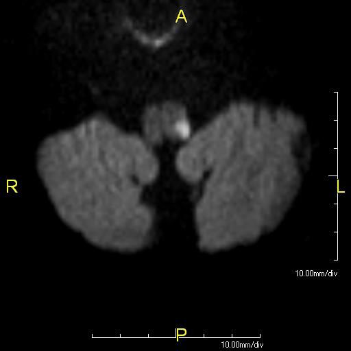
Overview
The eponymous brainstem syndromes are enduring because they map precisely onto the mediolateral perforator anatomy. The table below lists each classic syndrome with its level, offending artery, the structures involved and the resulting deficits. Textbooks vary somewhat on the Claude/Benedikt distinction and on whether Raymond spares the face; those caveats are noted.
| Syndrome | Level / region | Artery | Structures | Deficits |
|---|---|---|---|---|
| Wallenberg (lateral medullary) | Dorsolateral medulla | Intracranial vertebral (or PICA) | Spinal trigeminal nucleus/tract, spinothalamic tract, nucleus ambiguus (IX/X), vestibular nuclei, descending sympathetic fibres, inferior cerebellar peduncle, nucleus solitarius | Ipsilateral facial pain/temperature loss + contralateral body pain/temperature loss (crossed); ipsilateral Horner; dysphagia, hoarseness, dysarthria (palatal/vocal cord palsy); vertigo, nystagmus, lateropulsion; ipsilateral limb ataxia; loss of taste; hiccups. No limb weakness (pyramid spared) |
| Dejerine (medial medullary) | Medial medulla | Anterior spinal artery / vertebral paramedian | Pyramid (corticospinal tract), medial lemniscus, hypoglossal nucleus/nerve (XII) | Contralateral hemiparesis (face spared); contralateral vibration/proprioception loss; ipsilateral tongue weakness with deviation toward the lesion |
| Babinski–Nageotte | Hemimedulla (medial + lateral) | Vertebral | Combined medial + lateral medullary structures | Wallenberg features plus contralateral hemiparesis and posterior-column loss |
| Weber | Ventral midbrain (basis pedunculi) | PCA/basilar peduncular perforators | CN III fascicle + cerebral peduncle (corticospinal/corticobulbar) | Ipsilateral oculomotor palsy (ptosis, mydriasis, eye "down and out") + contralateral hemiparesis (including face) |
| Claude | Dorsal midbrain tegmentum | Paramedian midbrain perforators (basilar/P1) | CN III fascicle + red nucleus / superior cerebellar peduncle (decussation) | Ipsilateral III palsy + contralateral ataxia and incoordination (predominantly cerebellar, less hyperkinetic than Benedikt) |
| Benedikt | Midbrain tegmentum (deeper than Claude) | Paramedian perforators | CN III fascicle + red nucleus + spinothalamic tract ± medial lemniscus/SCP | Ipsilateral III palsy + contralateral involuntary movements (tremor, chorea/athetosis, ataxia) ± contralateral sensory loss |
| Nothnagel | Dorsal midbrain (tectum/tegmentum) | Perforators / mass lesions | CN III (uni/bilateral) + superior cerebellar peduncles / quadrigeminal plate | Uni- or bilateral III palsy with gaze paresis and cerebellar ataxia |
| Parinaud (dorsal midbrain / tectal) | Pretectum, posterior commissure | Top-of-basilar / posterior choroidal (or pineal mass) | Rostral interstitial MLF, posterior commissure, pretectal nuclei | Upgaze palsy, convergence–retraction nystagmus, light–near dissociation, lid retraction (Collier sign), impaired convergence |
| Millard–Gubler | Ventral (basal) pons | Basilar paramedian/short circumferential | CN VI and VII fascicles + corticospinal tract | Ipsilateral abducens palsy (esotropia) + ipsilateral peripheral facial palsy + contralateral hemiparesis |
| Foville | Dorsal caudal pontine tegmentum | Basilar paramedian perforators | CN VI/VII, PPRF/abducens nucleus, MLF, corticospinal tract (± descending sympathetic) | Ipsilateral conjugate horizontal gaze palsy + ipsilateral facial palsy + contralateral hemiparesis (± ipsilateral Horner) |
| Raymond | Ventral pons | Basilar paramedian | CN VI fascicle + corticospinal tract | Ipsilateral abducens palsy + contralateral hemiparesis (classically with facial sparing; some descriptions include contralateral central facial weakness) |
| Raymond–Cestan (upper pontine) | Rostral dorsal pontine tegmentum | Long circumferential (SCA territory) / basilar | Medial lemniscus, spinothalamic tract, MLF, superior/middle cerebellar peduncle ± CST | Contralateral hemisensory loss, ipsilateral limb ataxia, ± contralateral hemiparesis and INO ("rubral/upper pontine" picture) |
| Marie–Foix | Lateral pons | Long circumferential / AICA | Corticospinal tract, spinothalamic tract, middle cerebellar peduncle | Ipsilateral limb ataxia + contralateral hemiparesis + contralateral pain/temperature loss |
| Locked-in syndrome | Bilateral ventral pons | Basilar trunk occlusion | Bilateral corticospinal and corticobulbar tracts (basis pontis); tegmentum/reticular formation spared | Quadriplegia and anarthria/aphonia with preserved consciousness; communication only through preserved vertical gaze and blinking |
| Top-of-the-basilar | Rostral brainstem, thalami, occipitotemporal cortex | Embolic occlusion of the distal basilar | Midbrain tegmentum, medial thalami, PCA territories | Somnolence/coma, vertical gaze palsy and pupillary abnormalities, hemianopia/cortical blindness, peduncular hallucinosis, amnesia and confusion; often fluctuating |
| One-and-a-half syndrome | Dorsal pontine tegmentum | Paramedian perforator | Ipsilateral PPRF/abducens nucleus + ipsilateral MLF | Ipsilateral conjugate gaze palsy + INO; only preserved horizontal movement is abduction of the contralateral eye. Add ipsilateral facial palsy → "eight-and-a-half" |
Clinical pearls
- Wallenberg is the commonest brainstem stroke and is frequently caused by vertebral artery disease/dissection, not PICA occlusion itself; the striking preservation of limb strength distinguishes it from medial medullary stroke.
- Any oculomotor (III) palsy plus a contralateral long-tract sign is a midbrain (Weber/Benedikt/Claude) localisation; whether ataxia (Claude), hyperkinesia (Benedikt) or hemiparesis (Weber) dominates depends on how ventral vs dorsal the lesion sits.
- A ventral pontine picture with abducens ± facial palsy and crossed hemiparesis (Millard–Gubler/Foville/Raymond) should trigger urgent basilar imaging — these can herald evolving basilar occlusion and locked-in syndrome.
- Preserved vertical gaze and blink in an unresponsive quadriplegic patient is locked-in syndrome, not coma — always test voluntary vertical eye movements before declaring unresponsiveness.
Key references: Brazis PW, Masdeu JC, Biller J. Localization in Clinical Neurology, 7th ed. Caplan LR. Caplan's Stroke, 5th ed. Kim JS. Pure lateral medullary infarction: clinical–radiological correlation (Brain 2003). Bassetti C, et al. Medial medullary infarction (Neurology 1997). Silverman IE, et al. The crossed paralyses (Arch Neurol 1995).
Skull Base & Cavernous Sinus Syndromes; the Cerebellopontine Angle
Cavernous sinus — anatomy
The cavernous sinus is a paired dural venous channel flanking the sella. Running in its lateral wall, from superior to inferior, are cranial nerves III, IV, V1 and V2; passing within the sinus lumen, next to the cavernous ICA, are CN VI and the peri-carotid sympathetic plexus. This intimate arrangement explains the syndrome and its localising value.
Cavernous sinus syndrome
Lesions produce a painful ophthalmoplegia: variable palsies of III, IV and VI (often multiple and with pupil involvement), sensory loss over V1 ± V2, and — because the postganglionic sympathetic fibres travel on the ICA here — an ipsilateral Horner syndrome. The combination of a third-nerve palsy with a Horner (miosis) on the same side is a strong localiser to the cavernous sinus, since a compressive III palsy elsewhere would dilate, not constrict, the pupil. Proptosis, chemosis and an audible bruit suggest a carotid–cavernous fistula. Causes include cavernous ICA aneurysm, carotid–cavernous fistula, septic or aseptic cavernous sinus thrombosis, pituitary apoplexy, tumour (meningioma, metastasis, nasopharyngeal invasion) and the granulomatous Tolosa–Hunt syndrome.
Superior orbital fissure and orbital apex
| Syndrome | Nerves involved | Distinguishing feature |
|---|---|---|
| Cavernous sinus | III, IV, V1, (V2), VI + sympathetic | V2 involvement and Horner point here; vision usually spared early |
| Superior orbital fissure | III, IV, V1, VI (+ superior ophthalmic vein) | V2 spared (exits via foramen rotundum); optic nerve spared |
| Orbital apex (Rochon–Duvigneaud) | SOF contents + CN II | Visual loss distinguishes it from an isolated SOF syndrome |
Cerebellopontine angle (CPA) syndrome
The CPA is the cistern between the lateral pons/cerebellum and the petrous bone, traversed by cranial nerves V, VII and VIII (with IX below and VI medially) and by the AICA/labyrinthine artery. A mass or infarct here produces a recognisable, roughly sequential syndrome:
- VIII first — progressive unilateral sensorineural hearing loss, tinnitus and imbalance/vertigo (the earliest and commonest feature of a vestibular schwannoma).
- V — ipsilateral facial numbness and a depressed corneal reflex.
- VII — ipsilateral peripheral facial weakness and loss of taste (relatively late, as the nerve is resilient).
- Cerebellar/brainstem — ipsilateral limb ataxia and gaze-evoked nystagmus from peduncle/brainstem compression; late obstructive hydrocephalus with headache and papilloedema.
The commonest CPA masses are vestibular schwannoma (~80%), meningioma and epidermoid; the vascular counterpart is AICA territory infarction, which causes an acute CPA-like picture with deafness, vertigo, facial palsy, ipsilateral ataxia and Horner.
Other skull-base foramen syndromes
| Syndrome | Site | Nerves | Clinical clue |
|---|---|---|---|
| Gradenigo | Petrous apex | V (Gasserian ganglion) + VI | Retro-orbital pain + abducens palsy + otorrhoea (apical petrositis) |
| Vernet (jugular foramen) | Jugular foramen | IX, X, XI | Dysphagia, hoarseness, loss of gag, trapezius/SCM weakness |
| Collet–Sicard | Posterior retroparotid space | IX, X, XI, XII | Vernet + tongue hemiatrophy |
| Villaret | Retroparotid/retropharyngeal | IX, X, XI, XII + sympathetic | Collet–Sicard + Horner syndrome |
| Foster Kennedy | Olfactory groove / medial sphenoid | I, II | Ipsilateral optic atrophy + anosmia + contralateral papilloedema |
Clinical pearls
- Painful ophthalmoplegia + ipsilateral Horner = cavernous sinus until proven otherwise; image the sinus and carotid urgently (fistula, thrombosis, apoplexy, aneurysm).
- An acute stroke that causes deafness is almost always AICA (labyrinthine artery) — a crucial exception to the teaching that central lesions spare hearing.
- Progressive unilateral SNHL with an absent corneal reflex should prompt dedicated internal-auditory-canal MRI for a CPA schwannoma.
- V2 involvement separates a cavernous sinus lesion from a superior-orbital-fissure lesion; optic-nerve involvement marks the orbital apex.
Key references: Brazis PW, Masdeu JC, Biller J. Localization in Clinical Neurology, 7th ed. Kline LB, Hoyt WF. The Tolosa–Hunt syndrome (J Neurol Neurosurg Psychiatry 2001). Lee JH, et al. Cavernous sinus syndromes: imaging (RadioGraphics). Amarenco P, et al. AICA territory infarcts (Neurology).
Collateral Cerebral Circulation & Its Assessment
A tiered collateral system
Cerebral collaterals are recruited in a hierarchy determined by anatomic proximity and the time available. They determine how much brain survives per unit time after a large-vessel occlusion, and are now central to patient selection for reperfusion therapy.
| Tier | Pathway | Speed / character |
|---|---|---|
| Primary | Circle of Willis — anterior communicating (right ⇄ left ACA) and posterior communicating (carotid ⇄ vertebrobasilar) | Instantaneous; the main safeguard against a single ICA or proximal vertebral occlusion |
| Secondary — ophthalmic | ECA (facial/internal maxillary) → ophthalmic artery → ICA, with reversed ophthalmic flow | Recruited over minutes–hours in ICA occlusion; reversed flow is a marker of significant proximal disease |
| Secondary — leptomeningeal (pial) | Distal cortical anastomoses across the ACA–MCA and MCA–PCA borderzones; transdural (ECA→pial) connections | The decisive collateral in MCA occlusion; retrograde pial filling maintains penumbral cortex |
| Tertiary | Angiogenesis / neovascular "moyamoya" networks | Develops over weeks–months in chronic steno-occlusion |
Assessment and grading
Collateral status is inferred from vascular and perfusion imaging:
- Catheter angiography — ASITN/SIR collateral grade (0–4): 0 = none; 1 = slow collaterals to the periphery of the ischaemic bed; 2 = rapid collaterals to the periphery with persistent defect; 3 = slow but complete collateral filling by the late venous phase; 4 = complete and rapid retrograde collateral filling. This remains the reference standard.
- Single-phase CTA — regional leptomeningeal scores (e.g., Tan: good if >50% of the MCA territory fills; Maas; Miteff) comparing collateral vessels to the normal side.
- Multiphase CTA (mCTA, Menon/Calgary score, 0–5) — captures the temporal dynamics of retrograde filling across arterial, peak-venous and late-venous phases, adding delay information that single-phase imaging misses.
- CT perfusion — good collaterals manifest as a favourable core–penumbra mismatch (small rCBF<30% core, large Tmax>6s penumbra), the physiological correlate of robust pial flow.
- Hypoperfusion intensity ratio (Tmax>10s ÷ Tmax>6s volumes) is a surrogate — a low ratio indicates good collaterals.
Clinical relevance
- Robust collaterals slow infarct growth, sustain a "slow progressor" phenotype, enlarge the treatable time window (the anatomic basis of DAWN and DEFUSE-3 late-window selection), and predict better recanalisation, smaller final infarct and less haemorrhagic transformation after thrombectomy.
- Poor collaterals predict rapid core growth ("fast progressor"), futile reperfusion and higher haemorrhage risk.
- Congenital circle-of-Willis incompleteness (hypoplastic A1 or PComm) removes the primary collateral and raises the risk of borderzone and haemodynamic infarction during carotid occlusion or hypotension.
- In chronic ICA/MCA steno-occlusion, collateral adequacy (assessed with acetazolamide-challenge perfusion or breath-hold BOLD reactivity) helps identify exhausted cerebrovascular reserve and candidacy for revascularisation.
Key references: Liebeskind DS. Collateral circulation (Stroke 2003). Higashida RT, et al. ASITN/SIR collateral grading (Stroke 2003). Menon BK, et al. Multiphase CTA (Radiology 2015). Albers GW, et al. DEFUSE-3 (NEJM 2018). Nogueira RG, et al. DAWN (NEJM 2018).
Regulation of Cerebral Blood Flow & Autoregulation
Baseline flow and metabolic demand
The brain is ~2% of body mass yet receives ~15–20% of cardiac output and consumes ~20% of resting oxygen. Global cerebral blood flow (CBF) averages about 50 mL/100 g/min, partitioned as roughly 80 mL/100 g/min in grey matter and 20 mL/100 g/min in white matter. Perfusion is driven by the cerebral perfusion pressure (CPP = mean arterial pressure − intracranial pressure) and governed by cerebrovascular resistance, which the brain actively controls.
Ischaemic thresholds — the penumbra concept
| CBF (mL/100 g/min) | State | Consequence |
|---|---|---|
| ~50 | Normal | Full function |
| ~20–35 | Oligaemia | Reduced protein synthesis; still functional |
| ~15–20 | Electrical failure (penumbra threshold) | Loss of evoked potentials/EEG; neurons silent but viable — salvageable tissue |
| ~10–12 or below | Membrane/ionic failure | Loss of ion homeostasis, cytotoxic oedema, irreversible infarction (the core) |
Between the electrical-failure and membrane-failure thresholds lies the ischaemic penumbra — functionally silent but structurally intact tissue whose fate depends on the depth and duration of hypoperfusion, and which is the therapeutic target of reperfusion.
Autoregulation
Cerebral autoregulation maintains a nearly constant CBF across a plateau of CPP/MAP of roughly 60–150 mmHg by adjusting arteriolar tone: pressure rises cause reflex vasoconstriction and falls cause vasodilatation. Below the lower limit, vessels are maximally dilated and flow becomes pressure-passive (ischaemia); above the upper limit, forced dilatation causes hyperperfusion, blood–brain-barrier breakdown and oedema (hypertensive encephalopathy, PRES). Three interacting mechanisms mediate the plateau:
- Myogenic (the Bayliss effect) — smooth muscle contracts in response to increased transmural pressure/stretch; the fastest component.
- Metabolic — local accumulation of CO2/H+, adenosine, K+, and nitric oxide couples flow to neuronal activity (neurovascular coupling at the neurovascular unit of neuron–astrocyte–pericyte–endothelium).
- Neurogenic — perivascular sympathetic, parasympathetic and sensory innervation modulate tone and shift the autoregulatory limits.
Chemical regulation — CO2 and O2 reactivity
CBF is exquisitely sensitive to arterial CO2: within the physiological range flow changes roughly 3–4% per mmHg PaCO2 — hypercapnia is a potent vasodilator (raising CBF and, transiently, ICP) and hypocapnia a vasoconstrictor (the rationale, and the hazard, of hyperventilation for acute intracranial hypertension). Hypoxaemia increases CBF only below a PaO2 of roughly 50–60 mmHg. These responses underpin cerebrovascular reactivity testing (acetazolamide or breath-hold challenge) to assess reserve.
Autoregulation in disease and its stroke relevance
- Chronic hypertension shifts the entire autoregulatory curve to the right, so chronically hypertensive patients tolerate higher pressures but decompensate (become ischaemic) at "normal" pressures that would be well tolerated by others — a caution against rapid blood-pressure lowering.
- In and around acute infarction autoregulation is impaired or abolished; perfusion of the penumbra becomes pressure-passive, which is the physiological justification for permissive hypertension (avoiding hypotension that would collapse collateral flow) and for cautious, gradual BP reduction.
- After reperfusion, loss of autoregulation predisposes to hyperperfusion injury and haemorrhagic transformation; this drives post-thrombectomy/post-endarterectomy blood-pressure targets.
- The Cushing response (hypertension, bradycardia, irregular respiration) is a late reflex to critically raised ICP as CPP is defended.
Clinical pearls
- CPP, not MAP alone, drives perfusion — a "normal" blood pressure with a high ICP can be ischaemic; manage both.
- Do not aggressively normalise blood pressure in acute ischaemic stroke with a persisting occlusion or tight stenosis — the ischaemic bed is pressure-passive and collateral-dependent.
- Hyperventilation lowers ICP through CO2-mediated vasoconstriction but at the cost of reduced CBF; use it only as a brief bridge to definitive treatment.
Key references: Astrup J, Siesjö BK, Symon L. Thresholds in cerebral ischemia — the ischemic penumbra (Stroke 1981). Lassen NA. Cerebral blood flow and oxygen consumption in man (Physiol Rev 1959). Paulson OB, Strandgaard S, Edvinsson L. Cerebral autoregulation (Cerebrovasc Brain Metab Rev 1990). Powers WJ. Cerebral hemodynamics in ischemic cerebrovascular disease.
Subarachnoid Cisterns & the Cerebral Ventricular System
The subarachnoid space and its cisterns
Cerebrospinal fluid (CSF) circulates in the subarachnoid space between the arachnoid and pia. Where the brain's surface falls away from the arachnoid, the space widens into cisterns — CSF reservoirs that also transmit the major arteries and cranial nerves, so their anatomy is inseparable from aneurysm location and subarachnoid-haemorrhage (SAH) patterns.
| Cistern | Location | Key contents |
|---|---|---|
| Interpeduncular | Between the cerebral peduncles | Basilar apex and PCA/SCA origins, CN III, posterior perforating arteries |
| Suprasellar / chiasmatic | Above the sella, around the optic chiasm | Optic chiasm/nerves, ICA termini, ACA (A1) and AComm complex, pituitary stalk |
| Prepontine | Anterior to the pons | Basilar trunk, CN VI |
| Cerebellopontine (angle) | Lateral pontomedullary junction | CN V, VII, VIII; AICA |
| Cisterna magna (cerebellomedullary) | Between the medulla and cerebellum, below the vermis | Tonsils, vertebral arteries, PICA; common site for cisternal puncture |
| Ambient (perimesencephalic) | Lateral to the midbrain | PCA (P2), SCA, basal vein of Rosenthal, CN IV |
| Quadrigeminal (superior) | Behind the tectal plate | Vein of Galen, pineal, quadrigeminal plate; PCA/SCA branches |
| Sylvian (lateral) | Within the lateral fissure | MCA (M1–M2), middle cerebral vein |
| Lamina terminalis | Anterior to the third ventricle | AComm complex; a fenestration target in hydrocephalus surgery |
Because ~85% of saccular aneurysms arise on the anterior circle of Willis (AComm, PComm origin, MCA bifurcation), aneurysmal SAH fills the basal cisterns in a recognisable, often lateralising pattern. A peri-mesencephalic (pretruncal) pattern of blood confined to the interpeduncular/ambient cisterns with a negative angiogram is the benign, presumed-venous, non-aneurysmal variant. Blood in the cisterns also obstructs CSF flow and arachnoid resorption, producing acute or communicating hydrocephalus.
The ventricular system
CSF flows through four communicating ventricular cavities:
- Paired lateral ventricles, each with a frontal (anterior) horn, body, atrium/trigone, occipital (posterior) horn and temporal (inferior) horn — the largest reservoirs, housing the bulk of the choroid plexus.
- Each drains through an interventricular foramen of Monro into the midline third ventricle (bounded by the thalami, with the massa intermedia).
- The cerebral aqueduct (of Sylvius) conducts CSF through the midbrain to the fourth ventricle (between pons/medulla and cerebellum).
- From the fourth ventricle, CSF exits into the subarachnoid space through the paired lateral foramina of Luschka and the median foramen of Magendie.
CSF dynamics
Total CSF volume is ~150 mL; it is produced mainly by the choroid plexus at ~500 mL/day (turned over several times daily) and absorbed chiefly at the arachnoid granulations into the superior sagittal sinus, with a contribution from meningeal/glymphatic lymphatic pathways. Obstruction anywhere along the pathway causes non-communicating (obstructive) hydrocephalus (e.g., aqueductal stenosis, a fourth-ventricular mass); impaired resorption at the granulations causes communicating hydrocephalus (e.g., post-SAH, meningitis).
Stroke-specific relevance
- Cerebellar infarction or haemorrhage can compress the fourth ventricle and aqueduct, causing acute obstructive hydrocephalus and upward/tonsillar herniation — an emergency requiring EVD and/or suboccipital decompression.
- Intraventricular haemorrhage (from deep hypertensive ICH or aneurysm rupture) obstructs CSF flow and coats the granulations, causing hydrocephalus; casting of the fourth ventricle is especially dangerous.
- Aneurysmal SAH causes both early obstructive and delayed communicating hydrocephalus; serial ventricular size guides EVD and shunting.
- Recognising the perimesencephalic pattern spares patients repeated angiography and predicts an excellent prognosis.
Key references: Rhoton AL. The cerebral veins / the lateral and third ventricles / the posterior fossa cisterns (Neurosurgery 2002). Standring S. Gray's Anatomy, 42nd ed. van Gijn J, et al. Perimesencephalic nonaneurysmal SAH (Lancet Neurol). Blumenfeld H. Neuroanatomy through Clinical Cases, 3rd ed.
Blood Supply of the Spinal Cord
Longitudinal arterial trunks
Three longitudinal arteries run the length of the cord. A single midline anterior spinal artery (ASA), formed by paired branches of the intracranial vertebral arteries that unite near the foramen magnum, descends in the anterior median fissure and supplies the anterior two-thirds of the cord via central (sulcal) branches. The paired posterior spinal arteries (PSA), arising from the vertebral arteries or PICA, run along the posterolateral sulci and supply the posterior one-third — critically, the dorsal columns. A pial vasocorona anastomoses the anterior and posterior systems around the cord surface.
Segmental reinforcement and the great radicular artery
The longitudinal trunks are too small to perfuse the whole cord alone and are reinforced by radicular (segmental medullary) arteries derived from the vertebral, ascending/deep cervical, intercostal and lumbar arteries. The largest and most important is the artery of Adamkiewicz (arteria radicularis magna), which most often arises on the left between T9 and T12 and provides the dominant supply to the lumbar enlargement. The sulcal (central) arteries branching from the ASA supply alternate (right/left) halves of the cord and are functional end-arteries.
Watershed vulnerability
Because segmental reinforcement is unevenly distributed, the cord has a mid-thoracic watershed (approximately T4–T8) between the territory of the upper (cervical/upper-intercostal) feeders and the Adamkiewicz-dependent lower cord. This zone is the most vulnerable to global hypoperfusion — aortic cross-clamping/surgery, aortic dissection, profound hypotension and cardiac arrest — producing thoracic cord infarction. The anterior (ASA) territory is far more susceptible than the posterior, both because sulcal arteries are end-arteries and because a single ASA supplies a large territory.
Clinical spinal cord vascular syndromes
| Syndrome | Territory | Deficits |
|---|---|---|
| Anterior spinal artery syndrome | Anterior two-thirds | Acute para-/quadriplegia (corticospinal tracts), loss of pain and temperature below the level (spinothalamic), and autonomic dysfunction (bladder, bowel, blood pressure); dorsal columns spared — vibration and proprioception preserved (dissociated sensory loss). Often preceded by acute interscapular/back pain |
| Posterior spinal artery syndrome (rare) | Posterior one-third | Loss of vibration and proprioception below the level, sensory ataxia; relative sparing of strength and pain/temperature |
| Central (sulcal) / watershed cord infarct | Mid-thoracic anterior cord | Level-dependent paraparesis with dissociated sensory loss; classic after aortic surgery/hypotension |
Causes of cord ischaemia include aortic disease and surgery (the commonest), atherosclerosis and thromboembolism, vertebral/aortic dissection, systemic hypotension, vasculitis, decompression sickness, fibrocartilaginous embolism and, iatrogenically, aortic stent-grafting and interventional embolisation. MRI shows a longitudinally extensive anterior "owl's-eye" (or "snake-eye") T2 signal in the anterior horns on axial imaging; DWI restriction supports acute infarction.
Venous drainage (brief)
The cord drains via anterior and posterior spinal veins into radicular veins and the epidural venous plexus. Venous congestion (as in spinal dural arteriovenous fistula) produces a progressive myelopathy that is a stroke mimic and an important, treatable differential of subacute cord dysfunction.
Clinical pearls
- An acute painful para-/tetraplegia with preserved vibration/joint-position sense but lost pain/temperature is anterior spinal artery syndrome until proven otherwise — think aortic pathology and hypotension.
- The mid-thoracic cord is the "critical zone" for perioperative cord infarction; intraoperative MAP support and CSF drainage are protective during aortic surgery.
- The Adamkiewicz artery's variable (usually left, T9–T12) origin must be identified before thoracoabdominal aortic and spinal interventional procedures.
- Consider spinal dural AV fistula in a progressive lower-cord myelopathy with dilated perimedullary flow voids — venous, not arterial, but a mimic of cord ischaemia.
Key references: Novy J, et al. Spinal cord ischemia: clinical and imaging patterns (Arch Neurol 2006). Cheshire WP, et al. Spinal cord infarction (Neurology 1996). Standring S. Gray's Anatomy, 42nd ed. Vuong SM, et al. Vascular anatomy of the spinal cord.
Embryology of the Cerebral Circulation (Brief)
Origins from the aortic arches
The cranial arterial supply derives from the paired dorsal aortae and the pharyngeal (aortic) arch arteries. The third aortic arch and the cranial dorsal aorta form the common carotid and the proximal internal carotid; the fourth arch forms the arch of the aorta on the left and the proximal right subclavian on the right; the sixth arch gives the pulmonary arteries and the ductus arteriosus. The vertebral arteries develop from longitudinal anastomoses (post-costal anastomoses) linking the cervical intersegmental arteries, most of which regress leaving the seventh cervical intersegmental artery as the definitive origin from the subclavian. The transient stapedial artery (from the second/hyoid arch) normally regresses; its territory is assumed by the middle meningeal artery, and its persistence is a rare middle-ear variant.
Formation of the vertebrobasilar system and circle of Willis
Early in the fourth week the hindbrain is supplied by paired longitudinal neural arteries, which progressively fuse in the midline to form the single basilar artery; incomplete fusion leaves the fenestrations seen in adults. The circle of Willis is assembled between roughly the fifth and seventh weeks as the anterior and posterior communicating arteries connect the developing carotid and vertebrobasilar trees; asymmetric involution of its components accounts for the frequent adult hypoplasias and the strong link between A1/PComm hypoplasia and aneurysm formation.
The transient carotid–basilar anastomoses
Before the posterior communicating and vertebral arteries mature, the primitive internal carotid supplies the hindbrain circulation through four temporary channels, which normally regress in a cranial-to-caudal sequence. Their persistence produces the named variants encountered on adult angiography:
| Embryonic vessel | Connection | Persistent adult variant |
|---|---|---|
| Primitive trigeminal artery | Cavernous ICA → basilar | Persistent trigeminal artery (the commonest persistent anastomosis) |
| Primitive otic (acoustic) artery | Petrous ICA → basilar | Persistent otic artery (rarest) |
| Primitive hypoglossal artery | Cervical ICA → basilar (via hypoglossal canal) | Persistent hypoglossal artery |
| Proatlantal intersegmental artery | Cervical ICA/ECA → vertebral artery | Persistent proatlantal artery |
Development of the PCA and the fetal variant
The posterior cerebral artery is initially fed by the internal carotid through the posterior communicating artery; only later does the definitive P1 segment develop from the basilar apex to become the dominant supply. Failure of this transition explains the common adult fetal-type PCA, in which the carotid retains dominant supply to the occipital lobe through a large PComm with a hypoplastic P1 — anatomically a "retained embryonic pattern."
Clinical relevance
- Persistent carotid–basilar anastomoses are usually incidental but matter for aneurysm risk, for interpreting collateral flow, and as hazards during skull-base and endovascular procedures.
- Basilar and vertebral fenestrations — footprints of incomplete embryonic fusion — are prone to aneurysm formation at the proximal fusion point.
- The embryonic PCA-from-carotid pattern (fetal PCA) redistributes occipital stroke risk to the anterior circulation.
- Asymmetric involution underlies the everyday circle-of-Willis variants that govern collateral reserve and aneurysm distribution.
Key references: Padget DH. The development of the cranial arteries in the human embryo (Contrib Embryol 1948). Luh GY, et al. Persistent fetal carotid–vertebrobasilar anastomoses (AJR 1999). Menshawi K, et al. A functional perspective on cerebral arterial embryology. Standring S. Gray's Anatomy, 42nd ed.
Oculomotor Nerve (CN III) — Palsy, Pupil-Involving vs Pupil-Sparing
Anatomy
The oculomotor nuclear complex lies in the rostral midbrain at the level of the superior colliculus, ventral to the periaqueductal grey and cerebral aqueduct, dorsomedial to the red nucleus. Its internal organisation is decisive for localisation. The paired somatic subnuclei innervate the ipsilateral medial rectus (MR), inferior rectus (IR) and inferior oblique (IO); the superior rectus (SR) subnucleus is crossed, its axons decussating to innervate the contralateral SR; and a single midline central caudal nucleus supplies both levator palpebrae superioris muscles. Rostrally sit the Edinger–Westphal (visceral) subnuclei, whose preganglionic parasympathetic fibres drive pupillary constriction and accommodation. Consequently a discrete nuclear lesion produces bilateral ptosis and bilateral SR weakness superimposed on an ipsilateral third-nerve pattern — a signature no peripheral lesion reproduces.
Fascicles sweep ventrally through the red nucleus, substantia nigra and medial cerebral peduncle before emerging in the interpeduncular fossa. The nerve then runs forward in the subarachnoid space between the posterior cerebral and superior cerebellar arteries, lateral and parallel to the posterior communicating artery (PCoM) — the anatomical basis of aneurysmal compression. It pierces the dura, traverses the lateral wall of the cavernous sinus (above CN IV), and enters the orbit through the superior orbital fissure, dividing into a superior division (SR, levator) and an inferior division (MR, IR, IO, and the parasympathetic root to the ciliary ganglion).
Function
CN III mediates elevation (SR, IO), depression (IR), adduction (MR), incyclotorsion counterbalance, upper-lid elevation (levator) and, via the parasympathetic outflow, pupillary constriction and lens accommodation. Loss produces the classic “down-and-out” eye from unopposed lateral rectus (CN VI) and superior oblique (CN IV), with complete ptosis and, when the pupil is involved, a fixed dilated pupil.
Examination
Assess lid position, resting ocular alignment, ductions in all cardinal directions, and pupillary size/reactivity. The pupillomotor fibres run in the superomedial periphery of the nerve, supplied by pial vessels, whereas the somatic core is nourished by penetrating vasa nervorum. This microanatomy underlies the pupil rule: external compression tends to involve the pupil early, while microvascular ischaemia infarcts the core and spares it. A partial or evolving palsy demands serial pupil checks, since compressive lesions can declare pupil involvement over hours.
Lesion localization & causes
| Site | Localising features | Representative causes |
|---|---|---|
| Nuclear | Ipsilateral CN III palsy + bilateral ptosis + contralateral SR weakness | Midbrain infarct, haemorrhage, tumour |
| Fascicular | Ipsilateral CN III + crossed long-tract/cerebellar signs (named syndromes below) | Paramedian midbrain infarct, demyelination |
| Subarachnoid | Isolated CN III, often pupil-involving; pain frequent | PCoM aneurysm, uncal herniation, meningitis |
| Cavernous sinus / superior orbital fissure | CN III with IV, VI, V1 ± Horner; painful | Carotid–cavernous fistula, pituitary apoplexy, Tolosa–Hunt, thrombosis, metastasis |
| Orbital apex | Above + optic neuropathy (CN II) | Orbital pseudotumour, fungal (mucor/aspergillus), tumour |
Midbrain fascicular syndromes: Weber (CN III + contralateral hemiparesis, cerebral peduncle); Benedikt (CN III + contralateral tremor/involuntary movements, red nucleus); Claude (CN III + contralateral cerebellar ataxia, red nucleus/superior cerebellar peduncle); Nothnagel (CN III + gaze palsy + ataxia, tectum).
| Feature | Compressive (aneurysm, herniation, tumour) | Ischaemic (diabetic/hypertensive microvascular) |
|---|---|---|
| Pupil | Typically involved (dilated, poorly reactive) | Typically spared — core infarcted, pial fibres perfused |
| Pain | Often present, can be severe (aneurysm) | Mild peri-orbital ache possible |
| Onset/course | Progressive or apoplectic; may worsen | Acute, then recovers over 8–12 weeks |
| Aberrant regeneration | Can occur (compressive/congenital) | Never — if present, exclude ischaemia |
| Imperative action | Urgent CTA/MRA/DSA for aneurysm | Vascular risk-factor control; image if atypical |
Clinical pearls
- Pupil-involving CN III palsy is a neurosurgical emergency until a PCoM aneurysm is excluded, regardless of how “typical” the ptosis and gaze deviation appear.
- The pupil rule is not absolute: up to a fifth of compressive palsies are initially pupil-sparing and a minority of ischaemic palsies show partial pupil involvement. In any patient <50, or with incomplete/partial motor deficit, image regardless of pupil.
- Aberrant regeneration (lid elevation on downgaze/adduction, pupil constriction on adduction) indicates a chronic compressive or congenital lesion and excludes an ischaemic aetiology — a red flag for meningioma or slowly-expanding aneurysm.
- A dilated pupil with an otherwise normal eye and no ptosis or motility deficit is almost never CN III palsy — think tonic (Adie) pupil or pharmacological mydriasis.
Key references: Miller & Newman, Walsh & Hoyt's Clinical Neuro-Ophthalmology; Kerr & Hollenhorst pupillomotor topography; Leigh & Zee, The Neurology of Eye Movements (6th ed.).
Trochlear Nerve (CN IV)
Anatomy
The trochlear nerve is unique in three respects that dominate its clinical behaviour: it is the only cranial nerve to emerge from the dorsal brainstem, the only one that is fully decussated (each nucleus innervates the contralateral superior oblique), and the thinnest and longest-coursing cranial nerve. Its nucleus lies in the caudal midbrain at the inferior colliculus, just below the CN III complex, ventral to the aqueduct. Fascicles course dorsally and caudally around the periaqueductal grey, decussate in the anterior medullary velum, and exit dorsally below the inferior colliculus. The nerve then wraps around the midbrain, runs beneath the tentorial edge, enters the lateral wall of the cavernous sinus (below CN III, above V1), and passes through the superior orbital fissure outside the annulus of Zinn to reach the superior oblique.
Function
The superior oblique, acting through the trochlea, produces intorsion (its primary action in primary gaze), depression (greatest in adduction) and abduction. Depression in adduction is the action most readily tested clinically. Palsy therefore causes vertical and torsional diplopia that is worst on downgaze and on gaze away from the affected side — classically noticed when reading or descending stairs.
Examination
The hallmark is a hypertropia of the affected eye that increases on contralateral gaze and on ipsilateral head tilt. The Parks–Bielschowsky three-step test localises a vertical-muscle palsy: (1) which eye is higher; (2) is the hypertropia worse on left or right gaze; (3) is it worse on left or right head tilt. A superior oblique palsy gives a hyper that worsens on contralateral gaze and ipsilateral tilt. Patients characteristically adopt a compensatory contralateral head tilt with chin depression to fuse; an old photograph showing a longstanding head tilt suggests a decompensated congenital palsy. Measured excyclotorsion (double Maddox rod) supports acquired bilateral palsies.
Lesion localization & causes
| Site | Clues | Causes |
|---|---|---|
| Nuclear/fascicular (dorsal midbrain) | Contralateral SO palsy; may accompany Horner (descending sympathetic) or contralateral RAPD (brachium of superior colliculus) | Midbrain infarct, haemorrhage, demyelination |
| Subarachnoid (around tentorial edge) | Isolated palsy; vulnerable to head trauma given long course | Closed head injury (often bilateral), tumour, hydrocephalus |
| Cavernous sinus | With CN III, VI, V1, Horner | Fistula, thrombosis, tumour, inflammation |
The two commonest acquired causes are closed head trauma (the dorsal exit and long subarachnoid course make it exquisitely vulnerable; contrecoup against the tentorium may produce bilateral palsy) and microvascular ischaemia in older vasculopaths. Congenital/decompensated palsy is the single most common overall and is confirmed by large vertical fusional amplitudes and old photographs.
Differential diagnosis
Other causes of vertical diplopia must be excluded before attributing it to CN IV: skew deviation (central, part of the ocular tilt reaction — comitant, with excyclotorsion of the lower eye and other posterior-fossa signs), thyroid eye disease (restrictive IR, positive forced ductions), myasthenia gravis (fatigable, variable), orbital fracture with IR entrapment, and Brown syndrome. Bilateral SO palsy produces a “V-pattern” esotropia, right hyper on left gaze/left hyper on right gaze, and >10° of excyclotorsion.
Clinical pearls
- An isolated SO palsy that reverses on ipsilateral vs contralateral tilt and shows large vertical fusional range is congenital until proven otherwise — hunt for old photographs.
- Trauma severe enough to cause a unilateral CN IV palsy is often severe enough to cause a bilateral one; always test the fellow eye's torsion.
- A comitant vertical deviation with torsion of the hypotropic (lower) eye points to skew deviation, not CN IV — look for INO, gaze palsy or other brainstem signs and image the posterior fossa.
Key references: Leigh & Zee, The Neurology of Eye Movements; von Noorden, Binocular Vision and Ocular Motility; Brazis, Localization in Clinical Neurology.
Abducens Nerve (CN VI)
Anatomy
The abducens nucleus lies in the caudal pons beneath the floor of the fourth ventricle, encircled by the genu of the facial nerve (the facial colliculus). Critically, the nucleus is not a simple lower motor pool: it contains both motor neurons to the ipsilateral lateral rectus and internuclear neurons whose axons cross and ascend in the contralateral medial longitudinal fasciculus (MLF) to the medial rectus subnucleus of CN III. The abducens nucleus is therefore the final common pathway for conjugate horizontal gaze: a nuclear lesion causes an ipsilateral gaze palsy (both eyes fail to look toward the side of the lesion), not merely an abduction deficit. Fascicles pass ventrally through the pontine tegmentum and corticospinal tract to exit at the pontomedullary junction.
The nerve then ascends the clivus in the subarachnoid space, makes a sharp bend over the petrous apex, passes beneath the petroclinoid (Gruber's) ligament through Dorello's canal, and enters the cavernous sinus. Uniquely, within the cavernous sinus it runs free in the body of the sinus, lateral to the internal carotid artery, rather than embedded in the lateral wall — hence it is often the first nerve affected by cavernous pathology and lies immediately adjacent to the carotid sympathetic plexus (explaining CN VI palsy with a postganglionic Horner).
Function & examination
The lateral rectus abducts the eye; palsy produces an esotropia worse at distance and on ipsilateral gaze, with horizontal binocular diplopia and a compensatory face turn toward the paretic side. Test abduction, quantify the deviation at near and distance, and look for accompanying signs (facial weakness, hearing loss, Horner, papilloedema) that localise beyond the nerve itself.
Lesion localization & causes
| Site | Associated signs | Causes |
|---|---|---|
| Nuclear (caudal pons) | Ipsilateral horizontal gaze palsy (not isolated abduction loss) ± ipsilateral facial palsy | Pontine infarct, MS, tumour |
| Fascicular | Foville (+ ipsilateral VII, Horner, gaze palsy); Millard–Gubler (+ ipsilateral VII, contralateral hemiparesis) | Paramedian pontine infarct, glioma |
| Subarachnoid / clivus | Isolated palsy; a classic false-localising sign of raised ICP | Raised ICP, meningitis, clival tumour, aneurysm |
| Petrous apex | Gradenigo syndrome: VI + ipsilateral facial pain (V) + otorrhoea/deafness | Petrous apicitis, complicated otitis media, cholesteatoma |
| Cavernous sinus | With III, IV, V1, Horner; VI often affected first | Carotid–cavernous fistula, thrombosis, tumour, apoplexy |
| Orbit | Proptosis, restriction (mimics: forced ductions positive) | Tumour, thyroid eye disease, pseudotumour |
Differential diagnosis
Pseudo-abducens palsy and non-neurogenic esotropia must be excluded: thyroid eye disease with a tight medial rectus (positive forced ductions, restriction rather than weakness), myasthenia gravis (fatigable, variable, may mimic any pattern), medial orbital wall fracture with MR entrapment, spasm of the near reflex, Duane retraction syndrome (congenital, with globe retraction and palpebral narrowing on adduction), and convergence spasm in functional presentations.
Clinical pearls
- Isolated CN VI palsy is the great false-localising sign: raised ICP of any cause (IIH, tumour, CVST, post-LP intracranial hypotension) stretches the nerve over the petrous ridge. Check for papilloedema and image with venography in mind.
- A pontine gaze palsy is not a CN VI palsy. If both eyes fail to move past midline toward one side, the lesion is at the abducens nucleus/PPRF, not the nerve.
- A microvascular CN VI palsy in a vasculopath should recover in 8–12 weeks. Failure to recover, progression, or additional signs mandates imaging (MRI with contrast ± MRV) — do not accept “diabetic sixth” uncritically, especially in children (pontine glioma) or with any red flags.
- Bilateral CN VI palsies suggest raised ICP, clival/skull-base disease, Wernicke encephalopathy, or a paraneoplastic/inflammatory process.
Key references: Leigh & Zee, The Neurology of Eye Movements; Brazis, Localization in Clinical Neurology; Miller & Newman, Walsh & Hoyt.
Approach to Diplopia & the Ocular Motor Cranial Nerves
Framework
Diplopia is a localising symptom that can be dissected at the bedside with a disciplined sequence. First establish whether it is monocular (persists with the fellow eye covered — almost always ocular/refractive: dry eye, astigmatism, cataract, rarely cortical polyopia) or binocular (resolves when either eye is covered — an ocular misalignment). Binocular diplopia is then characterised by direction (horizontal, vertical, oblique/torsional), the gaze position of maximal separation (the image is most peripheral in the field of action of the paretic muscle, and the outer image belongs to the paretic eye), variability/fatigability, and accompanying neurological signs.
Localisation logic
Work anatomically from muscle → neuromuscular junction → nerve → nucleus → supranuclear pathways. Three questions rapidly narrow the field: (1) Is a single muscle/nerve implicated, or a pattern crossing nerve territories (suggesting orbit, cavernous sinus, or NMJ)? (2) Is the pupil, lid, or proptosis involved? (3) Are there fatigability, pain, or other cranial-nerve/long-tract signs? Multiple non-contiguous ocular motor nerves without a clear anatomical grouping should raise myasthenia gravis — the universal mimic that respects no anatomical boundary and spares the pupil.
| Pattern | Deviation | Key discriminators | Localisation |
|---|---|---|---|
| CN III palsy | Down-and-out, ptosis | ± Pupil (compressive vs ischaemic); aberrant regeneration = compressive | Nucleus → orbit; PCoM aneurysm if pupil-involving |
| CN IV palsy | Hyper, worse downgaze & contralateral gaze | Three-step test; ipsilateral tilt worsens; excyclotorsion; head tilt | Dorsal midbrain, long subarachnoid course (trauma) |
| CN VI palsy | Eso, worse distance & ipsilateral gaze | Face turn; false-localising with raised ICP | Pons → Dorello's canal → cavernous sinus |
| Internuclear ophthalmoplegia | Adduction lag + abducting nystagmus | Convergence may be spared; often bilateral in MS | MLF (pons/midbrain) |
| Gaze palsy | Both eyes fail to move to one side | Overcome by VOR (supranuclear) vs not (pontine) | FEF/PPRF/abducens nucleus |
| Skew deviation | Vertical, often comitant | Part of ocular tilt reaction; other brainstem signs | Otolithic/graviceptive pathway, posterior fossa |
| Myasthenia gravis | Any/variable | Fatigable, variable, pupil-spared; ptosis, orbicularis weakness | Neuromuscular junction |
| Thyroid eye disease | Restrictive (often IR then MR) | Positive forced ductions, proptosis, lid retraction/lag | Orbit (restrictive myopathy) |
Red flags mandating urgent imaging
- Any pupil-involving CN III palsy (aneurysm) → emergent CTA/MRA/DSA.
- Multiple ocular motor nerves in one orbit/cavernous sinus, or with V1–V2 or optic-nerve involvement.
- Diplopia with papilloedema, severe/thunderclap headache, proptosis, or systemic signs of GCA in an older patient.
- Progressive or non-resolving deficit, or any deficit in a child.
Clinical pearls
- The outer image belongs to the paretic eye, and separation is maximal in the direction of the weak muscle — a red-glass or cover test formalises this at the bedside.
- Comitant deviations (equal in all directions) suggest longstanding strabismus or skew; incomitant deviations suggest a discrete nerve or muscle lesion.
- Fatigable, variable, or bizarrely combined diplopia that spares the pupil is myasthenia until proven otherwise — ice pack, rest, edrophonium (historical), acetylcholine-receptor and MuSK antibodies, and repetitive stimulation/single-fibre EMG.
- Painful ophthalmoplegia narrows to cavernous sinus/orbital apex pathology (fistula, thrombosis, Tolosa–Hunt, tumour, invasive fungal infection in the immunocompromised or diabetic) — a genuine emergency.
Key references: Kline & Bajandas, Neuro-Ophthalmology Review Manual; Leigh & Zee; AAO Basic and Clinical Science Course, Neuro-Ophthalmology.
Optic Nerve (CN II) — Anatomy & Visual Field Localization
Anatomy
The afferent visual pathway begins at retinal photoreceptors, whose signal is relayed through bipolar cells to retinal ganglion cells whose axons form the retinal nerve fibre layer, converge at the optic disc, and become the optic nerve. The nerve has four segments — intraocular (the myelinated portion begins behind the lamina cribrosa), intra-orbital (25–30 mm, slack to allow eye movement), intracanalicular (fixed within the optic canal, vulnerable to trauma and compression), and intracranial — before reaching the chiasm. As a central-nervous-system tract it is invested by meninges and CSF (hence disc swelling with raised ICP) and, once damaged, does not regenerate.
At the chiasm, nasal retinal fibres (temporal visual field) decussate while temporal retinal fibres (nasal field) remain uncrossed. Inferonasal fibres historically described as looping into the contralateral optic nerve (Wilbrand's knee — an anatomically debated structure) underlie the junctional scotoma. Beyond the chiasm the optic tract carries contralateral hemifield information to the lateral geniculate nucleus (LGN); post-geniculate optic radiations split into Meyer's loop (temporal lobe, superior contralateral field) and the parietal projection (inferior contralateral field), terminating in the striate cortex (V1) around the calcarine fissure, where the macula is represented posteriorly at the occipital pole with a large cortical magnification.
Function & examination
Test visual acuity, colour vision (disproportionately affected in optic neuropathy — red desaturation, Ishihara plates), pupillary responses (a relative afferent pupillary defect is the objective sign of asymmetric optic-nerve/retinal disease), confrontation and formal (automated or Goldmann) perimetry, and fundoscopy for disc pallor, swelling, or cupping. The optic nerve conveys the afferent limb of the pupillary light reflex; a purely afferent lesion produces an RAPD without anisocoria.
Lesion localization & causes
| Location | Visual field defect | Representative causes |
|---|---|---|
| Retina/nerve fibre layer | Arcuate scotoma, altitudinal defect, central scotoma; respects horizontal raphe | Glaucoma, AION, BRAO, retinal detachment |
| Optic nerve | Monocular loss + RAPD; central scotoma or altitudinal defect | Optic neuritis, ION, compression, toxic/nutritional, hereditary |
| Optic nerve–chiasm junction | Junctional scotoma: ipsilateral central scotoma + contralateral superotemporal defect | Parasellar/optic-nerve compressive lesion |
| Chiasm | Bitemporal hemianopia (superior-predominant with inferior compression, e.g. pituitary) | Pituitary adenoma, craniopharyngioma, meningioma, aneurysm |
| Optic tract | Incongruous homonymous hemianopia + contralateral RAPD + “bow-tie” disc atrophy | Tumour, demyelination, infarct |
| LGN | Incongruous / sectoranopic homonymous defects (lateral choroidal vs anterior choroidal supply) | Thalamic/choroidal-artery infarct, AVM |
| Temporal radiation (Meyer's loop) | Contralateral superior quadrantanopia (“pie in the sky”) | Temporal lobe tumour/surgery, infarct |
| Parietal radiation | Contralateral inferior quadrantanopia (“pie on the floor”) | Parietal tumour, MCA infarct |
| Occipital cortex | Congruous homonymous hemianopia, often macular-sparing; keyhole/cerebral blindness if bilateral | PCA infarct, watershed, PRES, tumour |
Clinical pearls
- Congruity increases posteriorly: highly congruous homonymous defects localise to the occipital cortex; incongruous ones to the tract/LGN.
- Macular sparing favours an occipital (cortical) lesion — explained by dual MCA/PCA supply to the occipital pole and the vast cortical macular representation.
- A homonymous hemianopia with an RAPD localises to the optic tract (or, rarely, is a pre-existing optic neuropathy); post-geniculate lesions do not cause an RAPD.
- An isolated bitemporal defect that respects the vertical meridian is chiasmal — image the sella; ask about endocrine symptoms and apoplexy.
- Defects that respect the horizontal meridian (altitudinal, arcuate) are retinal/optic-nerve (nerve-fibre-layer anatomy); defects that respect the vertical meridian are retrochiasmal.
Key references: Miller & Newman, Walsh & Hoyt; Horton & Hoyt retinotopy of the human visual cortex; Kline & Bajandas, Neuro-Ophthalmology Review Manual.
Facial Nerve (CN VII) — Central vs Peripheral (Bell vs Stroke)
Anatomy
The facial motor nucleus lies in the caudal ventrolateral pontine tegmentum. Its axons take a remarkable intrapontine course: they ascend dorsomedially, loop around the abducens nucleus to form the internal genu (facial colliculus), then turn ventrolaterally to exit at the pontomedullary junction. The nerve crosses the cerebellopontine angle with CN VIII, enters the internal acoustic meatus, traverses the labyrinthine segment to the geniculate ganglion (external genu), gives off the greater petrosal nerve (lacrimation), runs the tympanic and mastoid segments giving the nerve to stapedius and the chorda tympani (taste to anterior two-thirds of tongue, submandibular/sublingual secretion), and exits the stylomastoid foramen to divide within the parotid into five branches supplying the muscles of facial expression. The nervus intermedius carries the sensory/parasympathetic components.
The clinically decisive fact is supranuclear innervation: the portion of the nucleus supplying the upper face (frontalis, orbicularis oculi) receives bilateral corticobulbar input, whereas the lower-face portion receives predominantly crossed (contralateral) input. This asymmetry is the anatomical basis for distinguishing central from peripheral weakness.
Central vs peripheral — the core distinction
| Feature | Central (supranuclear / UMN) | Peripheral (nuclear / infranuclear / LMN) |
|---|---|---|
| Forehead / eye closure | Spared (bilateral cortical input to upper face) | Involved — cannot wrinkle forehead or close eye |
| Emotional vs volitional movement | Dissociation possible (spared emotional smile in volitional palsy, or vice versa) | Both impaired equally |
| Associated signs | Hemiparesis, aphasia/neglect, gaze deviation (cortical); crossed signs (pontine) | Hyperacusis, taste loss, dry eye, ear/mastoid pain, vesicles |
| Bell phenomenon on attempted closure | Eye closes | Eye rolls up with visible sclera (incomplete closure) |
| Common cause | MCA infarct/haemorrhage, tumour | Bell palsy, Ramsay Hunt, otitis, parotid tumour, trauma |
A caveat: a pontine lesion involving the facial nucleus or fascicle produces a peripheral-pattern (whole-hemiface) palsy despite being “central” anatomically, typically accompanied by an ipsilateral abduction/gaze deficit (facial colliculus) and contralateral hemiparesis (Millard–Gubler, Foville). Thus a complete lower-motor-neuron facial palsy with crossed long-tract signs still localises to the brainstem.
Lesion localization along the nerve
- Nucleus/fascicle (pons): peripheral facial palsy + CN VI/gaze palsy + contralateral hemiparesis.
- Cerebellopontine angle: facial weakness + hearing loss/tinnitus (VIII) + ipsilateral ataxia ± V, VI (vestibular schwannoma, meningioma).
- Geniculate/labyrinthine: weakness + hyperacusis (stapedius) + impaired lacrimation + taste loss; vesicles in the ear canal suggest Ramsay Hunt (herpes zoster oticus).
- Distal to chorda tympani/stylomastoid: pure motor facial weakness, taste and lacrimation intact.
Differential diagnosis of an acute peripheral facial palsy
Although Bell palsy (idiopathic, likely HSV-mediated) accounts for the majority, exclude Ramsay Hunt (worse prognosis, treat with aciclovir plus steroids), Lyme disease (especially bilateral or in endemic areas — test before steroid-only therapy), sarcoidosis (Heerfordt), Guillain–Barré (often bilateral), diabetes, otitis media/cholesteatoma, parotid malignancy (slowly progressive, segmental), and leptomeningeal disease. Bilateral facial palsy is rarely Bell and demands a systematic search (GBS, Lyme, sarcoid, HIV, leukaemia/lymphoma).
Management pearls (Bell palsy)
- Oral corticosteroids within 72 hours improve the rate of complete recovery (Sullivan/Scottish Bell's Palsy trial); routine addition of antivirals confers marginal benefit and is generally reserved for severe (House–Brackmann IV–VI) palsy or suspected zoster.
- Eye protection is mandatory — lubricants, taping at night — to prevent exposure keratopathy when the eye will not close.
- Red flags against Bell: gradual onset, no recovery by 3–4 months, facial twitching/spasm preceding weakness, recurrent or bilateral palsy, or additional cranial neuropathies — image and reconsider.
Clinical pearls
- Forehead sparing = central; forehead involvement = peripheral — the single most useful bedside sign, but remember the pontine exception.
- Emotional–volitional dissociation localises to distinct pathways (volitional via corticobulbar tracts; emotional via extrapyramidal/limbic routes) — a preserved spontaneous smile with volitional lower-face weakness suggests a cortical lesion.
- Aberrant regeneration after peripheral palsy causes synkinesis (e.g. eye closure with smiling) and crocodile tears (gustatory lacrimation).
Key references: Sullivan et al., NEJM 2007 (Bell palsy corticosteroids/antivirals); AAN practice guideline on Bell palsy (Gronseth & Paulson); Brazis, Localization in Clinical Neurology.
Trigeminal Nerve & Nucleus (Onion-Skin Sensory Pattern)
Anatomy
The trigeminal nerve carries sensation from the face, cornea, oral and nasal mucosa, and dura, and supplies the muscles of mastication. Its three divisions — ophthalmic (V1, superior orbital fissure), maxillary (V2, foramen rotundum) and mandibular (V3, foramen ovale) — converge on the trigeminal (Gasserian) ganglion in Meckel's cave. Central processes enter the mid-pons at the root entry zone and distribute to an extensive brainstem nuclear column:
- Principal (pontine) sensory nucleus — discriminative touch.
- Spinal trigeminal nucleus — pain and temperature; a long column descending from the pons through the medulla to the upper cervical cord (C2–C4), where it becomes continuous with the dorsal horn. Its accompanying spinal trigeminal tract is somatotopically laminated.
- Mesencephalic nucleus — proprioception from muscles of mastication (primary sensory neurons uniquely housed within the CNS; jaw-jerk afferents).
- Motor nucleus — masseter, temporalis, pterygoids, tensor tympani, tensor veli palatini, mylohyoid.
The onion-skin (Sölder line) pattern
Within the spinal trigeminal nucleus, facial representation is arranged not by division but in a rostro-caudal, concentric “onion-skin” somatotopy. Perioral and central-face regions are represented rostrally (in the pons/rostral medulla), while progressively more peripheral facial zones (toward the ears and scalp margins) are represented caudally (in the lower medulla and upper cervical cord). Consequently a lesion of the caudal spinal tract/nucleus (e.g. lateral medullary infarct, syringobulbia, MS) produces sensory loss in the outer “rings” sparing the perioral area, whereas a rostral lesion affects the central face — a pattern that crosses divisional boundaries and thereby localises the lesion to the brainstem rather than to a peripheral division. This concentric, non-dermatomal loss is the clinical fingerprint of an intramedullary trigeminal lesion.
| Pattern of facial sensory loss | Localisation |
|---|---|
| Single division (V1/V2/V3), following peripheral territory | Peripheral branch, ganglion, or cavernous sinus/skull-base lesion |
| All three divisions on one side + weak mastication | Root entry zone / ganglion / whole nerve |
| Concentric “onion-skin”, perioral vs peripheral, dissociated (pain/temp > touch) | Spinal trigeminal nucleus/tract (brainstem) |
| Crossed: ipsilateral face + contralateral body pain/temperature loss | Lateral medulla (Wallenberg) — spinal trigeminal tract + spinothalamic tract |
Examination
Test light touch, pinprick and temperature in all three divisions and in a concentric fashion; the corneal reflex (afferent V1, efferent VII) is a sensitive early sign of trigeminal or pontine disease; assess masseter/temporalis bulk and jaw deviation (toward the weak pterygoid); and elicit the jaw jerk (brisk in bilateral supranuclear lesions).
Lesion localization & causes
- Supranuclear: mild contralateral facial sensory change, brisk jaw jerk (bilateral corticobulbar input means unilateral lesions cause little deficit).
- Pontine (principal/motor nucleus, root entry zone): ipsilateral facial numbness ± masticatory weakness, often with CN VI/VII, ataxia (MS, pontine infarct, tumour).
- Medullary/cervical (spinal nucleus): dissociated onion-skin loss (Wallenberg, syringobulbia, MS, foramen magnum lesions).
- Cerebellopontine angle: V + VII + VIII, ataxia (vestibular schwannoma, meningioma, epidermoid).
- Meckel's cave / cavernous sinus / skull base: divisional or full trigeminal loss ± ocular motor nerves (trigeminal schwannoma, perineural spread of skin/head-and-neck cancer, metastasis) — a chin/cheek numbness (“numb chin/cheek sign”) can herald malignancy.
Trigeminal neuralgia note
Classic trigeminal neuralgia is a paroxysmal, lancinating, trigger-evoked facial pain, usually V2/V3, most often from neurovascular compression at the root entry zone (superior cerebellar artery). Onset in a young patient, bilateral involvement, sensory deficit, or other cranial-nerve signs points to a secondary cause (MS plaque at the root entry zone, tumour) and mandates MRI. First-line therapy is carbamazepine/oxcarbazepine; refractory cases are considered for microvascular decompression or ablative procedures.
Clinical pearls
- Facial numbness that crosses divisional boundaries in a concentric pattern is a brainstem sign, not a peripheral one.
- An absent/depressed corneal reflex may be the earliest objective sign of a cerebellopontine-angle or trigeminal lesion, preceding overt numbness.
- Progressive isolated facial numbness (“numb cheek/chin”) warrants imaging of the entire nerve including the skull base — perineural tumour spread and mandibular metastases are classic.
Key references: Brazis, Localization in Clinical Neurology; Blumenfeld, Neuroanatomy through Clinical Cases; International Classification of Headache Disorders (ICHD-3) — trigeminal neuralgia.
Supranuclear Control of Eye Movements
Overview of the systems
Conjugate gaze is generated by several functional classes of movement, each with its own supranuclear circuitry converging on the brainstem gaze centres and, ultimately, the ocular motor nuclei: saccades (fast, foveating), smooth pursuit (tracking), the vestibulo-ocular reflex (VOR), optokinetic responses, vergence, and gaze-holding by the neural integrators.
Saccades
Voluntary saccades are commanded chiefly by the frontal eye field (FEF, Brodmann area 8), which drives contralateral saccades, and the supplementary eye field, with the superior colliculus as a key relay; the parietal eye field generates reflexive/visually-guided saccades. FEF output reaches the horizontal saccade generator in the paramedian pontine reticular formation (PPRF), which drives the ipsilateral abducens nucleus (and thence, via internuclear neurons and the MLF, the contralateral medial rectus) — producing an ipsilateral horizontal saccade. Vertical and torsional saccades are generated in the midbrain rostral interstitial nucleus of the MLF (riMLF). Burst neurons are held in check by omnipause neurons in the nucleus raphe interpositus.
Smooth pursuit
Pursuit is driven by a largely ipsilateral pathway: motion information from area MT/MST (temporo-occipital cortex) projects to the dorsolateral pontine nuclei, cerebellar vermis and flocculus/paraflocculus, then to the vestibular nuclei and gaze centres. Because the cortical command for pursuit to one side is ipsilateral, a large hemispheric lesion impairs pursuit toward the side of the lesion.
Gaze centres and neural integrators
- PPRF (pons) — horizontal saccade burst generator; lesion → ipsilateral horizontal gaze palsy (saccades lost, VOR may be preserved if abducens nucleus intact).
- Abducens nucleus (pons) — final common pathway for horizontal gaze; lesion → complete ipsilateral gaze palsy unaffected by VOR.
- riMLF (midbrain) — vertical/torsional saccade generator; bilateral lesions → vertical gaze palsy.
- Interstitial nucleus of Cajal (INC, midbrain) — vertical/torsional gaze-holding neural integrator; lesion → vertical gaze-evoked nystagmus, ocular tilt reaction.
- Nucleus prepositus hypoglossi + medial vestibular nucleus (pontomedullary) — horizontal gaze-holding integrator; dysfunction → gaze-evoked nystagmus.
- Flocculus/paraflocculus (cerebellum) — calibrates pursuit, VOR, and gaze-holding; lesions → impaired pursuit, gaze-evoked and rebound nystagmus, downbeat nystagmus.
Localising conjugate gaze deviations
| Lesion | Eye position / gaze | Overcome by VOR (doll's-eye/caloric)? |
|---|---|---|
| Destructive hemisphere (e.g. large MCA) | Eyes deviate toward the lesion (away from hemiparesis) | Yes — supranuclear |
| Hemispheric seizure (irritative focus) | Eyes deviate away from the focus (toward convulsing side) | N/A (ictal) |
| Thalamic haemorrhage | “Wrong-way” deviation — eyes away from lesion (toward hemiparesis) | Variable |
| Pontine (PPRF/abducens nucleus) | Eyes deviate away from the lesion (toward hemiparesis); gaze palsy toward lesion | No (nuclear) / partially (PPRF) |
| Dorsal midbrain (Parinaud) | Upgaze palsy, convergence-retraction nystagmus | Reflex upgaze may be preserved early |
Clinical pearls
- The pivotal bedside distinction between supranuclear and nuclear/infranuclear gaze palsy is whether the VOR overrides the deficit: a supranuclear gaze palsy is overcome by the oculocephalic manoeuvre or caloric stimulation; a pontine nuclear palsy is not.
- A patient who “looks at their own lesion” has a destructive hemispheric lesion; one who looks toward the hemiparesis has a pontine lesion (or is seizing/“wrong-way” thalamic).
- Progressive supranuclear palsy targets vertical saccades first (slowed then restricted downgaze) with preserved VOR early — a defining supranuclear signature; square-wave jerks and apraxia of eyelid opening accompany it.
- Selective slowing of saccades with preserved pursuit and VOR localises to the burst generators (PPRF/riMLF) or their inputs.
Key references: Leigh & Zee, The Neurology of Eye Movements (6th ed.); Sharpe & Wong, supranuclear ocular motor control; Pierrot-Deseilligny, cortical control of eye movements.
Internuclear Ophthalmoplegia (MLF) & One-and-a-Half Syndrome
Anatomy of the MLF
The medial longitudinal fasciculus (MLF) is a heavily myelinated paramedian tract linking the abducens nucleus in the pons with the contralateral medial rectus subnucleus of CN III in the midbrain. When the PPRF drives the abducens nucleus for a horizontal saccade, internuclear neurons from that nucleus cross and ascend in the contralateral MLF to yoke the medial rectus, producing conjugate horizontal gaze. Interruption of the MLF therefore decouples abduction of one eye from adduction of the other.
Internuclear ophthalmoplegia (INO)
An MLF lesion produces, on attempted horizontal gaze away from the side of the lesion: (1) failure or slowing of adduction of the ipsilateral eye (the eye on the side of the lesion), and (2) dissociated abducting nystagmus of the contralateral eye. Convergence is typically preserved when the lesion is caudal (below the CN III nucleus), since the medial rectus subnucleus and its convergence input are intact — helping distinguish INO from a medial rectus palsy. INO is named for the side of the adduction deficit (i.e. the side of the MLF lesion). Subtle INO may manifest only as slowed adducting saccades (best seen on rapid refixation) before any frank adduction limitation, and as skew deviation or vertical misalignment.
| Presentation | Typical cause | Notes |
|---|---|---|
| Bilateral INO, younger patient | Multiple sclerosis | Often with WEBINO (wall-eyed exotropia) if bilateral and rostral |
| Unilateral INO, older patient | Brainstem infarct (paramedian pons/midbrain) | Small penetrator disease; look for vascular risk factors |
| INO + other signs | Tumour, Wernicke, MS, trauma, drug toxicity | Wernicke and drug toxicity can be reversible |
One-and-a-half syndrome
A single paramedian pontine lesion involving the abducens nucleus/PPRF on one side (causing an ipsilateral conjugate horizontal gaze palsy — the “one”) plus the adjacent ipsilateral MLF (causing an INO on lateral gaze to the other side — the “half”) abolishes all horizontal movement except abduction of the contralateral eye. The one remaining movement often shows abducting nystagmus. In the acute phase the intact abducting eye may sit exotropic (“paralytic pontine exotropia”).
- Eight-and-a-half syndrome = one-and-a-half + ipsilateral facial (CN VII) palsy (fascicle looping around the abducens nucleus). One-and-a-half (7) + facial (1) + a half.
- Sixteen-and-a-half, thirteen-and-a-half and related eponyms extend the concept when additional adjacent structures (e.g. CN VIII, or contralateral findings) are involved.
- WEBINO (wall-eyed bilateral INO): bilateral adduction failure with exotropia in primary position from rostral bilateral MLF involvement.
Differential diagnosis
Distinguish INO from: a partial CN III (medial rectus) palsy (adduction fails and convergence fails; other CN III features present); myasthenia gravis (“pseudo-INO” — fatigable, variable, may perfectly mimic uni- or bilateral INO, pupil-sparing; a crucial mimic to test for); orbital restriction; and thyroid eye disease. A pseudo-INO from myasthenia should be suspected whenever an “INO” fluctuates, is bilateral without other brainstem signs, or coexists with ptosis and orbicularis weakness.
Clinical pearls
- The abducting eye carries the nystagmus; the adducting eye carries the deficit — and the INO is named for the side of the adduction failure (the lesion side).
- Bilateral INO in a young adult = MS; unilateral INO in an older adult = infarct, until proven otherwise.
- Preserved convergence with impaired adduction on versions supports a caudal MLF lesion over a medial rectus palsy — but convergence is an unreliable test (many normals converge poorly).
- Always test for fatigable myasthenia before committing to a structural INO, especially when brainstem imaging is clean.
Key references: Leigh & Zee, The Neurology of Eye Movements; Wall & Wray, one-and-a-half syndrome; Frohman et al., INO in multiple sclerosis.
Parinaud (Dorsal Midbrain) Syndrome
Anatomy
Parinaud (dorsal midbrain, pretectal, or Sylvian aqueduct) syndrome results from damage to the pretectal region and posterior commissure at the level of the superior colliculi. Key structures include the rostral interstitial nucleus of the MLF (riMLF) and interstitial nucleus of Cajal (vertical gaze), the posterior commissure (carrying fibres for vertical gaze and the pupillary light reflex), the pretectal olivary nuclei (afferent pupillary light-reflex integration), and fibres to the Edinger–Westphal nuclei. The compact dorsal location abutting the pineal region and the aqueduct explains the characteristic causes.
The clinical tetrad
| Sign | Mechanism | Bedside feature |
|---|---|---|
| Vertical gaze palsy (upgaze > downgaze) | riMLF/posterior commissure damage | Restricted voluntary upgaze; VOR/Bell may be preserved early (supranuclear) |
| Convergence-retraction nystagmus | Co-contraction of extra-ocular muscles on attempted upward saccades | Elicited by upward-rotating optokinetic drum; globes retract/converge |
| Light-near dissociation (pupils) | Pretectal afferent light-reflex fibres damaged; near response spared | Mid-dilated pupils, poor light reaction, better near constriction |
| Lid retraction (Collier sign) | Posterior commissure/central caudal region | Bilateral upper-lid retraction; “setting-sun” sign in infants |
Additional features include skew deviation, accommodation paresis, and (in infants with hydrocephalus) the setting-sun sign (forced downgaze with lid retraction).
Lesion localization & causes
- Pineal region tumours (germinoma, pineoblastoma) compressing the tectum — the classic cause in children and young adults; may also cause obstructive hydrocephalus.
- Obstructive hydrocephalus / aqueductal stenosis / shunt malfunction — distension of the aqueduct/third ventricle stretches the pretectum; a shunt-dependent patient with new upgaze palsy has a blocked shunt until proven otherwise.
- Midbrain infarction or haemorrhage (top-of-the-basilar syndrome) — the commonest vascular cause in older adults, often with other brainstem/thalamic signs.
- Demyelination (MS), trauma, arteriovenous malformation, and infection (e.g. tuberculoma, neurocysticercosis).
Differential diagnosis
Isolated vertical gaze limitation also occurs in progressive supranuclear palsy (downgaze affected early, slow vertical saccades, square-wave jerks, axial rigidity, falls), Niemann–Pick type C (vertical supranuclear gaze palsy in a young patient with organomegaly/cognitive decline), Whipple disease (oculomasticatory myorhythmia), and thyroid eye disease/restrictive myopathy (positive forced ductions). Light-near dissociation additionally occurs in Argyll Robertson pupils (neurosyphilis), Adie tonic pupil, and severe afferent visual loss.
Clinical pearls
- Convergence-retraction nystagmus is best unmasked with a downward-moving OKN target (the drum rotated so stripes move down forces repeated upward refixation saccades), producing the retractory jerks.
- In a patient with a ventriculoperitoneal shunt, a new dorsal-midbrain syndrome signals shunt failure and raised ICP — image urgently.
- Upgaze-predominant palsy with preserved reflex (VOR) upgaze confirms a supranuclear, pretectal localisation; loss of reflex upgaze implicates the ocular motor nuclei/nerves.
- Pineal-region Parinaud in a young man warrants tumour-marker testing (β-hCG, AFP) and contrast MRI.
Key references: Leigh & Zee, The Neurology of Eye Movements; Keane, dorsal midbrain syndrome series; Brazis, Localization in Clinical Neurology.
Horner Syndrome (Localization & Pharmacologic Testing)
Anatomy of the oculosympathetic pathway
The pupillary sympathetic supply is a three-neuron chain. The first-order (central) neuron descends from the posterolateral hypothalamus through the brainstem tegmentum and lateral cord to the ciliospinal centre of Budge–Waller (C8–T2). The second-order (preganglionic) neuron exits via the T1 (and adjacent) ventral roots, arches over the lung apex and subclavian artery, and ascends the sympathetic chain to synapse in the superior cervical ganglion. The third-order (postganglionic) neuron travels with the internal carotid artery through the carotid canal and cavernous sinus; sudomotor/vasomotor fibres to the face largely follow the external carotid, while pupillomotor and Müller's-muscle fibres continue with the internal carotid to the orbit.
Clinical features
The triad is miosis (weak dilator, with dilation lag — the affected pupil dilates slowly in darkness, so anisocoria is greater at 5 seconds than at 15), partial ptosis (Müller's muscle) with “upside-down ptosis” (mild elevation of the lower lid), and anhidrosis whose distribution helps localise. Iris heterochromia (lighter on the affected side) indicates a congenital Horner. Anisocoria is greater in dim light (a sympathetic/dilator defect), in contrast to a parasympathetic defect which is worse in bright light.
Localization by the anhidrosis pattern & company kept
| Order | Anhidrosis | Associated features | Representative causes |
|---|---|---|---|
| First-order (central) | Ipsilateral hemibody (whole half of body) | Brainstem/cord signs; often Wallenberg pattern | Lateral medullary (Wallenberg) infarct, hypothalamic/thalamic lesion, cord tumour, syrinx, MS |
| Second-order (preganglionic) | Ipsilateral face and neck | Arm pain, brachial plexopathy, hoarseness | Pancoast (apical lung) tumour, thyroid/neck mass, first-rib/brachial-plexus trauma, mediastinal disease |
| Third-order (postganglionic) | Little/none (or only medial forehead/nose) | Pain (dissection), cluster headache; CN VI palsy if cavernous | Internal carotid dissection, cavernous sinus lesion, cluster headache, skull-base tumour, herpes zoster |
Pharmacologic testing
Step 1 — confirm the diagnosis. Apraclonidine 0.5–1% (a weak α1/α2 agonist) has become the practical first-line agent: in a denervated Horner pupil, dilator-muscle denervation supersensitivity causes the affected pupil to dilate and the ptosis to reverse, producing a reversal of anisocoria (the smaller pupil becomes larger). It has little effect on the normal pupil. Caveats: supersensitivity takes days to develop, so apraclonidine can be falsely negative in acute (<1–2 week) Horner; and it should be avoided in infants <1 year (risk of CNS depression/apnoea). The historical gold-standard cocaine 4–10% blocks noradrenaline reuptake: a normal pupil dilates but a Horner pupil (of any order) fails to dilate because little noradrenaline is being released — confirming the diagnosis but not the level. (Cocaine metabolite in urine may confound drug screens.)
Step 2 — localise the lesion. Hydroxyamphetamine 1% releases noradrenaline from intact postganglionic terminals: it dilates first- and second-order (preganglionic) Horner pupils (terminal intact) but fails to dilate a third-order (postganglionic) Horner pupil (terminal degenerated). This distinguishes postganglionic from central/preganglionic lesions but cannot be performed on the same day as cocaine.
| Agent | Purpose | Horner pupil response |
|---|---|---|
| Apraclonidine 0.5–1% | Confirm (first-line) | Dilates (reversal of anisocoria); may be negative if acute |
| Cocaine 4–10% | Confirm (classic) | Fails to dilate |
| Hydroxyamphetamine 1% | Localise (pre- vs postganglionic) | Dilates if pre/central; fails to dilate if postganglionic |
Imaging strategy
An acutely painful Horner syndrome is carotid dissection until excluded — obtain urgent vascular imaging (CTA or MRA neck with fat-suppressed axial T1, or contrast MRA) and start antithrombotic therapy. When the order can be localised pharmacologically, tailor imaging: central → MRI brain/brainstem/cord; preganglionic → chest apex/neck imaging (CT chest for Pancoast tumour, brachial plexus); postganglionic → carotid and cavernous-sinus/skull-base imaging. When localisation is uncertain, image the whole pathway from hypothalamus to carotid bifurcation and chest apex.
Clinical pearls
- Painful Horner = dissection; do not wait for pharmacologic testing to image the carotid.
- Anisocoria greater in the dark (with dilation lag) = sympathetic (Horner); greater in the light = parasympathetic (CN III/tonic pupil).
- A Horner with an ipsilateral CN VI palsy localises to the cavernous sinus; with a crossed sensory/cerebellar Wallenberg pattern, to the lateral medulla.
- Congenital Horner shows iris heterochromia and may follow birth trauma; a newly acquired Horner in a child mandates exclusion of neuroblastoma (urinary catecholamines, chest/abdomen imaging).
- Apraclonidine's false-negative window in acute Horner is a common trap — a negative test soon after symptom onset does not exclude the diagnosis.
Key references: Miller & Newman, Walsh & Hoyt; Kardon, apraclonidine testing in Horner syndrome; Digre & Corbett, Practical Neuro-Ophthalmology.
Assessment of Oculomotor Function & Nystagmus (Central vs Peripheral)
Systematic oculomotor examination
A structured bedside examination interrogates each functional subsystem in turn: fixation (looking for square-wave jerks, nystagmus, ocular flutter/opsoclonus), range of ductions and versions, saccades (latency, velocity, accuracy — hypo/hypermetria), smooth pursuit (gain, saccadic intrusions), the VOR and its suppression (fixation of a target moving with the head), optokinetic responses, vergence, and gaze-holding in eccentric positions. Cover/uncover and alternate-cover testing quantify phorias and tropias; the head-impulse test probes the horizontal VOR.
Characterising nystagmus
Define the waveform (jerk — slow drift with corrective fast phase, named for the fast phase; vs pendular — equal-velocity oscillation), the plane (horizontal, vertical, torsional, mixed), direction and whether it changes with gaze, the effect of fixation (peripheral vestibular nystagmus is suppressed by fixation and enhanced by removing it, e.g. with Frenzel goggles; central nystagmus is not), and provoking manoeuvres (positional testing, head-shaking, Valsalva). Physiological end-point nystagmus (few unsustained beats at extreme gaze, symmetric) is normal.
| Feature | Peripheral (labyrinth/CN VIII) | Central (brainstem/cerebellum) |
|---|---|---|
| Direction | Unidirectional, horizontal-torsional; fast phase away from lesion | May be purely vertical, purely torsional, or direction-changing |
| Effect of fixation | Suppressed by fixation | Not suppressed (may increase) |
| Gaze dependence (Alexander's law) | Obeys Alexander's law; single direction of fast phase | Gaze-evoked, changes direction with gaze |
| Head-impulse test | Abnormal (corrective saccade) — supports peripheral | Normal in acute central AVS — a worrying finding |
| Skew deviation | Absent | May be present |
| Accompanying signs | Hearing loss, tinnitus, severe vertigo, fatigable | Diplopia, dysarthria, ataxia, long-tract/cranial signs |
HINTS in acute vestibular syndrome
In a patient with an acute vestibular syndrome (continuous vertigo, nystagmus, nausea, gait instability lasting days), the HINTS battery outperforms early MRI-DWI for detecting stroke: Head-Impulse, Nystagmus, Test-of-Skew. A reassuring, peripheral pattern is a positive (abnormal) head impulse, unidirectional horizontal nystagmus, and no skew. A central/dangerous pattern — “INFARCT” — is a normal head-Impulse, direction-changing (Fast-phase Alternating) nystagmus, or Refixation on Cover Test (skew); any one of these mandates posterior-fossa workup. HINTS applies only to continuous AVS with nystagmus, not to episodic or positional symptoms, and is most reliable when performed by an experienced examiner. Adding new hearing loss (“HINTS-plus”) increases sensitivity for AICA territory stroke.
Named central nystagmus patterns
- Downbeat nystagmus — cervicomedullary junction/flocculus; Chiari malformation, degeneration, drugs (lithium, anticonvulsants), Wernicke; worse on lateral and downgaze.
- Upbeat nystagmus — medulla or pontomesencephalic tegmentum.
- Periodic alternating nystagmus — direction reverses cyclically (~90–120 s); nodulus/uvula; treat with baclofen.
- Seesaw nystagmus — parasellar/diencephalic lesions.
- Bruns nystagmus — coarse gaze-paretic toward a cerebellopontine-angle mass plus fine vestibular nystagmus away from it.
- Gaze-evoked nystagmus — neural-integrator failure (cerebellar, drug/alcohol intoxication).
Clinical pearls
- In acute continuous vertigo, a normal head-impulse test is a red flag — it suggests a central (posterior-circulation) lesion, the opposite of intuition.
- Fixation suppresses peripheral, not central, nystagmus — examine with and without fixation (cover one eye and view the other, or use Frenzel goggles/ophthalmoscopy).
- Pure vertical or pure torsional nystagmus is central until proven otherwise; peripheral nystagmus is mixed horizontal-torsional and never purely vertical.
- Positional nystagmus that is immediate, non-fatiguing, direction-changing, or without latency suggests central positional vertigo rather than BPPV.
Key references: Kattah et al., Stroke 2009 (HINTS); Newman-Toker, acute vestibular syndrome; Leigh & Zee, The Neurology of Eye Movements.
Central Retinal Artery Occlusion (CRAO) — A Stroke Equivalent
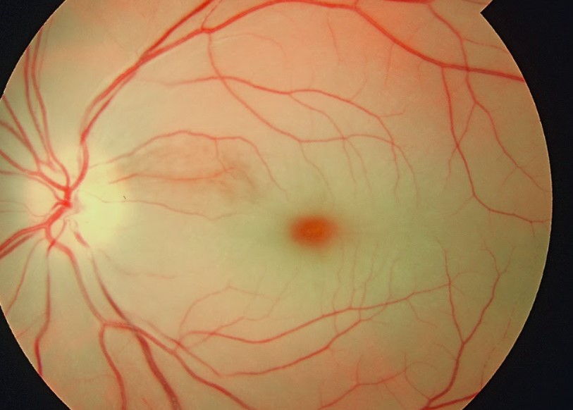
Definition & pathophysiology
CRAO is acute ischaemic infarction of the inner retina from occlusion of the central retinal artery, the first intra-orbital branch of the ophthalmic artery (itself the first branch of the internal carotid). It is, in effect, an ischaemic stroke of the eye — sharing embolic and atherosclerotic mechanisms with cerebral infarction — and is now managed within acute-stroke pathways. Mechanisms include carotid or aortic atheroembolism, cardioembolism, in-situ thrombosis on retinal atherosclerosis, small-vessel disease, and, critically in the older patient, giant cell arteritis (GCA). The inner retina tolerates ischaemia poorly; experimental primate data suggest substantial irreversible damage within roughly 90–240 minutes, underpinning the “time is retina” urgency.
Clinical features
Sudden, painless, severe monocular visual loss (counting-fingers to light-perception), with a profound RAPD. Fundoscopy shows a pale, oedematous retina with a cherry-red spot at the fovea (thin foveal retina overlying intact choroidal circulation), attenuated “boxcar” arterioles, and sometimes a visible embolus (bright, refractile Hollenhorst cholesterol plaque at a bifurcation). A cilioretinal artery (present in ~15–30%) may spare central vision. Branch RAO produces sectoral field loss. A history of preceding amaurosis fugax is common.
Acute treatment — current status of thrombolysis
The 2021 AHA/ASA Scientific Statement reframed CRAO as an ischaemic stroke requiring emergent evaluation, ideally in an emergency department with stroke expertise and rapid access to neuro-imaging and stroke consultation. Conservative “ocular” therapies— ocular massage, anterior-chamber paracentesis, IOP-lowering drops/acetazolamide, hyperbaric oxygen, inhaled carbogen, and vasodilators—have no proven benefit and are not recommended as standard of care.
Intravenous thrombolysis remains investigational and off-label. Observational data and an individual-participant meta-analysis (Shahjouei/Dumitrascu et al., 2023) suggested higher rates of visual recovery when tPA is given within ~4.5 hours of onset, generating equipoise. Randomised evidence is now emerging but not yet definitive: the French THEIA trial (reported 2025) comparing IV alteplase with aspirin within 4.5 hours found more visual improvement with alteplase but the difference was not statistically significant and the trial was underpowered by slow recruitment and unexpectedly high improvement in both arms. Two further RCTs — REVISION (German, alteplase vs placebo) and TenCRAOS (Norwegian, tenecteplase vs aspirin) — are ongoing/awaited. Historic intra-arterial thrombolysis (the EAGLE trial, 2010) showed no benefit over standard care and more complications, and is not recommended. Bottom line (2026): treat CRAO as a hyperacute stroke — emergent triage, exclude GCA, offer carefully selected off-label IV thrombolysis within 4.5 hours at experienced centres while awaiting definitive RCT data.
| Intervention | Evidence / status |
|---|---|
| IV thrombolysis (alteplase/tenecteplase) ≤4.5 h | Investigational, off-label; meta-analysis signal, THEIA neutral/underpowered; REVISION & TenCRAOS pending |
| Intra-arterial thrombolysis | Not recommended (EAGLE negative, higher complications) |
| Ocular massage, paracentesis, IOP-lowering, hyperbaric O₂, carbogen | Unproven; not standard of care |
| High-dose corticosteroids | Only if arteritic (GCA) — start immediately, do not await biopsy |
Workup — treat as a stroke
- Immediate exclusion of GCA in patients ≥50: ESR, CRP, platelets; ask about jaw claudication, scalp tenderness, headache, PMR, constitutional symptoms — arteritic CRAO threatens the fellow eye and demands emergent corticosteroids.
- Urgent carotid imaging (duplex, CTA or MRA) — CRAO carries a high risk of ipsilateral carotid stenosis and subsequent cerebral stroke; symptomatic high-grade stenosis warrants expedited revascularisation.
- Cardioembolic evaluation: ECG/telemetry for atrial fibrillation, echocardiography.
- Brain MRI-DWI — a substantial minority of CRAO patients have concurrent silent acute cerebral infarcts.
- Vascular risk-factor optimisation and antiplatelet therapy (secondary prevention as for ischaemic stroke); anticoagulate if a cardioembolic source is found.
Clinical pearls
- CRAO is a stroke — it is a neurological emergency, not merely an ophthalmic one; the highest-yield acute action is often the urgent search for a treatable embolic source and for GCA.
- Always exclude GCA first in the older patient — a missed diagnosis costs the second eye within days.
- A Hollenhorst plaque mandates carotid imaging even if vision recovers.
- Do not delay stroke-standard secondary prevention while debating thrombolysis; the embolic workup changes long-term outcome more reliably than acute ocular manoeuvres.
Key references: Mac Grory et al., AHA/ASA Scientific Statement on Management of CRAO, Stroke 2021; Shahjouei/Dumitrascu et al., IPD meta-analysis of thrombolysis for CRAO, 2023; THEIA trial report 2025; EAGLE study, Ophthalmology 2010.
Retinal Vein Occlusion
Definition & classification
Retinal vein occlusion (RVO) is venous outflow obstruction of the retinal circulation and is the second commonest retinal vascular disorder after diabetic retinopathy. It is classified anatomically into central RVO (CRVO) — obstruction at or posterior to the lamina cribrosa — and branch RVO (BRVO) — obstruction at an arteriovenous crossing where a thickened arteriole compresses the vein; hemiretinal vein occlusion is an intermediate variant. Each is further divided physiologically into non-ischaemic (perfused) and ischaemic (non-perfused) subtypes, a distinction that drives prognosis and neovascular risk.
Pathophysiology
Unlike arterial occlusion, RVO reflects venous thrombosis favoured by Virchow's triad at the arteriovenous crossing (BRVO) or lamina cribrosa (CRVO): endothelial change from arteriosclerosis (shared hypertensive/atherosclerotic risk factors), stasis, and hypercoagulability. Raised intraluminal pressure produces intraretinal haemorrhage, macular oedema (the main cause of vision loss), and, if capillary non-perfusion is extensive, retinal/optic-disc/iris neovascularisation with the feared complication of neovascular glaucoma.
Clinical features
Painless, usually less severe and more variable visual loss than CRAO. Fundoscopy of CRVO shows the classic “blood-and-thunder” appearance: haemorrhages in all four quadrants, dilated tortuous veins, disc oedema, cotton-wool spots and macular oedema. BRVO shows a sectoral, wedge-shaped distribution respecting the horizontal raphe. An RAPD and extensive non-perfusion on fluorescein angiography mark the ischaemic subtype, which carries high neovascular risk.
| Feature | Non-ischaemic RVO | Ischaemic RVO |
|---|---|---|
| Vision | Better (often >20/200) | Poor (often ≤20/200) |
| RAPD | Absent/mild | Present, marked |
| Capillary non-perfusion (FA) | Limited | Extensive |
| Neovascular risk | Low | High — iris/angle neovascularisation, neovascular glaucoma |
Systemic associations & workup
RVO shares risk factors with cerebrovascular disease — hypertension, diabetes, hyperlipidaemia, smoking, and raised intraocular pressure/glaucoma — and is a marker of systemic vascular risk. Evaluation targets modifiable vascular risk factors and (selectively) other contributors: in younger patients or those without vascular risk factors, consider hypercoagulable states, hyperviscosity (myeloproliferative disease, paraproteinaemia), inflammatory/vasculitic causes, and oral contraceptive use. Unlike CRAO, RVO is a venous event and is not an acute arterial-embolic stroke equivalent; nevertheless the shared risk-factor burden warrants cardiovascular risk assessment and control.
Management
- Intravitreal anti-VEGF (ranibizumab, aflibercept, bevacizumab) is first-line for macular oedema (evidence from BRAVO/CRUISE, VIBRANT and related trials) with intravitreal corticosteroids (dexamethasone implant) as an alternative.
- Panretinal photocoagulation for neovascularisation/ischaemic CRVO to prevent neovascular glaucoma; sector laser for ischaemic BRVO.
- Control blood pressure, glucose, lipids; treat glaucoma; manage identified systemic contributors.
Clinical pearls
- “Blood-and-thunder fundus” = CRVO; a sectoral, raphe-respecting haemorrhage pattern = BRVO.
- An RAPD in RVO signals the ischaemic subtype and mandates close surveillance for neovascular glaucoma.
- RVO is a venous occlusion — treat it as a marker of systemic vascular risk, but reserve thrombophilia testing for the young or otherwise unexplained case.
- A combined CRAO+CRVO or bilateral/recurrent RVO should prompt evaluation for hyperviscosity, vasculitis, or hypercoagulability.
Key references: Central Vein Occlusion Study (CVOS) and Branch Vein Occlusion Study (BVOS); BRAVO/CRUISE and VIBRANT anti-VEGF trials; AAO Preferred Practice Pattern, Retinal Vein Occlusions.
Ischemic Optic Neuropathy (Arteritic AION vs Non-Arteritic)
Definition & anatomy
Ischaemic optic neuropathy (ION) is infarction of the optic nerve. It is subclassified by location — anterior (AION, involving the optic-nerve head, supplied by the short posterior ciliary arteries; produces disc swelling) versus posterior (PION, retrolaminar, initially normal disc) — and, crucially, by mechanism: arteritic (from giant cell arteritis, GCA) versus non-arteritic (NAION). Distinguishing arteritic from non-arteritic ION is one of the true emergencies of neuro-ophthalmology, because untreated GCA blinds the fellow eye within days to weeks.
Clinical features & the critical distinction
| Feature | Arteritic AION (GCA) | Non-arteritic AION (NAION) |
|---|---|---|
| Age | Usually >70 | 50–70 |
| Vision loss | Severe (often counting-fingers/worse), may be preceded by amaurosis fugax | Moderate; altitudinal defect common |
| Systemic symptoms | Jaw claudication, scalp tenderness, headache, PMR, weight loss, fever | Absent |
| Disc appearance | Pallid (chalky-white) swelling, may be cupped later | Hyperaemic swelling, crowded “disc at risk”, small cup in fellow eye |
| ESR / CRP / platelets | Elevated ESR & CRP; thrombocytosis | Normal |
| Fluorescein angiography | Choroidal filling delay | Normal choroidal filling |
| Fellow-eye risk | High and rapid without treatment | Lower; contralateral over years (“pseudo-Foster-Kennedy”) |
NAION is the commonest acute optic neuropathy over 50: painless, often on waking (implicating nocturnal hypotension), producing an altitudinal (usually inferior) field defect, RAPD, and hyperaemic disc swelling. It occurs on a structurally predisposed “disc at risk” (small, crowded optic disc with little/no cup). Associations include hypertension, diabetes, hyperlipidaemia, obstructive sleep apnoea, nocturnal hypotension, phosphodiesterase-5 inhibitors (temporal association), and amiodarone. There is no proven acute therapy; management is risk-factor modification and treating sleep apnoea (the IONDT trial found optic-nerve-sheath decompression ineffective and possibly harmful).
Arteritic ION — GCA workup & treatment
In any patient ≥50 with acute ION, GCA must be actively excluded. Send ESR, CRP and full blood count (platelets) immediately; CRP is more sensitive and less age-dependent, and the combination of raised ESR and CRP maximises sensitivity, with thrombocytosis adding specificity. Modern practice favours fast-track GCA pathways to minimise the interval to diagnosis and treatment:
- Temporal artery ultrasound looking for the non-compressible “halo” sign (hypoechoic circumferential wall oedema) and the compression sign is now a first-line diagnostic tool in many fast-track clinics (recommended first-line by EULAR); high specificity when positive by experienced operators, though sensitivity is operator-dependent.
- Temporal artery biopsy (ideally ≥1–2 cm to mitigate skip lesions) remains the diagnostic gold standard, especially in the US; corticosteroids should NOT be delayed for the biopsy, which stays informative for at least 1–2 weeks after starting steroids.
- Large-vessel involvement can be assessed with vascular ultrasound, MRA/CTA, or PET where available.
Treatment is immediate high-dose corticosteroid on clinical suspicion, before biopsy: for visual involvement, many use IV methylprednisolone ~1 g/day for 3 days followed by high-dose oral prednisolone (~1 mg/kg/day), then a slow taper over 12–18 months guided by symptoms and inflammatory markers. Tocilizumab (IL-6 receptor antagonist; GiACTA trial) is an effective steroid-sparing agent reducing relapse and cumulative steroid exposure. Low-dose aspirin is often added. The goal is protection of the fellow eye and prevention of GCA-related cerebral/systemic ischaemia.
Clinical pearls
- Pallid (chalky-white) disc swelling with severe vision loss in an elderly patient is arteritic until proven otherwise — start steroids first, investigate second.
- Normal ESR does not exclude GCA; check CRP and platelets, and treat on strong clinical suspicion regardless.
- Do not delay corticosteroids to obtain the temporal artery biopsy — the biopsy remains positive for one to two weeks after treatment begins.
- NAION on a “disc at risk” has no proven acute therapy; counsel on the ~15% fellow-eye risk and evaluate for sleep apnoea and nocturnal hypotension.
- Cupping that develops after ION favours an arteritic mechanism; a hyperaemic crowded disc favours NAION.
Key references: Hayreh, ischaemic optic neuropathies; Ischemic Optic Neuropathy Decompression Trial (IONDT); GiACTA trial (Stone et al., NEJM 2017); EULAR 2018 recommendations for large-vessel vasculitis imaging; ACR/EULAR GCA classification.
Transient Monocular Vision Loss (Amaurosis Fugax)
Definition
Amaurosis fugax is transient monocular visual loss (TMVL), most often lasting seconds to minutes with full recovery. When embolic or haemodynamic in origin it constitutes a retinal transient ischaemic attack and carries the same urgency and prognostic weight as a hemispheric TIA — a warning of impending stroke and a mandate for rapid vascular evaluation. The classic description is a painless “curtain or shade” descending or ascending across the vision of one eye.
Mechanisms & differential diagnosis
| Category | Clues | Examples |
|---|---|---|
| Embolic (retinal TIA) | Abrupt curtain, seconds–minutes; vascular risk factors; Hollenhorst plaque | Carotid atheroembolism (commonest), cardioembolism, aortic arch |
| Haemodynamic / low-flow | Provoked by posture, bright light, exercise; dimming/“brown-out” | Critical carotid stenosis/occlusion, ocular ischaemic syndrome |
| Arteritic (GCA) | Age >50, jaw claudication, headache, raised ESR/CRP; may precede CRAO/AION | Giant cell arteritis — emergency |
| Vasospastic (“retinal migraine”) | Younger, recurrent stereotyped, may be followed by headache; diagnosis of exclusion | Retinal vasospasm/migraine |
| Ocular / local | Positional, transient obscurations with papilloedema; anterior-segment causes | Raised ICP (transient visual obscurations), impending vein occlusion, hyphaema, tear-film/dry eye |
| Haematologic | Hyperviscosity, thrombophilia, sickle cell | Polycythaemia, paraproteinaemia, antiphospholipid syndrome |
Evaluation — treat embolic TMVL as a TIA
- Urgent carotid imaging (duplex, CTA or MRA) to detect operable ipsilateral internal-carotid stenosis; symptomatic high-grade (70–99%) stenosis warrants expedited carotid endarterectomy/stenting.
- Cardioembolic workup: ECG and prolonged rhythm monitoring for atrial fibrillation, echocardiography.
- Brain MRI and vascular risk-factor assessment (lipids, glucose, blood pressure).
- In any patient ≥50, check ESR/CRP and screen for GCA symptoms — TMVL can be the harbinger of arteritic infarction of the second eye.
- Start antiplatelet therapy and optimise vascular risk factors, as for cerebral TIA; anticoagulate if a cardioembolic source is confirmed.
Distinguishing features at the bedside
The pattern of loss helps: a horizontal “curtain” over one eye suggests retinal ischaemia; a homonymous field disturbance affecting both eyes (patients often mislabel it “one eye”) suggests a retrochiasmal/occipital cause — ask the patient to test each eye separately during an event. A slowly spreading scintillating scotoma with fortification spectra over 20–30 minutes is migrainous visual aura (usually binocular/homonymous). Light-induced transient dimming that takes seconds to recover suggests ocular ischaemic syndrome from severe carotid disease.
Clinical pearls
- Embolic amaurosis fugax is a retinal TIA — work it up as urgently as a hemispheric TIA; it predicts both cerebral stroke and permanent visual loss.
- Always exclude GCA in the older patient presenting with TMVL — missing it risks bilateral blindness.
- Confirm the event was truly monocular (tested one eye at a time); apparent “monocular” loss is frequently a homonymous hemianopia.
- TMVL provoked by bright light or posture points to haemodynamic critical carotid stenosis rather than embolism.
Key references: Amaurosis Fugax Study Group; NASCET/ECST carotid endarterectomy trials; AHA/ASA TIA and secondary-prevention guidelines; Biousse & Newman, Neuro-Ophthalmology Illustrated.
Papilledema & Raised ICP
Definition
Papilloedema denotes optic-disc swelling caused specifically by raised intracranial pressure; it is by definition bilateral (though it may be asymmetric). The term should be reserved for this mechanism — disc swelling from local optic-nerve disease (optic neuritis, ION, infiltration, compression) is more precisely called optic disc oedema or “disc swelling”. Raised CSF pressure is transmitted along the subarachnoid sheath of the optic nerve, obstructing axoplasmic flow at the lamina cribrosa and producing axonal swelling and secondary vascular changes at the disc.
Clinical features
Symptoms of raised ICP: headache (worse on waking, lying flat, Valsalva), pulsatile tinnitus, transient visual obscurations (brief, seconds-long greying of vision often provoked by posture change), diplopia from a false-localising CN VI palsy, and nausea/vomiting. Crucially, central acuity and colour vision are often preserved until late in papilloedema (contrast with optic neuritis), and the earliest field defect is an enlarged blind spot; progressive disease causes nasal and then generalised constriction and, ultimately, blindness from optic atrophy.
Fundoscopic signs (graded by the Frisén scale I–V): blurring of the disc margins (nasal first), obscuration of the peripapillary retinal nerve-fibre layer and of vessels crossing the disc, disc hyperaemia and elevation, loss of spontaneous venous pulsations, peripapillary haemorrhages and cotton-wool spots, and (chronic) optic atrophy with disc gliosis. Loss of spontaneous venous pulsation is a useful but imperfect sign (absent in ~10–20% of normals).
Differential diagnosis — papilloedema vs pseudopapilloedema vs optic disc oedema
| Entity | Laterality | Acuity early | Key discriminators |
|---|---|---|---|
| Papilloedema (raised ICP) | Bilateral | Preserved | Headache, TVOs, CN VI palsy; raised opening pressure |
| Optic disc oedema (ON, ION, compression) | Usually unilateral | Reduced, with RAPD | Pain (ON), altitudinal defect (ION), dyschromatopsia |
| Pseudopapilloedema (disc drusen, crowded disc, hyperopia) | Bilateral/unilateral | Normal | No true swelling; drusen autofluoresce, seen on OCT/ultrasound; anomalous vessels |
Workup
- Urgent neuro-imaging first: MRI brain with MR venography to exclude a mass, hydrocephalus, and cerebral venous sinus thrombosis before lumbar puncture.
- Lumbar puncture with opening pressure (measured in the lateral decubitus position) and CSF analysis once a mass/obstruction is excluded.
- Formal perimetry and OCT of the retinal nerve-fibre layer to quantify and monitor.
- When no structural or venous cause is found and CSF constituents are normal with a raised opening pressure, the diagnosis is idiopathic intracranial hypertension (IIH) — typically an obese woman of childbearing age; the modified Dandy criteria apply.
Management principles
Treat the underlying cause. In IIH: weight loss, acetazolamide (evidence from the IIHTT trial) ± topiramate; discontinue causative agents (tetracyclines, vitamin A/retinoids, some hormonal therapies). Fulminant or vision-threatening papilloedema is a surgical emergency — optic-nerve-sheath fenestration to protect vision, or CSF shunting; venous sinus stenting is increasingly used for selected patients with a significant transverse-sinus stenosis and pressure gradient. Serial perimetry guides urgency — visual field, not headache, is the organ at risk.
Clinical pearls
- Bilateral disc swelling with normal acuity and colour = papilloedema until imaging (with venography) proves otherwise — the field, not the acuity, is the metric of danger.
- Always image before LP, and always include venography to catch CVST masquerading as IIH.
- Distinguish disc drusen (pseudopapilloedema) with OCT/B-scan ultrasound/autofluorescence to avoid an unnecessary invasive workup.
- Rapidly progressive field loss overrides headache in triaging papilloedema toward surgery.
Key references: Friedman & Jacobson, diagnostic criteria for IIH (modified Dandy); IIH Treatment Trial (IIHTT, acetazolamide); Frisén papilloedema grading; NORDIC/consensus IIH management reviews.
Relative Afferent Pupillary Defect (RAPD)
Principle
The RAPD (Marcus Gunn pupil) is the single most valuable objective sign of asymmetric anterior visual-pathway dysfunction. Because the pupillary light reflex has a consensual limb — light in either eye drives equal, bilateral constriction via bilateral pretectal projections to both Edinger–Westphal nuclei — a difference in afferent input between the two eyes is unmasked by the swinging-flashlight test. It reflects a difference in conducted light signal, not the absolute status of either eye alone.
The swinging-flashlight test
In a dimly lit room with the patient fixating a distant target (to relax accommodation), swing a bright light briskly and rhythmically from one eye to the other, pausing ~1–3 seconds on each. Normally both pupils hold a stable, symmetric constriction. With an RAPD, when the light moves to the affected eye the reduced afferent signal produces a net paradoxical dilation of both pupils (the eye “escapes”); swinging back to the healthy eye reconstricts them. The defect is relative — it grades the difference between the two eyes and can be quantified in log units with neutral-density filters placed over the better eye.
Causes & localization
| Produces an RAPD | Does NOT produce an RAPD |
|---|---|
| Optic neuritis, ischaemic optic neuropathy | Cataract, refractive error, corneal/media opacity |
| Optic-nerve compression/infiltration/glaucoma (asymmetric) | Functional (non-organic) visual loss |
| CRAO/large retinal detachment, extensive retinal disease | Bilateral symmetric optic neuropathy (defects cancel) |
| Optic tract lesion (contralateral RAPD, modest) | Amblyopia (usually none or trivial) |
| Asymmetric chiasmal disease | Isolated macular disease with mild acuity loss (small if any) |
Key localising points: an RAPD indicates optic nerve or extensive retinal disease on the side of the defect; media opacities (cataract) and refractive error never cause an RAPD (a useful discriminator in unexplained visual loss). An optic tract lesion causes a contralateral RAPD (more crossed than uncrossed fibres) accompanied by a homonymous hemianopia and “bow-tie” atrophy; post-geniculate lesions cause no RAPD. In midbrain (pretectal) disease a contralateral RAPD can occur without visual loss from damage to afferent light-reflex fibres.
Clinical value
- An RAPD provides objective evidence of an optic neuropathy even when the patient cannot cooperate with acuity/field testing, and helps confirm organic disease in suspected functional visual loss.
- The absence of an RAPD in a patient claiming severe unilateral visual loss with a normal fundus argues strongly for non-organic loss or bilateral symmetric disease.
- It cannot be masked by an efferent pupillary defect: even if one pupil is fixed (e.g. CN III palsy, pharmacologically dilated, or synechiae), the RAPD can still be assessed by watching the consensual response in the reactive fellow pupil (a “reverse” RAPD test).
Clinical pearls
- An RAPD localises to the optic nerve (or extensive retina), never to cataract or refractive error — it is the objective fingerprint of an optic neuropathy.
- A dense homonymous hemianopia with an RAPD points to the optic tract; without an RAPD, to the post-geniculate radiations/cortex.
- Assess the RAPD by the consensual response when one pupil is unreactive — a fixed efferent pupil does not prevent detection.
- Grade and record it (e.g. in neutral-density-filter log units) to track optic neuritis or compressive disease over time.
Key references: Kardon & Thompson, the pupil and pupillary testing; Miller & Newman, Walsh & Hoyt; Levatin, swinging-flashlight test.
Differential Diagnosis of Acute Visual Impairment
A two-axis framework
Acute visual loss is most efficiently triaged along two axes: monocular vs binocular (which requires each eye to be tested separately — patients routinely mislabel a homonymous hemianopia as “one eye”) and transient vs persistent. Monocular loss localises anterior to the chiasm (eye, retina, optic nerve); binocular homonymous loss localises retrochiasmally; binocular simultaneous loss suggests chiasmal, bilateral, or cortical disease. Cross-tabulating these axes generates a disciplined differential.
| Transient (recovers) | Persistent | |
|---|---|---|
| Monocular | Amaurosis fugax (retinal TIA), GCA, ocular ischaemic syndrome, retinal migraine, papilloedema (TVOs), impending vein occlusion, intermittent angle closure | CRAO/BRAO, CRVO, ION (arteritic/NAION), optic neuritis, retinal detachment, vitreous haemorrhage, acute angle-closure glaucoma, endophthalmitis |
| Binocular | Migraine with aura, occipital TIA/seizure, bilateral papilloedema TVOs, hypotension/hypoperfusion, PRES | Bilateral occipital infarct (cortical blindness), chiasmal compression/apoplexy, bilateral optic neuropathy (toxic, nutritional, hereditary, inflammatory), functional visual loss |
Anchoring features by mechanism
- Painless + RAPD + monocular: optic-nerve or retinal ischaemia — CRAO (cherry-red spot), ION (disc swelling), optic neuritis (young, pain on eye movement, often after).
- Painful red eye: acute angle-closure glaucoma (haloes, mid-dilated fixed pupil, nausea), keratitis, uveitis, endophthalmitis — ophthalmological emergencies.
- Floaters/photopsia then a curtain: retinal detachment or vitreous haemorrhage.
- Bitemporal defect, endocrine features, sudden headache: pituitary apoplexy (chiasmal) — urgent.
- Homonymous field loss, macular-sparing, older vasculopath: occipital (PCA) infarct.
- Scintillating fortification spectra evolving over 20–30 min, binocular: migraine aura (a diagnosis of exclusion in first presentations).
- Age ≥50 with visual loss + systemic symptoms + raised ESR/CRP: GCA until excluded — treat immediately.
- Preserved pupils and optokinetic response with dense binocular loss: consider cortical blindness (Anton syndrome if denied) or functional loss.
Immediate actions & red flags
- Any acute visual loss in a patient ≥50: check ESR/CRP and screen for GCA.
- Monocular ischaemic loss (CRAO/amaurosis): stroke-pathway workup — carotid imaging, cardiac evaluation, antithrombotic therapy.
- Binocular homonymous or cortical loss: image as an acute stroke (MRI-DWI, vessel imaging); consider thrombolysis eligibility.
- Painful red eye or trauma: emergent ophthalmology referral.
- Bilateral disc swelling: image with venography and measure CSF pressure.
Clinical pearls
- Test each eye separately before accepting “monocular” — the commonest error is mislabelling a homonymous hemianopia.
- The presence or absence of an RAPD, pain, and the pattern of field loss localises the great majority of cases at the bedside before imaging.
- In older patients, GCA is the diagnosis you cannot afford to miss in any acute visual loss, transient or persistent, monocular or evolving to bilateral.
- Vertical-meridian-respecting defects are retrochiasmal; horizontal-meridian-respecting (altitudinal) defects are retinal/optic-nerve.
Key references: Biousse & Newman, Neuro-Ophthalmology Illustrated; Miller & Newman, Walsh & Hoyt; AAO Preferred Practice Patterns.
Pupillary Anatomy & Abnormal Pupils
The light-reflex arc & near response
The afferent limb runs from retinal ganglion cells through the optic nerve, chiasm and tract, leaving before the LGN to reach the pretectal olivary nuclei of the midbrain; these project bilaterally (via the posterior commissure) to both Edinger–Westphal nuclei, giving the consensual response. The efferent (parasympathetic) limb travels with CN III to the ciliary ganglion, then via short ciliary nerves to the sphincter pupillae (and ciliary muscle for accommodation). The near response (accommodation–convergence–miosis triad) is driven supranuclearly and reaches the Edinger–Westphal nuclei by a more ventral route than the light reflex — the anatomical basis of light-near dissociation (near response preserved while light reflex is lost). Sympathetic dilator control follows the three-neuron oculosympathetic pathway (see Horner syndrome).
Approach to anisocoria
First decide which pupil is abnormal by comparing anisocoria in light and dark: greater in dark = the small pupil is abnormal (sympathetic/Horner or physiological); greater in light = the large pupil is abnormal (parasympathetic — CN III palsy, tonic pupil, or pharmacological). Physiological (simple) anisocoria is common (≤1 mm difference, equal in light and dark, brisk reactions). Then examine the light and near reactions, lid and motility, and use targeted pharmacology.
| Pupil | Signature | Mechanism / test | Association |
|---|---|---|---|
| Adie (tonic) pupil | Large, poorly reactive to light, slow tonic near response and slow redilation; sector/vermiform iris movements | Postganglionic ciliary-ganglion denervation; cholinergic supersensitivity — constricts to dilute pilocarpine 0.1% | Holmes–Adie (with areflexia); usually benign, young women |
| Argyll Robertson pupils | Small, irregular, bilateral; no light reaction, brisk near response; poor dilation | Light-near dissociation from peri-aqueductal/pretectal damage | Neurosyphilis (also diabetes); “prostitute's pupil” |
| CN III palsy (efferent) | Fixed dilated pupil with ptosis & motility deficit | Loss of parasympathetic outflow; constricts to standard pilocarpine 1% | Compressive (aneurysm) vs ischaemic (see CN III) |
| Pharmacological mydriasis | Very large, fixed, no ptosis/motility deficit | Atropinic block: fails to constrict to pilocarpine 1% | Scopolamine patch, nebulised ipratropium, plant alkaloids, accidental instillation |
| Horner (sympathetic) | Small pupil, dilation lag, mild ptosis; anisocoria worse in dark | Apraclonidine reversal; hydroxyamphetamine localises | Dissection, Pancoast, brainstem (see Horner) |
Light-near dissociation — causes
Loss of the light reflex with a preserved near response localises broadly and has a characteristic differential: dorsal midbrain (Parinaud) syndrome, Argyll Robertson pupils (neurosyphilis), Adie tonic pupil (near response is tonic), severe bilateral afferent visual loss (no light input but intact near drive), aberrant regeneration of CN III, and diabetes. The pattern — bilateral small irregular (Argyll Robertson) vs unilateral large tonic (Adie) vs midbrain signs (Parinaud) — distinguishes them.
Pharmacologic testing summary
- Dilute pilocarpine 0.1%–0.125%: constricts an Adie (denervation supersensitivity) but not a normal pupil — confirms tonic pupil.
- Pilocarpine 1%: constricts a neurogenic CN III palsy pupil but not a pharmacologically (atropine) blocked pupil — distinguishes them.
- Apraclonidine / hydroxyamphetamine: confirm and localise Horner syndrome (small-pupil anisocoria).
Clinical pearls
- A large fixed pupil with no ptosis and full motility is not a CN III palsy — think tonic (Adie) or pharmacological pupil; dilute pilocarpine and the pilocarpine-1% test separate them.
- Bilateral small irregular pupils with light-near dissociation = Argyll Robertson — test for syphilis.
- Anisocoria greater in the dark = abnormal small pupil (Horner); greater in the light = abnormal large pupil (parasympathetic).
- An Adie pupil is initially large but becomes miotic over years (“little old Adie”); vermiform iris movements on slit-lamp are diagnostic.
Key references: Kardon & Thompson, pupillary physiology and testing; Loewenfeld, The Pupil; Miller & Newman, Walsh & Hoyt.
Pulsatile Tinnitus (Vascular Causes: dAVF, Venous, Carotid)
Definition & significance
Pulsatile tinnitus (PT) is a rhythmic sound synchronous with the heartbeat, arising from turbulent or transmitted vascular flow near the cochlea. Unlike continuous (non-pulsatile) tinnitus, PT frequently has an identifiable, sometimes dangerous, and often treatable structural cause, so it warrants a directed vascular investigation. It is subdivided into objective PT (audible to the examiner with a stethoscope over the ear/mastoid/orbit/neck — implying a true acoustic source such as a bruit) and subjective PT; and, mechanistically, into arterial, arteriovenous, and venous causes.
Vascular causes
| Category | Cause | Clues |
|---|---|---|
| Arteriovenous | Dural arteriovenous fistula (dAVF) | Objective bruit; can be abolished by ipsilateral carotid/jugular compression; red-flag — risk of haemorrhage if cortical venous reflux |
| Carotid–cavernous fistula | Proptosis, chemosis, ophthalmoplegia, orbital bruit; often post-traumatic | |
| Arteriovenous malformation | Focal deficits, headache, seizures | |
| Arterial | Carotid atherosclerotic stenosis | Older vasculopath; neck bruit; other vascular disease |
| Carotid/vertebral dissection or fibromuscular dysplasia | Neck pain, Horner, TIA; younger patient | |
| Aberrant internal carotid, persistent stapedial artery | Retrotympanic mass on otoscopy; congenital course anomaly | |
| Glomus (paraganglioma) tumours | Pulsatile retrotympanic mass; conductive hearing loss; Brown sign (blanches with pneumatic pressure) | |
| Venous | Idiopathic intracranial hypertension | Obese woman, headache, papilloedema, transient visual obscurations; relieved by jugular compression/lying down |
| Transverse/sigmoid sinus stenosis or diverticulum; high-riding/dehiscent jugular bulb | Low-pitched venous hum; positional; abolished by ipsilateral jugular compression or head turn | |
| Cerebral venous sinus thrombosis | Headache, papilloedema, focal signs, seizures |
Bedside maneuvers
- Auscultate over the mastoid, external auditory canal, periauricular region, orbit and neck for a bruit (objective PT).
- Light ipsilateral internal-jugular compression or head turn that abolishes the sound suggests a venous source (sinus stenosis, jugular anomaly, IIH); an arterial/AV source is typically unaffected by jugular compression but a carotid source may change with carotid compression.
- Otoscopy for a retrotympanic vascular mass (glomus tumour, aberrant carotid, high jugular bulb) — do not biopsy a pulsatile middle-ear mass.
- Fundoscopy for papilloedema (IIH, CVST).
Imaging workup
- MRI/MRA and MR venography of the brain and skull base — the workhorse for dAVF, sinus stenosis/thrombosis, and mass lesions; look for flow voids and venous anomalies.
- CT/CTA and CT venography and high-resolution temporal-bone CT — for bony anomalies (dehiscent jugular bulb, sigmoid diverticulum, aberrant carotid, otospongiosis).
- Catheter digital subtraction angiography (DSA) — the gold standard when a dAVF or fistula is suspected, and required to characterise venous drainage/cortical reflux and to guide endovascular treatment.
- Where IIH is suspected, proceed to LP with opening pressure after imaging (with venography).
Clinical pearls
- Objective pulsatile tinnitus with a bruit is a dural AV fistula until proven otherwise — and a dAVF with cortical venous reflux carries a real haemorrhage risk, making DSA and treatment mandatory.
- PT that disappears with ipsilateral jugular compression or lying flat points to a venous cause — think IIH/sinus stenosis and check for papilloedema.
- A pulsatile retrotympanic mass should never be biopsied — image it (glomus tumour, aberrant carotid, high jugular bulb).
- New PT with proptosis, chemosis and an orbital bruit is a carotid–cavernous fistula — urgent neurointerventional referral.
- PT can be the presenting symptom of raised ICP; conversely, treating a transverse-sinus stenosis (stenting) may cure both the tinnitus and the intracranial hypertension in selected patients.
Key references: Hofmann et al. and Sismanis, pulsatile tinnitus reviews; Signorelli et al., diagnostic imaging algorithm for pulsatile tinnitus; consensus reviews on dural AV fistula classification (Borden/Cognard) and venous-sinus stenting for IIH.
Disorders of Consciousness — Overview
Definition
Consciousness has two orthogonal components: arousal (wakefulness, level of consciousness, driven by the ascending reticular activating system and thalamus) and awareness (the content of consciousness, generated by widespread thalamocortical and cortico–cortical networks). Disorders of consciousness (DoC) are classified by dissociating these two axes: a patient may be aroused yet unaware (vegetative state), or transiently unaware because arousal itself has failed (coma). Precise nosology matters because prognosis, ethical decisions and communication with families hinge on the distinction.
The syndromes
| State | Arousal (eyes/sleep–wake) | Awareness | Defining features | Typical substrate |
|---|---|---|---|---|
| Coma | Absent (eyes closed, no sleep–wake cycles) | Absent | Unarousable unresponsiveness; no eye opening to stimulation; usually evolves within 2–4 weeks | Bilateral hemispheric injury or paramedian upper brainstem (ARAS) lesion |
| Vegetative state / unresponsive wakefulness syndrome (UWS) | Present (spontaneous eye opening, sleep–wake cycles) | Absent | Wakefulness without awareness; reflexive movements, roving eyes, no reproducible command following | Diffuse cortical / thalamic injury with preserved brainstem |
| Minimally conscious state (MCS) | Present | Partial, fluctuating | Inconsistent but reproducible purposeful behaviour (visual pursuit, command following, localisation, contingent emotion). MCS+ = language; MCS− = non-language responses | Partially preserved thalamocortical networks |
| Akinetic mutism | Present | Reduced (abulia) | Profound apathy; wakeful, visually tracking, but near-absent spontaneous movement or speech despite intact motor pathways | Bilateral mesial frontal / anterior cingulate or paramedian thalamic damage |
| Locked-in syndrome | Present | Intact | Quadriplegia + anarthria with preserved consciousness; communication via vertical eye movements and blinking. Not a disorder of consciousness | Ventral pons (basilar occlusion, central pontine myelinolysis) |
Emergence and covert consciousness
Emergence from MCS is signalled by reliable functional communication (accurate yes/no) or functional object use. A critical modern concept is cognitive motor dissociation (CMD) (also called covert consciousness): patients who appear behaviourally vegetative or minimally conscious yet demonstrate reproducible command following on task-based fMRI (e.g., motor-imagery paradigms) or EEG. CMD is present in roughly 15–25% of behaviourally unresponsive patients and predicts better long-term recovery, underscoring that the bedside examination systematically underestimates awareness.
Clinical pearls
- Serial standardised assessment with the Coma Recovery Scale–Revised (CRS-R) markedly reduces misclassification of MCS as vegetative — a single examination misses fluctuating awareness in up to 40% of cases.
- The 2018 AAN/ACRM/NIDILRR guideline abandoned the term “permanent vegetative state,” substituting “chronic” VS/UWS because late recovery (especially post-traumatic) occurs.
- Always exclude a locked-in patient masquerading as comatose: ask the patient to look up and blink. Preserved vertical gaze with quadriplegia is basilar occlusion until proven otherwise.
Key references: Giacino et al. AAN/ACRM/NIDILRR Practice Guideline on Disorders of Consciousness (Neurology 2018); Posner, Saper, Schiff & Plum, Diagnosis of Stupor and Coma (5th ed.); Schnakers et al. CRS-R validation; Owen et al. and Schiff, cognitive motor dissociation.
Neuroanatomy of Consciousness
The two pillars: arousal and awareness
Consciousness requires an intact arousal system (the ascending reticular activating system, ARAS, and its thalamic relays) driving two functioning cerebral hemispheres that generate awareness. Coma therefore results only from (1) a lesion of the paramedian brainstem tegmentum from the rostral pons through the midbrain, (2) bilateral diencephalic (thalamic) injury, or (3) diffuse/bilateral cortical dysfunction. A single unilateral hemispheric lesion does not cause coma unless it produces enough mass effect to compress the contralateral hemisphere or the brainstem.
The ascending reticular activating system
The ARAS is not a single tract but a constellation of nuclei in the rostral pontine and midbrain tegmentum projecting rostrally by two routes:
- Dorsal (thalamic) pathway: cholinergic input from the pedunculopontine (PPN) and laterodorsal tegmental (LDT) nuclei to the intralaminar and midline thalamic nuclei and the thalamic reticular nucleus, which then project diffusely to cortex.
- Ventral (extrathalamic) pathway: bypasses the thalamus through the lateral hypothalamus and basal forebrain (cholinergic nucleus basalis of Meynert) to cortex.
Neurochemically the system is a summation of ascending modulatory populations: cholinergic (PPN/LDT), noradrenergic locus coeruleus, serotonergic raphe nuclei, dopaminergic ventral tegmental area, histaminergic tuberomammillary nucleus, and glutamatergic parabrachial nucleus/precoeruleus. Orexin (hypocretin) neurons of the lateral hypothalamus stabilise wakefulness. Because these are anatomically compact in the paramedian midbrain–upper pons, a small strategically placed lesion (basilar tip embolus, central pontine myelinolysis, midbrain haemorrhage) can abolish consciousness disproportionate to its size.
Thalamus and the cortical networks of awareness
The thalamus is both a relay of the ARAS and an intrinsic hub: bilateral paramedian thalamic infarction (e.g., artery of Percheron occlusion) produces acute coma or hypersomnolence. Awareness itself depends on integrity and integration across two large-scale cortical systems — the default-mode network (medial prefrontal, posterior cingulate/precuneus, lateral parietal) mediating internal awareness, and the lateral frontoparietal executive/external-awareness network. The mesocircuit hypothesis (Schiff) frames recovery of consciousness as restoration of thalamostriatal–cortical loop activity, and explains paradoxical arousal responses to zolpidem and the rationale for central thalamic deep brain stimulation.
Clinical pearls
- The precuneus/posterior cingulate is the most consistently hypometabolic region in the vegetative state and among the first to recover with returning awareness.
- Coma from a brainstem lesion always implicates the paramedian upper pons–midbrain; medullary lesions cause cardiorespiratory failure, not coma.
- The artery of Percheron (a single trunk supplying both paramedian thalami ± rostral midbrain) is the classic cause of “unexplained” bithalamic coma with vertical gaze palsy.
Key references: Parvizi & Damasio, brainstem and consciousness; Saper et al., ascending arousal system (Nature 2005); Schiff, mesocircuit model; Edlow et al., neuroanatomic connectivity of human arousal (J Neuropathol Exp Neurol 2012).
Topical Localisation & Etiology of Coma
The core dichotomy: structural versus metabolic/diffuse
The first bedside decision in coma is whether the process is structural (demanding urgent imaging and often neurosurgery) or metabolic/toxic/diffuse (demanding systemic correction). The pattern of the neurological examination — symmetry, focality, pupils and the trajectory over time — localises the lesion.
| Feature | Structural coma | Metabolic / diffuse coma |
|---|---|---|
| Findings | Focal, asymmetric; localising signs | Symmetric, non-focal |
| Pupils | May be asymmetric / fixed (localising) | Usually small & reactive (light reflex preserved until late) |
| Eye movements | Gaze palsy, dysconjugate, skew | Roving conjugate, preserved oculocephalics |
| Motor | Focal weakness, asymmetric posturing | Tremor, asterixis, multifocal myoclonus, symmetric tone |
| Course | Progressive, orderly rostrocaudal deterioration | Waxing and waning; level fluctuates |
The reactive pupil is the key discriminator: because pupillary pathways are relatively resistant to metabolic insult, preserved light reflexes in a deeply comatose patient point to a metabolic cause (important exceptions: opioids give pinpoint but reactive pupils; anticholinergics/atropine give fixed dilated; glutethimide and severe anoxia/hypothermia can fix pupils).
Structural coma: supratentorial versus infratentorial
- Supratentorial (mass, haemorrhage, large infarct, subdural): coma develops secondarily as the mass causes lateral and downward herniation compressing the diencephalon and brainstem — hence an orderly rostrocaudal march of signs, often preceded by a hemispheric deficit and a depressed but initially arousable state.
- Infratentorial (pontine haemorrhage, basilar occlusion, cerebellar mass/stroke): coma is abrupt because the ARAS is struck directly, and is accompanied by cranial nerve, gaze and “crossed” signs, abnormal breathing patterns and early pupillary/oculomotor abnormalities. A cerebellar mass is a neurosurgical emergency — it compresses the fourth ventricle (hydrocephalus) and brainstem and can herniate upward and downward.
Etiologic workup
Immediate: point-of-care glucose, airway/oxygenation/perfusion, temperature, and empirical thiamine/dextrose/naloxone when indicated. Then: full metabolic and toxicology panel (Na, glucose, Ca, ammonia, osmolar gap, ABG, lactate, renal/hepatic, TSH, cortisol), CT head (± CTA for basilar occlusion), and — if the cause remains unexplained or infection/seizure is possible — lumbar puncture and EEG (non-convulsive status epilepticus is a readily missed, treatable cause of coma). MRI resolves brainstem stroke, encephalitis and osmotic demyelination not seen on CT.
Clinical pearls
- Any comatose patient with a normal CT and no metabolic explanation needs an EEG — assume non-convulsive status until excluded.
- Sudden coma with pinpoint reactive pupils and quadriparesis is pontine haemorrhage or basilar thrombosis, not opioid overdose, until imaged.
- Never lumbar-puncture a comatose patient with a posterior fossa mass or obstructive hydrocephalus before imaging — risk of fatal tonsillar herniation.
Key references: Posner, Saper, Schiff & Plum, Diagnosis of Stupor and Coma (5th ed.); Wijdicks, The Comatose Patient; Edlow et al., diagnosis of reversible causes of coma (Lancet 2014).
Glasgow Coma Scale & FOUR Score
Glasgow Coma Scale (GCS)
The GCS (Teasdale & Jennett, 1974) grades three responses; the total (3–15) is the sum of the best response in each domain. It is the lingua franca of acute neurology, drives trauma triage (severe ≤8, moderate 9–12, mild 13–15) and anchors intubation decisions.
| Score | Eye opening (E) | Verbal response (V) | Motor response (M) |
|---|---|---|---|
| 6 | — | — | Obeys commands |
| 5 | — | Oriented | Localises to pain |
| 4 | Spontaneous | Confused | Withdraws (normal flexion) |
| 3 | To speech/sound | Inappropriate words | Abnormal flexion (decorticate) |
| 2 | To pressure/pain | Incomprehensible sounds | Extension (decerebrate) |
| 1 | None | None | None |
Limitations: the verbal score is uninterpretable in intubated (score reported as “T”), aphasic or non-native-speaking patients; periorbital swelling confounds eye opening; sedation and paralysis invalidate the whole score. The GCS says nothing about brainstem function or pupils, so guidelines increasingly report GCS-Pupils (GCS-P) = GCS minus a pupil reactivity score (0, 1 or 2 non-reactive pupils), extending the range to 1–15 and improving prognostic granularity at the low end. Record the components (E, V, M) separately, never only the sum.
FOUR Score (Full Outline of UnResponsiveness)
The FOUR score (Wijdicks et al., 2005) was designed to overcome the GCS’s blind spots. It has four components each scored 0–4 (total 0–16), omits the verbal response (so it is fully applicable to intubated patients) and adds brainstem and respiratory assessment — letting it detect locked-in syndrome, herniation and progression toward brain death.
| Score | Eye (E) | Motor (M) | Brainstem reflexes (B) | Respiration (R) |
|---|---|---|---|---|
| 4 | Eyelids open, tracking or blinking to command | Thumbs-up, fist or peace sign to command | Pupil and corneal reflexes present | Not intubated, regular breathing |
| 3 | Open but not tracking | Localising to pain | One pupil wide and fixed | Not intubated, Cheyne–Stokes |
| 2 | Open to loud voice | Flexion to pain | Pupil OR corneal absent | Not intubated, irregular |
| 1 | Open to pain | Extension to pain | Pupil AND corneal absent | Breathes above ventilator rate |
| 0 | Remain closed to pain | None / generalised myoclonus status | Absent pupil, corneal AND cough | Breathes at ventilator rate or apnea |
A FOUR score of 0 (all reflexes and respiration absent) flags a patient who should be evaluated for brain death. The eye component uniquely captures the locked-in patient (E4 through voluntary blinking/vertical tracking despite M0). The FOUR score has at least equivalent inter-rater reliability and outcome prediction to the GCS in ICU cohorts.
Clinical pearls
- Always test the motor response in the best limb; asymmetry is clinically important but the GCS records the best.
- “Localising” (M5) means reaching toward and crossing the midline to a central noxious stimulus; simple withdrawal (M4) does not localise.
- The FOUR score is the better tool in the intubated ICU patient and when tracking brainstem deterioration; the GCS remains the field/trauma standard.
Key references: Teasdale & Jennett (Lancet 1974); Wijdicks et al., FOUR score (Ann Neurol 2005); Brennan, Murray & Teasdale, GCS-Pupils score (J Neurosurg 2018).
Brainstem Reflexes in Coma
Why brainstem reflexes localise
Each brainstem reflex has a fixed anatomical arc, so their presence or loss maps deterioration along the neuraxis (midbrain → pons → medulla) and forms the physiological core of both coma localisation and brain death determination.
| Reflex | Afferent | Efferent | Level | Normal response |
|---|---|---|---|---|
| Pupillary light | CN II | CN III (parasympathetic) | Midbrain | Constriction, direct & consensual |
| Corneal | CN V1 | CN VII | Pons | Bilateral blink |
| Oculocephalic (doll’s eyes) | CN VIII | CN III, VI (via MLF) | Pons–midbrain | Eyes move opposite to head turn |
| Oculovestibular (cold caloric) | CN VIII | CN III, VI | Pons–midbrain | Tonic deviation toward cold ear |
| Gag | CN IX | CN X | Medulla | Palatal/pharyngeal elevation |
| Cough | CN X (tracheal) | CN X | Medulla | Cough to suctioning |
How to test
- Oculocephalic: only after the cervical spine is cleared. In the comatose patient with an intact brainstem, turning the head produces conjugate eye deviation opposite to the direction of rotation (“doll’s eyes present”). Absence indicates brainstem dysfunction (or, rarely, drug effect).
- Oculovestibular (caloric): confirm an intact tympanic membrane, elevate the head 30°, and irrigate with up to 50–60 mL ice-cold water. In a comatose patient with an intact brainstem but no cortical function, the eyes deviate tonically toward the cold ear with no corrective nystagmus (the fast cortical phase is lost). Mnemonic COWS (Cold Opposite, Warm Same) describes the fast phase and applies to the awake patient. No eye movement at all signals brainstem failure. This is the more definitive test and is required in brain death determination.
- Corneal / gag / cough: use a cotton wisp or saline drop for the cornea; test gag with a tongue depressor and cough with deep tracheal suctioning — the two medullary reflexes assessed before declaring brainstem death.
Clinical pearls
- Ocular movements in a comatose patient traverse the entire tegmentum from midbrain (III) to pontomedullary junction (VI) via the MLF; testing them is a rapid survey of brainstem integrity.
- Absent oculocephalics but present calorics is usually a technique problem or neck restriction — the caloric is the stronger stimulus.
- Reflex loss in brain death is rostrocaudal in reverse: pupils and corneals typically disappear before the medullary cough during evolving catastrophic injury, but all must be absent for the determination.
Key references: Posner, Saper, Schiff & Plum, Diagnosis of Stupor and Coma; Greer et al., 2023 AAN/AAP/CNS/SCCM brain death guideline; Wijdicks, The Comatose Patient.
Pupillary Responses & Automated Pupillometry (NPi)
The pupil as a localising sign
Pupillary size and reactivity integrate sympathetic (dilator) and parasympathetic (constrictor, via the Edinger–Westphal nucleus and CN III) tone, and each pattern localises.
| Pupillary pattern | Localisation / cause |
|---|---|
| Unilateral fixed, dilated (“blown”) | CN III compression — uncal herniation, posterior communicating aneurysm |
| Midposition (4–6 mm), fixed | Midbrain lesion (loss of both sympathetic & parasympathetic) |
| Pinpoint, reactive (need magnifier) | Pontine lesion (haemorrhage) or opioids |
| Small, reactive | Diencephalic/metabolic; also Horner (asymmetric) |
| Bilateral fixed, dilated | Severe midbrain injury, global anoxia, atropine/anticholinergics, hypothermia |
Because the light reflex is metabolically robust, a preserved pupillary response in deep coma argues for a metabolic rather than structural process (see coma localisation).
Automated pupillometry and the Neurological Pupil index (NPi)
Manual pupillary assessment is unreliable: inter-examiner agreement is poor, small changes are missed, and anisocoria <1 mm is frequently overlooked. Infrared automated pupillometry quantifies size, constriction velocity, latency and percentage constriction, and distils them into the Neurological Pupil index (NPi), a scalar from 0 to 5 derived from a normative algorithm. NPi ≥3 is normal; NPi <3 is abnormal; NPi = 0 denotes a non-reactive pupil. The index is largely independent of ambient light and observer, enabling reproducible trending.
Applications include: early, objective detection of evolving herniation/CN III compromise (a falling NPi can precede clinically apparent anisocoria), titration of osmotherapy, and multimodal neuroprognostication. In the prospective multicentre ORANGE study (Oddo et al., Lancet Neurology 2023) of acute brain injury, an abnormal NPi (<3) was independently associated with 6-month poor outcome (adjusted OR ~1.4 per 10% increase in abnormal readings) and with mortality (adjusted HR ~5.6), and progression toward NPi = 0 carried the strongest mortality signal. After cardiac arrest, an NPi at or near 0 is a highly specific predictor of poor neurological outcome and is being incorporated into multimodal prognostic algorithms.
Clinical pearls
- Trend the NPi rather than reacting to a single value; a unilateral drop toward 0 is an early, quantifiable herniation alarm.
- Automated pupillometry removes the “is it really reactive?” ambiguity that plagues manual examination in bright ICU lighting or with small pupils.
- Low NPi is specific but not sufficient for poor-prognosis decisions — combine with SSEP, EEG, NSE and imaging, and never within the first 72 h off sedation after arrest.
Key references: Oddo et al., ORANGE study (Lancet Neurol 2023); Chen et al. and Couret et al., pupillometry reliability; Sandroni et al., ERC/ESICM neuroprognostication (Intensive Care Med 2021).
Decorticate & Decerebrate Posturing
Definition and mechanism
Abnormal posturing is a reflex motor pattern released when descending cortical control is lost, revealing the level of the lesion by the balance between the (facilitatory) rubrospinal/red nucleus and (extensor) vestibulospinal/reticulospinal systems.
| Decorticate (flexor) | Decerebrate (extensor) | |
|---|---|---|
| Arms | Flexion, adduction at elbows & wrists, held to chest | Extension, adduction, internal rotation, forearm pronation |
| Legs | Extension, internal rotation, plantar flexion | Extension, plantar flexion |
| Lesion level | Above the midbrain red nucleus (cerebral white matter, internal capsule, thalamus) | Between the red nucleus and the vestibular nuclei (midbrain–upper pons) |
| Mechanism | Rubrospinal (flexor) drive to arms preserved; corticospinal lost | Rubrospinal lost; unopposed vestibulospinal/reticulospinal extensor drive |
| GCS motor | M3 | M2 |
| Prognosis | Less ominous | More ominous |
Clinical significance
The key clinical value is dynamic: progression from decorticate to decerebrate posturing marks caudal (rostrocaudal) deterioration as a mass or oedema drives the diencephalon and then the midbrain downward. Further descent produces flaccidity and no response (lower pontine/medullary failure), the terminal motor pattern. Posturing may be spontaneous, provoked by noxious stimulation, or unilateral (indicating asymmetric brainstem or hemispheric involvement). It must be distinguished from spinal reflex movements, dystonia and non-epileptic events; genuine posturing is stereotyped, tonic and typically bilateral in symmetrical brainstem compromise.
Clinical pearls
- “Decorticate” — arms flex toward the core/cord; a useful memory aid pairing flexion with the higher (cortical) lesion.
- New decerebrate posturing in a previously decorticate patient is a herniation emergency until proven otherwise — escalate ICP management and reimage.
- Beware bilateral extensor posturing from a treatable metabolic catastrophe (hepatic coma, hypoglycaemia) or a large posterior fossa lesion, not only supratentorial herniation.
Key references: Posner, Saper, Schiff & Plum, Diagnosis of Stupor and Coma; classic decerebrate rigidity physiology (Sherrington); Wijdicks, The Comatose Patient.
Rostrocaudal Deterioration & Herniation Syndromes
Concept
Because the cranium is a rigid box partitioned by the falx and tentorium, an expanding mass forces brain tissue across these dural edges and through the foramen magnum. Herniation both shifts and ischaemically injures the displaced tissue, producing predictable, stepwise signs — the “rostrocaudal deterioration” described by Plum and Posner in central herniation.
| Syndrome | What herniates | Structures compressed | Cardinal signs |
|---|---|---|---|
| Uncal (lateral transtentorial) | Medial temporal uncus over the tentorial edge | Ipsilateral CN III, midbrain, PCA | Early ipsilateral fixed dilated pupil (“blown”), then ptosis & “down-and-out” eye; contralateral hemiparesis; PCA infarct; declining consciousness |
| Central (transtentorial) | Diencephalon displaced downward through the tentorial notch | Diencephalon → midbrain → pons → medulla, sequentially | Orderly rostrocaudal march: small reactive pupils + Cheyne–Stokes → midposition fixed pupils + decerebrate → apnea; Duret haemorrhages |
| Subfalcine (cingulate) | Cingulate gyrus under the falx cerebri | Contralateral cingulate; anterior cerebral artery | Often clinically silent early; ACA-territory infarction → contralateral leg weakness; may progress to central herniation |
| Tonsillar (downward, foramen magnum) | Cerebellar tonsils through the foramen magnum | Medulla | Neck stiffness, Cushing triad (hypertension, bradycardia, irregular respiration), apnea, sudden cardiorespiratory collapse |
| Upward transtentorial | Superior cerebellum/vermis up through the tentorial notch | Dorsal midbrain, aqueduct | Loss of upgaze, small poorly reactive pupils, obstructive hydrocephalus; may be precipitated by ventricular drainage of a posterior fossa mass |
Key mechanistic points
- Kernohan’s notch phenomenon: uncal herniation can push the midbrain against the opposite tentorial edge, compressing the contralateral cerebral peduncle and producing hemiparesis ipsilateral to the mass — a classic false localising sign. The dilated pupil (ipsilateral to the mass) remains the reliable localiser.
- Duret haemorrhages: small paramedian pontine/midbrain haemorrhages from stretching of perforating basilar branches during downward central herniation — usually a terminal finding.
- A unilateral fixed dilated pupil in a deteriorating patient is CN III compression from uncal herniation until proven otherwise and mandates immediate osmotherapy, hyperventilation as a bridge, and neurosurgical decompression.
Clinical pearls
- The earliest reliable sign of uncal herniation is the ipsilateral dilating pupil, often before major depression of consciousness — automated pupillometry can detect it sooner.
- Upward herniation is iatrogenically precipitated by draining CSF from above (EVD) when a posterior fossa mass is present — decompress the mass, do not simply drain.
- Cushing’s triad is a late, pre-terminal medullary sign of brainstem compression, not an early warning.
Key references: Plum & Posner, rostrocaudal deterioration; Posner, Saper, Schiff & Plum, Diagnosis of Stupor and Coma (5th ed.); Ropper, herniation syndromes (NEJM).
Brain Death — Determination & Prerequisites (2023 Consensus)
Definition
Brain death, or death by neurologic criteria (BD/DNC), is the complete and permanent cessation of all functions of the brain, including the brainstem. In the United States (Uniform Determination of Death Act) it is legally equivalent to circulatory death. The 2023 AAN/AAP/CNS/SCCM consensus practice guideline (Greer et al., Neurology 2023) replaced and harmonised the separate 2010 adult and 2011 pediatric guidelines into a single framework and clarified prerequisites, examiner requirements, the apnea test and ancillary testing.
Prerequisites (all must be satisfied before examination)
| Domain | Requirement |
|---|---|
| Established cause | Known, irreversible, catastrophic brain injury sufficient to explain the clinical picture, supported by neuroimaging |
| Observation period | Adequate time to exclude recovery; after cardiac arrest / anoxic injury wait ≥24 h before evaluation (allow for return of drug metabolism and evolution of injury) |
| Blood pressure | Systolic BP ≥100 mm Hg or MAP ≥60 mm Hg (vasopressors permitted) |
| Temperature | Core temperature ≥36°C (normothermia) |
| Metabolic/endocrine | Correct severe electrolyte, acid–base, glucose and endocrine derangements; pH within a normal range |
| Drugs / intoxicants | No confounding CNS depressants: wait ≥5 half-lives (adjust for organ dysfunction/hypothermia), consider drug levels; exclude alcohol and paralytics |
| Neuromuscular blockade | Confirm absence with train-of-four (4 twitches) |
The clinical examination
Three findings define BD/DNC on examination:
- Coma: no eye opening or motor response to noxious stimulation in all four limbs and cranially (spinally mediated reflexes such as triple flexion or the Lazarus sign may persist and do not preclude the diagnosis).
- Absent brainstem reflexes: no pupillary, corneal, oculocephalic, oculovestibular (caloric), gag or cough reflexes.
- Apnea: no respiratory drive on a formal apnea test (see separate topic).
Examiner and procedural requirements
Examiners must be competent and credentialed per institutional policy, and to avoid conflict of interest must not be involved in organ procurement/transplantation. The 2023 guideline specifies:
- Adults (≥18 y): a single, complete clinical examination including apnea test by one qualified clinician is sufficient (individual institutions may still require two).
- Children (<18 y): two examinations by different attending clinicians, separated by an observation interval (≥12 h; longer, up to 24 h, in neonates), with an apnea test at each.
Special situations requiring extra caution or ancillary testing include high cervical cord injury, severe facial trauma, pre-existing pupillary abnormalities, and inability to complete the apnea test.
Clinical pearls
- Brain death is a clinical diagnosis; ancillary tests are supplementary, not a substitute for a determinable examination.
- Spinal reflexes and the Lazarus sign frequently distress families — explain proactively that they arise below the foramen magnum.
- Targeted temperature management and its sedatives extend drug clearance times; err toward longer waiting periods and confirmatory levels.
Key references: Greer DM et al., 2023 AAN/AAP/CNS/SCCM Pediatric and Adult Brain Death/Death by Neurologic Criteria Consensus Practice Guideline (Neurology 2023); World Brain Death Project (Greer et al., JAMA 2020); Uniform Determination of Death Act.
Apnea Test Protocol
Rationale
The apnea test demonstrates absence of medullary respiratory drive by allowing PaCO2 to rise to a level that maximally stimulates the respiratory centres. It is the last and most hazardous component of brain death determination and is performed only after all prerequisites and the reflex examination are satisfied.
Step-by-step protocol
- Confirm readiness: normothermia (≥36°C), SBP ≥100 / MAP ≥60, euvolaemia, and a baseline arterial blood gas with PaCO2 normalised to 35–45 mm Hg and normal pH.
- Pre-oxygenate: ventilate with FiO2 1.0 (PEEP ~5 cm H2O) for ≥10 minutes to achieve PaO2 >200 mm Hg, reducing the risk of hypoxaemia during the apneic period.
- Disconnect the ventilator and deliver oxygen passively — e.g., 100% O2 at 6–10 L/min via a catheter at the carina or a T-piece/CPAP circuit (avoid high-flow insufflation that can cause barotrauma).
- Observe the chest and abdomen closely for any respiratory effort for approximately 8–10 minutes.
- Draw an arterial blood gas at the end (and repeat if needed).
Interpretation
| Result | Criteria |
|---|---|
| Positive (consistent with brain death) | No respiratory effort and PaCO2 ≥60 mm Hg, or a rise of ≥20 mm Hg above baseline (the latter accommodates chronic CO2 retainers) |
| Negative | Any respiratory effort → patient is not brain dead |
| Aborted / indeterminate | SpO2 falls <85–90%, systolic BP falls / haemodynamic instability, or significant arrhythmia — reconnect ventilator, draw ABG; if target PaCO2 already reached with no effort the test may still be interpretable, otherwise proceed to an ancillary test |
Cautions and pre-emptive measures
- Optimise haemodynamics and consider vasopressors before starting; hypotension is the commonest reason for abortion.
- In chronic hypercapnia or when a safe apnea test is not feasible (high cervical cord injury, severe hypoxaemia, haemodynamic fragility), substitute an ancillary blood-flow study.
- CO2 rises roughly 3 mm Hg per apneic minute, so ~8–10 minutes usually achieves the ≥60 mm Hg target from a normal baseline.
Clinical pearls
- Pre-oxygenation to PaO2 >200 buys the apneic time; skipping it invites early desaturation and an aborted test.
- A rising PaCO2 with a corresponding fall in pH but no breath is the physiological endpoint — document both the gas and the absence of effort.
- Never perform the apnea test before excluding residual paralytics (train-of-four) — a “false positive” apnea from neuromuscular blockade is catastrophic.
Key references: Greer DM et al., 2023 AAN/AAP/CNS/SCCM guideline (Neurology 2023); Wijdicks, apnea testing methodology; SCCM/AAN implementation resources and checklist.
Ancillary Tests for Brain Death
When ancillary testing is indicated
Ancillary tests are used only when the clinical examination or apnea test cannot be completed or is confounded (e.g., severe facial/ocular trauma, high cervical cord injury, unresolved drug effect, cardiopulmonary instability precluding apnea testing). The 2023 guideline is emphatic that ancillary tests supplement but never replace a determinable clinical examination, and that a positive ancillary result cannot override any evidence of retained brain function. Broadly, tests assess either cerebral blood flow or electrical activity; the guideline now prioritises blood-flow–based methods because they interrogate the whole brain, including the brainstem.
| Test | Finding in brain death | 2023 status / caveats |
|---|---|---|
| Radionuclide cerebral perfusion scintigraphy (99mTc-HMPAO, SPECT preferred) | “Hollow/empty skull” — absent intracranial tracer uptake, no brainstem or cerebellar flow | Preferred/accepted; lipophilic tracer with SPECT assesses brainstem flow, minimally confounded by sedatives |
| Four-vessel catheter (digital subtraction) cerebral angiography | No intracerebral filling at the level of carotid/vertebral entry despite patent external circulation | Accepted gold-standard flow study; invasive, requires transport & contrast |
| EEG | Electrocerebral silence | No longer recommended in the 2023 US guideline — assesses only cortical electrical activity, not the brainstem, and is confounded by drugs, hypothermia and ICU artefact |
| CT angiography / CT perfusion | Absent opacification of intracranial vessels | Not endorsed — insufficient validation and false-negative rates; not a recommended confirmatory test |
| Transcranial Doppler (TCD) | Reverberating/oscillating flow, small systolic spikes, or absent diastolic flow | Highly operator- and window-dependent; considered a lesser/adjunctive option and not a preferred US ancillary test |
Interpretation principles
- Blood-flow studies confirm the pathophysiology of brain death — intracranial pressure exceeding perfusion pressure produces cerebral circulatory arrest.
- A misleading ancillary result (e.g., preserved posterior fossa flow, TCD signal through a craniectomy defect or in infants with open fontanelles) must prompt re-evaluation, not a declaration.
- Electrical (EEG, evoked potentials) and CT-based methods have moved out of favour in the US precisely because they can be discordant with true whole-brain death.
Clinical pearls
- Reach for a radionuclide flow study first when an ancillary test is needed — it is the most robust to sedative confounding.
- Document explicitly why an ancillary test was required (which clinical component could not be completed).
- The recent de-emphasis of EEG and CTA is a notable change from older local protocols — verify your institution has updated its policy to the 2023 consensus.
Key references: Greer DM et al., 2023 AAN/AAP/CNS/SCCM guideline (Neurology 2023); World Brain Death Project (JAMA 2020); nuclear medicine practice reviews of BD/DNC perfusion imaging.
Specific Consciousness Disorders & Classification
Quantitative versus qualitative disturbances (Beneš–Drábek framework)
A useful heuristic, associated with the Czech neurological tradition (Beneš–Drábek), divides altered consciousness into two axes that mirror the arousal/awareness dichotomy:
- Quantitative disorders — reduced level/vigilance along a continuum: somnolence → sopor (stupor) → coma.
- Qualitative disorders — altered content with relatively preserved arousal: delirium, amentia (acute confusional/incoherent state), obnubilation (clouding), and twilight (dissociative/complex partial) states.
The framework is a bedside organiser rather than a formal diagnostic system, and terminology varies by region; DSM-5 and ICD nosology should be used for formal diagnosis.
Delirium
An acute, fluctuating disturbance of attention and awareness with an identifiable medical, toxic or metabolic precipitant — the prototypical qualitative disorder. Subtypes are hyperactive, hypoactive (frequently missed and prognostically worse), and mixed. Screen with the CAM / CAM-ICU or 4AT; management is cause-directed with attention to sedation minimisation, sleep–wake and sensory optimisation, and antipsychotics reserved for dangerous agitation. Delirium is an independent predictor of mortality and long-term cognitive decline.
Catatonia
A neuropsychiatric syndrome of motor dysregulation — stupor, mutism, negativism, catalepsy, waxy flexibility (cerea flexibilitas), posturing, echolalia/echopraxia and stereotypy — occurring with psychiatric, autoimmune (e.g., anti-NMDA-receptor encephalitis), metabolic and structural disease. Quantify with the Bush–Francis Catatonia Rating Scale; a lorazepam challenge is diagnostic and therapeutic, and ECT is definitive, particularly for malignant catatonia (with autonomic instability and hyperthermia, overlapping neuroleptic malignant syndrome).
Cotard syndrome
A nihilistic delusional state in which patients believe they are dead, do not exist, or have lost organs/the soul. It is a neuropsychiatric phenomenon of severe psychotic depression, schizophrenia or organic brain disease (and is not a disorder of arousal); treatment targets the underlying condition (antidepressants, antipsychotics, ECT).
Clinical pearls
- Hypoactive delirium is easily mistaken for depression or fatigue; routine CAM-ICU screening in the ICU prevents this.
- Consider autoimmune (anti-NMDA-R) encephalitis in young patients presenting with new catatonia, psychiatric symptoms and seizures — check antibodies, MRI, EEG and screen for teratoma.
- The Beneš–Drábek quantitative/qualitative split is a helpful teaching scaffold but should be translated into DSM-5/ICD terms when documenting.
Key references: DSM-5-TR; Inouye et al., delirium (Lancet 2014); Bush et al., Catatonia Rating Scale; Debruyne et al., Cotard syndrome reviews; Beneš & Drábek, classification of disorders of consciousness.
Prognostication after Coma & Cardiac Arrest
Principles
Neuroprognostication after hypoxic–ischaemic brain injury must be multimodal, deferred until confounders have cleared, and based on concordant predictors — no single test is sufficient to withdraw care. The ERC/ESICM 2021 guideline recommends assessment ≥72 h after return of spontaneous circulation (ROSC), off sedation and neuromuscular blockade, and normothermic, in a patient who remains comatose (M ≤3) with no reversible cause. Premature prognostication risks a self-fulfilling prophecy.
Predictors of poor outcome (used in combination)
| Modality | Poor-outcome finding | Timing |
|---|---|---|
| Clinical exam | Bilaterally absent pupillary and corneal reflexes; automated NPi at/near 0 | ≥72 h |
| Myoclonus | Early status myoclonus (with malignant EEG) | <72 h supportive, not alone |
| SSEP | Bilaterally absent N20 cortical response | ≥24–72 h |
| EEG | Suppression, burst-suppression, or unreactive/refractory status | ≥24–72 h |
| Biomarker | Neuron-specific enolase (NSE) markedly elevated or rising (e.g., >60 µg/L at 48–72 h) | 48–72 h |
| Imaging | Diffuse anoxic injury: loss of grey–white differentiation / reduced GWR on CT; extensive DWI restriction on MRI | >24 h (MRI 2–5 d) |
The guideline advises using ≥2 concordant predictors from different categories before predicting a poor outcome, acknowledging that even validated markers carry non-zero false-positive rates.
Clinical pearls
- Sedatives, hypothermia, renal failure (NSE), haemolysis (falsely elevated NSE) and seizures all confound — correct or account for each before prognosticating.
- Bilaterally absent N20 SSEPs and a malignant unreactive EEG together are among the most robust poor-outcome combinations.
- Late awakening occurs, especially with prolonged sedation clearance and post-arrest metabolic encephalopathy — when uncertain, wait and reassess rather than commit.
Key references: Nolan, Sandroni et al., ERC/ESICM Post-Resuscitation Care Guidelines (Resuscitation / Intensive Care Med 2021); Sandroni et al., prediction of poor outcome after cardiac arrest; Oddo et al., ORANGE (Lancet Neurol 2023).
Intracranial Hypertension — Pathophysiology
The Monro–Kellie doctrine
The intracranial compartment is a fixed-volume box containing three constituents — brain parenchyma (~80%), cerebrospinal fluid (~10%) and blood (~10%). Because the box cannot expand, any increase in one component (or addition of a mass) must be offset by displacement of another, or ICP rises. The first buffers are displacement of CSF into the spinal thecal sac and reduction of cerebral venous blood volume. Normal ICP in a supine adult is 7–15 mm Hg; sustained values >20–22 mm Hg generally warrant treatment.
Compliance and the pressure–volume curve
The relationship between added volume and ICP is exponential, not linear. Early on the system is compliant (the flat part of the curve): large volume additions cause little ICP change as compensatory reserves are recruited. Once reserves are exhausted, the curve turns steep and small further increments cause large, dangerous rises in ICP — the clinical “falling off the cliff.” Elastance (the inverse of compliance) rises accordingly. The clinical corollary is that a patient can deteriorate abruptly after appearing stable.
Cerebral perfusion and autoregulation
Cerebral perfusion pressure (CPP) = MAP − ICP. Cerebral autoregulation normally holds cerebral blood flow constant across a MAP range (~50–150 mm Hg), but after injury it is frequently impaired, making CBF pressure-passive and the brain vulnerable to both ischaemia (low CPP) and hyperaemic oedema (high CPP). A CPP target of 60–70 mm Hg is generally recommended; individualised “optimal CPP” can be estimated from the pressure-reactivity index (PRx).
ICP waveforms
The pulsatile ICP waveform has three arterial peaks:
- P1 (percussion wave) — arterial pulsation, relatively constant.
- P2 (tidal wave) — reflects intracranial compliance; when P2 exceeds P1, compliance is poor and reserves are nearly exhausted.
- P3 (dicrotic wave) — follows the dicrotic notch.
Lundberg waves describe slower trends: A waves (plateau waves) are sustained rises to 50–100 mm Hg lasting 5–20 minutes, always pathological and indicating critically reduced compliance with impending herniation; B waves are oscillations of 0.5–2/min associated with unstable ICP; C waves are low-amplitude and generally benign.
Clinical pearls
- A rising P2 (P2 > P1) on the ICP monitor is an early morphological warning that compliance is failing, often before mean ICP climbs.
- Plateau (A) waves may transiently reverse with brief hyperventilation, CSF drainage or MAP augmentation — treat the compliance problem, not just the number.
- Because CPP couples ICP and MAP, treating intracranial hypertension is as much about supporting MAP as lowering ICP.
Key references: Brain Trauma Foundation Guidelines (4th ed., 2016/2017); Czosnyka & Pickard, monitoring of cerebrospinal dynamics; Lundberg, continuous ICP recording.
Management of Intracranial Hypertension
A tiered approach
Contemporary management (exemplified by the Seattle International Severe TBI Consensus Conference, SIBICC 2019) is organised into escalating tiers, treating sustained ICP >20–22 mm Hg, with each higher tier carrying greater risk. Within a tier, therapies are considered broadly equivalent and are individualised.
| Tier | Interventions |
|---|---|
| Tier 0 (baseline, all patients) | Head of bed 30° and midline neck (optimise venous drainage), avoid hypotension/hypoxia, normocapnia (PaCO2 35–45), normothermia, treat pain/agitation, CPP 60–70, control glucose & sodium, seizure treatment as indicated |
| Tier 1 | Increase analgesia/sedation; intermittent CSF drainage via EVD; osmotherapy bolus (mannitol 0.25–1 g/kg or hypertonic saline); maintain PaCO2 at low-normal (35–38) |
| Tier 2 | Scheduled/repeated hyperosmolar therapy; mild hyperventilation (PaCO2 32–35, with brain-oxygen monitoring); trial of neuromuscular blockade; MAP challenge to assess autoregulation and set CPP |
| Tier 3 (highest risk) | Barbiturate/pentobarbital coma; secondary decompressive craniectomy; mild hypothermia (35–36°C) |
Osmotherapy: mannitol versus hypertonic saline
| Mannitol | Hypertonic saline (3%, or 23.4% bolus) | |
|---|---|---|
| Mechanism | Osmotic gradient + rheologic (early plasma expansion, reflex vasoconstriction) then osmotic diuresis | Osmotic gradient; volume-expanding rather than diuretic |
| Haemodynamics | Diuresis → hypovolaemia/hypotension risk | Preferred in the hypovolaemic/hypotensive patient |
| Monitoring | Serum osmolality & osmolar gap (hold if gap >~18–20); keep osmolality <320; renal function | Serum sodium (target-guided); avoid rapid overcorrection; central access for high concentrations |
| Cautions | Renal injury, rebound, avoid in hypovolaemia | Hyperchloraemic acidosis, volume overload / pulmonary oedema, phlebitis |
Both are effective; hypertonic saline is increasingly favoured, especially where perfusion pressure must be preserved.
Key do’s and don’ts
- Hyperventilation is only a short-term bridge to definitive therapy; prophylactic or aggressive hypocapnia (PaCO2 <30) causes cerebral vasoconstriction and ischaemia and is contraindicated as routine practice.
- Corticosteroids are not indicated for cytotoxic oedema of TBI or ischaemic stroke — the CRASH trial showed increased mortality with high-dose steroids in head injury. Steroids are reserved for vasogenic oedema around tumour or abscess.
- Prophylactic hypothermia and early routine decompressive craniectomy have not improved outcomes and can worsen them (see decompressive craniectomy topic); use them as targeted, later-tier measures.
Clinical pearls
- Escalate stepwise but do not delay a life-saving jump to Tier 3 (osmotherapy + surgery) in acute herniation with a blown pupil.
- Give a hyperosmolar bolus while arranging the CT/OR — buy time for definitive decompression.
- Track the osmolar gap (not just calculated osmolality) to dose mannitol safely and detect accumulation.
Key references: Hawryluk et al., SIBICC management algorithm (Intensive Care Med 2019); Brain Trauma Foundation Guidelines (4th ed.); CRASH trial (Lancet 2004/2005); Neurocritical Care Society guidelines on hyperosmolar therapy.
Decompressive Craniectomy
Rationale
Removing a large bone flap (and opening the dura) converts the closed cranium into an open one, allowing swollen brain to expand outward rather than herniate — a mechanical solution to refractory intracranial hypertension. It reliably lowers ICP; the clinical question in every setting is whether the survivors it produces have acceptable outcomes.
Malignant middle cerebral artery (MCA) infarction
Large hemispheric infarction with life-threatening oedema (“malignant” MCA syndrome) is the strongest indication. A pooled analysis of the three European trials (DECIMAL, DESTINY, HAMLET) of hemicraniectomy within 48 h in patients aged ≤60 showed a dramatic mortality reduction (roughly from ~70% to ~20–30%) and an increase in the proportion surviving with moderate disability, at the cost of more survivors with moderately severe disability. DESTINY II extended this to patients >60 y: surgery reduced mortality, but most survivors had substantial disability (mRS 4–5), making the decision highly values-dependent. Favourable factors: younger age, earlier surgery, and adequate flap size (≥12 cm diameter) to avoid external herniation.
Traumatic brain injury
| Trial | Setting | Key result |
|---|---|---|
| DECRA (2011) | Early bifrontal craniectomy for moderately raised ICP (>20 mm Hg for >15 min), a “neuroprotective” strategy | Lowered ICP and ICU stay but worse functional outcomes — do not decompress early for moderately elevated ICP |
| RESCUEicp (2016) | Last-tier craniectomy for refractory ICP (>25 mm Hg for 1–12 h despite tier 1–2 therapy) | At 6 months lower mortality (26.9% vs 48.9%) but more survivors in vegetative state or with severe disability; benefit consolidated by 12 months (favourable 45% vs 32%) |
The two trials frame the central trade-off: craniectomy converts deaths into survivors across a spectrum of disability. It is a Tier 3 option for refractory ICP, not a first-line manoeuvre.
Practical and later considerations
- Complications: haemorrhagic expansion, external cerebral herniation, subdural hygroma, infection, and later the syndrome of the trephined (“sinking skin flap”) with paradoxical neurological decline relieved by cranioplasty.
- Cranioplasty (autologous or synthetic flap) is typically performed weeks to months later once swelling resolves.
Clinical pearls
- In malignant MCA infarction, decide early (ideally within 48 h and before herniation) — waiting for a blown pupil forfeits the benefit.
- Frame the conversation honestly: surgery trades mortality for a chance of survival with meaningful, but often significant, disability — especially in older patients.
- Make the flap large; an undersized craniectomy causes venous infarction at the bony edge and negates the benefit.
Key references: Vahedi et al., pooled DECIMAL/DESTINY/HAMLET analysis (Lancet Neurol 2007); Jüttler et al., DESTINY II (NEJM 2014); Cooper et al., DECRA (NEJM 2011); Hutchinson et al., RESCUEicp (NEJM 2016).
Normal Pressure Hydrocephalus
Definition
Normal pressure hydrocephalus (NPH) is a communicating hydrocephalus with ventriculomegaly out of proportion to cortical atrophy and a normal mean CSF pressure on random measurement, presenting in older adults with a characteristic clinical triad. Idiopathic NPH (iNPH) is the common form; secondary NPH follows subarachnoid haemorrhage, meningitis or trauma. It is one of the few potentially reversible causes of dementia and gait disorder, hence the importance of recognition.
The Hakim–Adams triad
- Gait disturbance — the earliest and most shunt-responsive feature: a broad-based, shuffling, “magnetic” apraxic gait with reduced step height and turning en bloc (“feet stuck to the floor”).
- Cognitive impairment — a subcortical/frontal pattern with psychomotor slowing, inattention and executive dysfunction (distinct from the amnestic profile of Alzheimer disease).
- Urinary incontinence — urgency progressing to incontinence, typically later.
“Wet, wobbly and wacky” is the classic mnemonic; gait dominates and predicts shunt response best.
Diagnosis
- Imaging: ventriculomegaly with Evans index >0.3 (ratio of frontal horn width to maximal inner skull diameter); disproportionately enlarged subarachnoid-space hydrocephalus (DESH) with tight high convexity/medial sulci and dilated Sylvian fissures; a narrow (<90°) callosal angle; transependymal CSF.
- Large-volume lumbar puncture (“tap test”): remove 30–50 mL of CSF and assess objective gait (and cognition) before and after; improvement predicts shunt response but a negative test does not exclude it.
- Extended lumbar drainage (e.g., ~10 mL/h for 2–3 days) has higher sensitivity/positive predictive value for selecting shunt responders when the tap test is equivocal.
Management
Treatment is CSF shunting, usually a ventriculoperitoneal shunt with a programmable (adjustable) valve (± anti-siphon device) allowing non-invasive pressure titration to balance under- and over-drainage. Gait typically responds best and earliest; cognition and continence are less predictable. Shunt complications include subdural haematoma/hygroma (over-drainage), infection, obstruction and seizures. Careful selection (favourable when gait-predominant, shorter symptom duration, positive drainage trial, DESH morphology) improves outcomes; coexisting Alzheimer or vascular pathology dampens the cognitive benefit.
Clinical pearls
- Gait improvement after a high-volume tap is the single most useful predictor of shunt benefit — video and time the gait before and after.
- Ventriculomegaly plus a narrow callosal angle and DESH favours NPH over atrophy-related ex-vacuo dilatation.
- A new focal deficit or cognitive decline after shunting suggests a subdural collection from over-drainage — image and adjust the valve upward.
Key references: Adams & Hakim, original NPH description; International NPH Guidelines (Relkin et al.); Japanese iNPH Guidelines (3rd ed.); Halperin et al., AAN practice guideline on iNPH shunting.
Idiopathic Intracranial Hypertension
Definition
Idiopathic intracranial hypertension (IIH; pseudotumour cerebri) is raised intracranial pressure without a mass, hydrocephalus, structural or vascular cause, and with normal CSF composition. It classically affects women of childbearing age with obesity, and recent weight gain is a strong risk factor. The principal threat is permanent visual loss from chronic papilloedema, making it an ophthalmic as well as neurological emergency in its fulminant form.
Clinical features
- Headache (most common), often daily, worse in the morning or with Valsalva.
- Transient visual obscurations (seconds-long greyouts, often postural), progressive visual field loss (enlarged blind spot, nasal defects), and eventually central vision.
- Pulsatile tinnitus and horizontal diplopia from a non-localising sixth-nerve palsy (a false localising sign of raised ICP).
- Papilloedema — the cardinal sign; grade and follow with fundus photography and OCT.
Diagnosis (modified Dandy criteria)
- Symptoms/signs attributable only to raised ICP or papilloedema; normal neurological exam except CN VI.
- Elevated opening pressure on lumbar puncture: >25 cm H2O in adults (>28 in children), measured in the lateral decubitus position, with normal CSF constituents.
- Neuroimaging (MRI + MR venography mandatory) excludes mass, hydrocephalus and cerebral venous sinus thrombosis. Supportive MRI signs: empty/partially empty sella, posterior globe flattening, distended perioptic subarachnoid space with a tortuous optic nerve, and bilateral transverse sinus stenosis.
Management
- Weight loss is disease-modifying (even ~6–10% of body weight); bariatric surgery for the severely obese.
- Acetazolamide is first-line medical therapy (carbonic anhydrase inhibitor reducing CSF production). The IIH Treatment Trial (IIHTT) showed acetazolamide (titrated, up to ~4 g/day) plus a weight-loss diet improved visual field function versus diet alone in mild visual loss. Topiramate is an alternative (also promotes weight loss). Furosemide is adjunctive.
- Surgical/procedural options for threatened or failing vision: optic nerve sheath fenestration (protects vision, less effect on headache), CSF diversion (VP/LP shunt), and venous sinus stenting for patients with a significant transverse sinus stenosis and a measured pressure gradient (commonly ≥8 mm Hg), which can normalise pressure and papilloedema.
- Fulminant IIH (rapid, severe vision loss) demands urgent aggressive treatment — high-dose acetazolamide, sometimes temporising lumbar drainage, and expedited surgery/stenting.
Clinical pearls
- Always obtain MR venography — cerebral venous sinus thrombosis is the key mimic and is missed on plain MRI/CT.
- Serial automated perimetry and OCT of the retinal nerve fibre layer, not headache, drive escalation — vision can deteriorate silently.
- Measure the opening pressure properly: relaxed, lateral decubitus, legs extended; a falsely high reading from a tense, flexed patient over-diagnoses IIH.
Key references: Friedman, Liu & Digre, revised diagnostic criteria for pseudotumour cerebri (Neurology 2013); NORDIC IIH Treatment Trial (Wall et al., JAMA 2014); venous sinus stenting series and meta-analyses.
Hydrocephalus — Types & CSF Physiology
CSF physiology
CSF is produced predominantly by the choroid plexus (with a substantial extrachoroidal contribution) at roughly 0.3–0.4 mL/min (~500 mL/day); total CSF volume is ~150 mL, turning over several times daily. The classical bulk-flow path runs from the lateral ventricles → foramina of Monro → third ventricle → cerebral aqueduct (of Sylvius) → fourth ventricle → foramina of Luschka (lateral) and Magendie (median) → subarachnoid space, with absorption at the arachnoid granulations into the venous sinuses and, as increasingly recognised, along meningeal lymphatic and glymphatic routes. Hydrocephalus results from obstruction to flow, impaired absorption, or (rarely) overproduction.
Classification
| Type | Site of problem | Examples |
|---|---|---|
| Obstructive (non-communicating) | Blockage within the ventricular system, proximal to the arachnoid granulations | Aqueductal stenosis, colloid cyst at the foramen of Monro, posterior fossa tumour, intraventricular haemorrhage/clot |
| Communicating | CSF exits the ventricles but absorption/flow over the convexities or at the granulations is impaired | Post-subarachnoid haemorrhage, post-meningitis, carcinomatous meningitis, NPH |
| Ex-vacuo (not true hydrocephalus) | Ventricular enlargement from parenchymal loss | Atrophy (ageing, neurodegeneration, prior infarct) |
An additional physiologic descriptor: choroid plexus papilloma/carcinoma can cause overproduction hydrocephalus. Acute obstructive hydrocephalus is a neurosurgical emergency (rapid ICP rise, herniation).
Presentation and management
- Acute: headache, vomiting, depressed consciousness, upgaze palsy and “sunset eyes” (dorsal midbrain compression), papilloedema; treat urgently with an external ventricular drain (which also monitors ICP).
- Definitive options: ventriculoperitoneal (or atrial/pleural) shunt; endoscopic third ventriculostomy (ETV) is elegant for obstructive hydrocephalus (e.g., aqueductal stenosis), bypassing the block without hardware; treat the causative lesion (tumour, clot) where possible.
Clinical pearls
- Distinguish true hydrocephalus from ex-vacuo dilatation: true hydrocephalus shows temporal horn enlargement, transependymal oedema and effaced sulci; atrophy shows widened sulci commensurate with ventricular size.
- In posterior fossa masses, drain with caution — supratentorial EVD placement can precipitate upward herniation; definitive posterior fossa decompression is preferred.
- Consider ETV over shunting in non-communicating hydrocephalus to avoid lifelong shunt dependence and its complications.
Key references: Rekate, classification of hydrocephalus; Greenberg, Handbook of Neurosurgery; contemporary reviews of CSF dynamics and glymphatic clearance (Iliff, Nedergaard).
Invasive & Non-invasive ICP Monitoring
Why and when to monitor
ICP monitoring allows detection of intracranial hypertension before clinical herniation, computation of CPP, and titration of therapy. The Brain Trauma Foundation recommends monitoring in salvageable severe TBI (GCS 3–8) with an abnormal CT, or with a normal CT plus two of: age >40, uni-/bilateral posturing, or SBP <90. The BEST-TRIP randomised trial found that ICP-monitor–guided management was not superior to care guided by serial imaging and clinical examination (in a resource-limited setting), tempering — but not eliminating — enthusiasm; monitoring remains standard where available and is most valuable when integrated into multimodal, protocolised care.
Invasive monitors
| Device | Location | Strengths | Limitations |
|---|---|---|---|
| External ventricular drain (EVD) | Lateral ventricle | Gold standard: measures global ICP, allows therapeutic CSF drainage, can be re-zeroed/recalibrated, samples CSF | Ventriculitis/infection (~5–10%), tract haemorrhage, difficult placement in slit/shifted ventricles; cannot read while draining |
| Intraparenchymal microtransducer (fibre-optic or strain gauge, e.g., Camino/Codman) | Brain parenchyma | Easy, rapid; lower infection/haemorrhage risk; reliable waveform | Measures local pressure; zero-drift over days (cannot recalibrate in situ); no CSF drainage |
| Subdural / epidural | Extra-axial | Lower risk | Less accurate |
Multimodal invasive neuromonitoring may add brain tissue oxygen (PbtO2, Licox), cerebral microdialysis (lactate/pyruvate ratio, glucose, glutamate), and jugular venous oximetry to detect ischaemia not reflected by ICP alone.
Non-invasive methods
- Optic nerve sheath diameter (ONSD) on ultrasound — a dilated sheath (roughly >5–6 mm, 3 mm behind the globe) suggests raised ICP; quick and bedside but operator-dependent.
- Transcranial Doppler — a rising pulsatility index and falling diastolic velocity correlate with reduced CPP.
- Automated pupillometry (NPi) — objective detection of evolving compression/herniation.
- Fundoscopy/papilloedema, ONSD on CT/MRI, and tympanic membrane displacement — useful adjuncts.
None reliably replaces invasive measurement for continuous quantitative management, but they are valuable for triage, screening and settings where invasive monitoring is unavailable or contraindicated (e.g., coagulopathy).
Clinical pearls
- Choose an EVD when you need both to measure and to treat (drain CSF); choose a parenchymal monitor when ventricles are slit-like or drainage is not required — but remember it can drift and reads only local pressure.
- An EVD cannot simultaneously drain and give a true reading; interpret ICP with the drain closed to the transducer.
- Use non-invasive tools (ONSD, TCD, NPi) to decide who needs imaging or invasive monitoring, not as a substitute for it in the patient already being managed for high ICP.
Key references: Brain Trauma Foundation Guidelines (4th ed., 2016/2017); Chesnut et al., BEST-TRIP trial (NEJM 2012); Le Roux et al., multimodality neuromonitoring consensus (Neurocrit Care 2014); Robba et al., non-invasive ICP assessment.
Clinical Consequences of Stroke — Overview
Definition & epidemiology
Stroke is the leading global cause of acquired adult disability. Among survivors, roughly half retain a hemiparesis, about a third cannot walk independently, a fifth to a third have aphasia, and depression, cognitive impairment, fatigue and pain are each present in a substantial minority. The clinical consequences are best framed not as a single "deficit" but as a cascade running from the biological lesion to the patient’s lived participation in society. Understanding this hierarchy is what allows a neurologist to set realistic goals, choose outcome measures, and communicate prognosis.
The ICF framework
The WHO International Classification of Functioning, Disability and Health (ICF, 2001) replaced the older impairment–disability–handicap model with a biopsychosocial, bidirectional scheme. It separates three levels of consequence and adds contextual modifiers:
| ICF level (old term) | Meaning | Stroke examples | Typical measure |
|---|---|---|---|
| Body function/structure — impairment | Loss or abnormality of a physiological function or anatomical structure | Hemiparesis, hemianopia, aphasia, hemisensory loss, neglect | NIHSS, MRC power, MoCA |
| Activity — activity limitation (disability) | Difficulty executing a task or action | Cannot walk, dress, transfer, feed self | Barthel Index, Functional Independence Measure (FIM) |
| Participation — participation restriction (handicap) | Problem with involvement in life situations/roles | Unable to work, drive, resume social/family role | Stroke Impact Scale, mRS (blends activity+participation) |
| Contextual: environmental & personal factors | External and internal modifiers of function | Home layout, caregiver, mood, motivation, comorbidity | — |
The key clinical insight is that impairment and disability are only loosely coupled: two patients with identical hemiparesis can differ enormously in independence depending on cognition, mood, apraxia, neglect, social support and environment. Rehabilitation therefore targets every level, not just the motor deficit.
Natural history of recovery
- Most measurable recovery occurs in the first 3 months, driven early by penumbral salvage and resolution of diaschisis/oedema, then by neuroplastic reorganisation; gains continue at a slower rate for 6–12 months and function can improve for years through adaptation.
- The proportional recovery rule (patients tend to recover ~70% of their maximal possible impairment reduction over the first months) is a useful heuristic for upper-limb and aphasia recovery, but is now recognised to be partly a statistical (mathematical coupling/ceiling) artefact and does not apply to severe cases who "fail to recover proportionally."
- Early prognostic anchors: initial NIHSS, age, presence of any voluntary finger extension/shoulder abduction at 72 h (PREP2 algorithm for arm), lesion load on corticospinal tract, and continence.
Key references: WHO ICF (2001); Winstein CJ et al., AHA/ASA Guidelines for Adult Stroke Rehabilitation and Recovery, Stroke 2016;47:e98–e169; Stinear CM et al., PREP2, Ann Clin Transl Neurol 2017.
Stroke Rehabilitation — Principles & Timing
Neuroplasticity — the substrate of recovery
Rehabilitation exploits the brain’s capacity for experience-dependent reorganisation. Recovery has an early spontaneous phase (first days–weeks) driven by reperfusion of penumbra, resolution of oedema and remote functional depression (diaschisis), and a longer plasticity phase in which surviving networks remap. Mechanisms include unmasking of latent horizontal connections, dendritic sprouting and synaptogenesis, altered GABAergic inhibition, up-regulation of growth-promoting factors (BDNF), and cortical map reorganisation in perilesional and connected areas. Contralesional recruitment can be adaptive or, for the paretic hand, maladaptive (interhemispheric inhibition). Crucially, animal data show a time-limited critical period of heightened plasticity in the first weeks–months, arguing for well-timed, adequately dosed therapy.
Kleim & Jones’ principles of experience-dependent plasticity guide therapy design: use it or lose it; use it and improve it; specificity; repetition; intensity; timing; salience; age; transference; interference. Task-specific, high-repetition, salient practice drives more reorganisation than passive modalities.
Timing — the AVERT lesson
The Phase III AVERT trial (Lancet 2015) randomised 2104 patients to very early (<24 h), higher-dose out-of-bed mobilisation versus usual care. The very-early, high-dose arm had lower odds of a favourable outcome (mRS 0–2) at 3 months. The prespecified dose-response analysis (Neurology 2016) clarified the message: keeping sessions short and frequent early on was associated with better outcome, whereas larger total daily amounts (longer time out of bed) were harmful. The practical rule is: mobilise early but gently — avoid intensive, prolonged upright activity in the first 24 h — then escalate.
Intensity, dose and setting
- Dose matters: humans achieve only a fraction of the thousands of task repetitions used in animal recovery models; more therapy time generally yields better outcomes. Constraint-induced movement therapy (CIMT; EXCITE trial) and high-intensity task-specific training improve arm function in selected patients.
- Organised stroke-unit / inpatient rehabilitation care reduces death and dependency (strong evidence) — coordinated multidisciplinary team (neurology/rehab physician, PT, OT, SLT, nursing, neuropsychology, social work).
- Adjuncts with variable evidence: functional electrical stimulation, robotics, mirror therapy, virtual reality, mental practice, and non-invasive brain stimulation (rTMS/tDCS — still investigational).
- Pharmacological enhancement: routine fluoxetine to boost motor recovery is not recommended after the FOCUS/AFFINITY/EFFECTS trials (see Post-Stroke Depression). Avoid drugs that impair plasticity where possible (benzodiazepines, typical antipsychotics, some antiepileptics, alpha-2 agonists).
Key references: AVERT Trial Collaboration, Lancet 2015;386:46–55; AVERT dose-response, Neurology 2016;86:2138–45; Kleim JA, Jones TA, J Speech Lang Hear Res 2008; Winstein CJ et al., AHA/ASA Rehabilitation Guideline, Stroke 2016.
Post-Stroke Dysphagia
Definition & epidemiology
Swallowing impairment affects 40–70% of acute strokes. It is a major driver of aspiration pneumonia, dehydration, malnutrition and prolonged stay, and independently predicts death and poor outcome. Most improves within 1–2 weeks, but a significant minority have persistent dysphagia.
Pathophysiology
Swallowing depends on a bilaterally represented cortical network (insula, frontal operculum, primary sensorimotor cortex, anterior cingulate) projecting to the brainstem central pattern generator in the medulla (nucleus tractus solitarius and nucleus ambiguus). Hemispheric strokes cause dysphagia through disruption of this asymmetrically dominant swallowing cortex; recovery often depends on reorganisation of the intact hemisphere. Medullary (lateral medullary/Wallenberg) strokes cause severe dysphagia by directly damaging the pattern generator and can produce cricopharyngeal dysfunction. Deficits include impaired oral bolus control, delayed swallow trigger, reduced laryngeal elevation and airway closure, and poor pharyngeal clearance.
Screening & diagnostic workup
- Screen every patient before any oral intake (including oral medication) — keep NPO until a validated swallow screen is passed. This single step reduces pneumonia.
- Bedside screens: water-swallow test, Gugging Swallowing Screen (GUSS), Toronto Bedside Swallowing Screening Test (TOR-BSST), Yale Swallow Protocol. A failed screen triggers formal SLT assessment.
- Silent aspiration (aspiration without a cough) occurs in up to two-thirds of aspirators and is missed at the bedside — instrumental assessment is needed when clinical suspicion persists.
- Instrumental gold standards: videofluoroscopic swallow study (VFSS / modified barium swallow) and fibre-optic endoscopic evaluation of swallowing (FEES) — define mechanism, penetration/aspiration (Penetration-Aspiration Scale), and guide diet/manoeuvres.
Management
- Diet modification using the IDDSI framework (texture-modified foods, thickened fluids); note recent trials question the benefit of routinely thickened liquids — individualise via instrumental findings.
- Compensatory techniques: chin-tuck, head rotation to the weak side, effortful swallow, supraglottic swallow, Mendelsohn manoeuvre.
- Rehabilitative exercises: Shaker/head-lift, tongue-strengthening, expiratory muscle strength training; neuromuscular electrical stimulation and pharyngeal electrical stimulation remain investigational.
- Nutrition: if unsafe swallow, nasogastric tube early; if prolonged (>2–4 weeks) consider PEG. The FOOD trials showed no benefit to routine early supplementation, a non-significant trend favouring early tube feeding over avoidance, and better outcomes with NGT than early PEG in the first weeks.
- Oral hygiene and upright positioning reduce aspiration pneumonia; avoid unnecessary sedation.
Key references: FOOD Trial Collaboration, Lancet 2005; AHA/ASA Rehabilitation Guideline 2016; Dziewas R et al., ESO/ESSD dysphagia guideline, Eur Stroke J 2021.
Post-Stroke Depression
Definition & epidemiology
Post-stroke depression (PSD) affects approximately one-third of survivors, with peak incidence at 3–6 months but risk extending years. It is under-diagnosed, worsens functional recovery, cognition, quality of life and adherence, and independently increases mortality.
Pathophysiology
PSD is biopsychosocial. Biological contributors include disruption of frontal-subcortical and monoaminergic (serotonergic/noradrenergic) circuits, neuroinflammation with pro-inflammatory cytokines, and reduced neurotrophic signalling. The historical claim that left dorsolateral frontal/basal-ganglia lesions specifically cause PSD (Robinson) has not been robustly replicated in meta-analysis — lesion location is at most a weak factor. Psychosocial reaction to disability, poor social support and prior depression are strong predictors.
Screening & the key differential
Screen actively (e.g. PHQ-2 then PHQ-9, or HADS); aphasia confounds verbal screens — use the Aphasic Depression Rating Scale or caregiver-informant/observational tools. Distinguish three overlapping but distinct entities:
| Entity | Core feature | Mood | Response to SSRI |
|---|---|---|---|
| Depression | Pervasive low mood, anhedonia, guilt, hopelessness, neurovegetative signs | Sad/dysphoric | Yes |
| Apathy | Loss of motivation, initiative and emotional engagement without sadness; often frontal-subcortical/anterior cingulate/caudate lesions | Neutral (not distressed) | Poor — SSRIs may worsen; consider dopaminergics/stimulants |
| Emotionalism / pseudobulbar affect | Involuntary, brief, stereotyped crying (or laughing) incongruent with mood | Normal or mildly low | Yes — rapid low-dose response |
Management
- SSRIs are first line for PSD: sertraline 50–200 mg/day, escitalopram 10–20 mg/day, or citalopram (limit to ≤20 mg/day if age >60 or QT risk). Nortriptyline is effective but limited by anticholinergic/cardiac effects. Allow 4–6 weeks for response.
- Cautions: hyponatraemia (SIADH), falls and fracture (relevant given AFFINITY/EFFECTS fracture signal), and increased GI/intracranial bleeding when combined with antithrombotics.
- Psychological therapies (problem-solving, CBT) and structured exercise help; combine with pharmacotherapy in moderate-severe cases.
- Prophylaxis: routine antidepressant prophylaxis in all stroke patients is not recommended. Note that PSD (a mood indication) is separate from the failed use of fluoxetine to enhance motor recovery.
Key references: Towfighi A et al., AHA/ASA Scientific Statement on Post-Stroke Depression, Stroke 2017;48:e30–e43; Hackett ML, Pickles K, Int J Stroke 2014.
Vascular Cognitive Impairment & Dementia
Definition & epidemiology
Vascular cognitive impairment (VCI) is the spectrum of cognitive decline attributable to cerebrovascular disease, ranging from mild VCI to vascular dementia (VaD). It is the second commonest dementia after Alzheimer disease (AD), and mixed AD + vascular pathology is the most common substrate of late-life dementia. Cognitive impairment is present in up to a third of stroke survivors.
Subtypes & mechanisms
| Subtype | Substrate |
|---|---|
| Post-stroke dementia | New cognitive decline temporally linked to a clinical stroke |
| Subcortical ischaemic VCI | Small-vessel disease — lacunes + confluent white-matter hyperintensities (Binswanger); prominent dysexecutive/slowed profile |
| Multi-infarct | Multiple large/cortical infarcts, stepwise decline |
| Strategic infarct | Single infarct in a critical hub: thalamus (paramedian), caudate, genu of internal capsule, angular gyrus, medial temporal, basal forebrain |
| Hypoperfusion / haemorrhagic | Watershed injury; post-ICH; cerebral amyloid angiopathy (lobar microbleeds, superficial siderosis) |
| Hereditary | CADASIL, CARASIL, COL4A1, Fabry |
The characteristic profile contrasts with AD: executive dysfunction, slowed processing speed and impaired attention predominate early, with relative sparing of episodic memory encoding (retrieval-type memory deficit that improves with cueing). Gait disturbance, early urinary symptoms, mood/apathy and pseudobulbar features often accompany subcortical VCI.
Diagnostic criteria & workup
- Criteria: VASCOG (Sachdev 2014), AHA/ASA Statement (Gorelick 2011), NINDS-AIREN (VaD), and DSM-5 vascular neurocognitive disorder. All require (1) cognitive decline, (2) imaging or historical evidence of significant cerebrovascular disease, and (3) a plausible temporal/topographic link.
- MoCA is preferred over MMSE for its sensitivity to executive/subcortical dysfunction; use the 30/60-minute NINDS-CSN VCI harmonisation protocols for detailed assessment.
- MRI: rate white-matter change (Fazekas), lacunes, microbleeds (SWI/GRE), atrophy; look for strategic infarcts and CAA markers.
Management
There is no disease-specific therapy; the priorities are aggressive vascular risk-factor control (BP lowering — the SPRINT MIND substudy reduced incident MCI; glycaemia, lipids, antithrombotics as indicated) and treatment of comorbid AD. Cholinesterase inhibitors (donepezil, galantamine) and memantine give small symptomatic benefit in VaD/mixed disease. Treat depression, apathy and sleep-disordered breathing; provide caregiver support.
Key references: Sachdev P et al., VASCOG criteria, Alzheimer Dis Assoc Disord 2014; Gorelick PB et al., AHA/ASA VCI Statement, Stroke 2011;42:2672; SPRINT MIND, JAMA 2019.
Post-Stroke (Stroke-Related) Epilepsy
Definitions — early vs late seizures
Stroke is the commonest cause of new-onset epilepsy in older adults. The pivotal distinction (ILAE) is timing relative to stroke onset:
- Early (acute symptomatic / provoked) seizure: within 7 days. Reflects the acute injury and its metabolic milieu (ionic shifts, glutamate, blood products). Recurrence risk is relatively low and a single early seizure does not constitute epilepsy.
- Late (unprovoked / remote symptomatic) seizure: >7 days. Arises from the mature gliotic scar. A single late unprovoked seizure carries a >60% 10-year recurrence risk, which meets the ILAE definition of epilepsy after just one event — hence chronic treatment is generally warranted.
Risk factors for post-stroke seizures include cortical involvement, haemorrhagic stroke (especially lobar ICH and haemorrhagic transformation), larger infarct size and greater severity.
Prediction tools
| Score | Population | Components |
|---|---|---|
| SeLECT | Ischaemic stroke (late seizures) | Severity (NIHSS), Large-artery atherosclerosis, Early seizure, Cortical involvement, Territory (MCA) |
| CAVE | ICH (late seizures) | Cortical involvement, Age <65, Volume >10 mL, Early seizure |
| 2HELPS2B | Acutely EEG-monitored patients (seizure risk on cEEG) | See below |
The 2HELPS2B score (Struck 2017; validated 2020) predicts seizure risk in patients on continuous EEG and guides monitoring duration. One point each for: Sporadic epileptiform discharges; Lateralised periodic discharges/LRDA/BIPDs; Plus features (superimposed fast/rhythmic/sharp activity); Frequency >2 Hz of any periodic/rhythmic pattern; Prior clinical/subclinical seizure; and 2 points for Brief potentially Ictal Rhythmic Discharges (BIRDs). Seizure risk rises from ~5% at 0 points to >95% at ≥6; a score of 0 supports ~1 h screening EEG, whereas ≥1 warrants ≥24 h monitoring.
Management & drug selection
- Do not give primary prophylaxis — antiseizure medication to prevent a first seizure is not recommended after ischaemic stroke or ICH.
- Treat a single late unprovoked seizure (epilepsy-level risk); for an early seizure, short-term treatment is reasonable but long-term therapy is usually unnecessary.
- Prefer newer agents with minimal enzyme induction and few drug interactions (protects anticoagulant/statin/antiplatelet levels and reduces vascular/bone risk): levetiracetam, lamotrigine (excellent tolerability/efficacy in the elderly), and lacosamide. Avoid enzyme-inducing agents (phenytoin, carbamazepine) where possible.
- Consider non-convulsive status epilepticus (obtain cEEG) in any stroke patient with unexplained depressed consciousness.
Key references: Galovic M et al., SeLECT, Lancet Neurol 2018; Struck AF et al., 2HELPS2B, JAMA Neurol 2017 & validation JAMA Neurol 2020; Holtkamp M et al., ILAE post-stroke seizure recommendations, Eur Stroke J 2017.
Central Post-Stroke Pain (Dejerine-Roussy)
Definition & epidemiology
Central post-stroke pain (CPSP) is neuropathic pain arising directly from a lesion of the central somatosensory (spinothalamic–thalamocortical) pathway. It affects roughly 1–12% of stroke survivors. The eponymous thalamic pain syndrome of Dejerine and Roussy (1906) is the classic form, but CPSP can follow lesions anywhere along the pathway, including the lateral medulla (Wallenberg) and parietal cortex.
Pathophysiology
Injury to the spinothalamic system — classically the ventral posterolateral/posteromedial thalamus — produces central sensitisation, disinhibition and aberrant thalamic/cortical hyperexcitability. A near-universal feature is an underlying deficit of pain and temperature sensation in the painful territory, reflecting spinothalamic involvement; pure dorsal-column lesions rarely cause CPSP.
Clinical features
- Onset is often delayed weeks to months after the stroke, sometimes as motor recovery proceeds.
- Constant or paroxysmal burning, aching, freezing or lancinating pain within a region of sensory abnormality, frequently hemibody.
- Evoked features: allodynia (pain to light touch/cold), dysaesthesia, hyperpathia (delayed, exaggerated, after-lasting pain).
- Diagnosis is clinical and one of exclusion — differentiate from spasticity-related pain, hemiplegic shoulder pain, complex regional pain syndrome, and musculoskeletal pain.
Management
| Line | Agent | Dosing notes |
|---|---|---|
| First | Amitriptyline | Start 10–25 mg nocte, titrate to 75 mg; caution in cardiac disease, glaucoma, elderly (anticholinergic, QT) |
| First | Gabapentin / pregabalin | Gabapentin titrate to 1800–3600 mg/day; pregabalin 150–600 mg/day; renal dose adjustment |
| First/second | Lamotrigine | Slow titration (rash); RCT evidence in CPSP |
| Second | Duloxetine / venlafaxine (SNRI) | Useful if comorbid depression |
| Adjunct/refractory | Combination therapy; tramadol/opioids (cautiously); motor-cortex rTMS; deep brain / motor cortex stimulation |
CPSP is frequently refractory; set realistic expectations, aim for partial relief and improved function, and combine pharmacological with non-pharmacological approaches.
Key references: Klit H, Finnerup NB, Jensen TS, Lancet Neurol 2009;8:857; Finnerup NB et al., NeuPSIG neuropathic pain guideline, Lancet Neurol 2015.
Post-Stroke Spasticity
Definition & epidemiology
Spasticity is a velocity-dependent increase in tonic stretch reflexes (muscle tone) with exaggerated tendon jerks, resulting from loss of descending inhibitory control — part of the positive features of the upper motor neurone (UMN) syndrome. It emerges in up to a third of stroke survivors, typically over weeks–months; early hypertonia and severe initial weakness predict it. Untreated spasticity contributes to contracture, pain, impaired hygiene, poor positioning and disability.
Clinical features & assessment
The UMN syndrome combines positive features (spasticity, hyperreflexia, clonus, spastic dystonia, flexor/extensor spasms, co-contraction) and negative features (weakness, fatigability, loss of dexterity). Common patterns: adducted/internally rotated shoulder, flexed elbow/wrist/fingers, and equinovarus foot. Assessment tools:
- Modified Ashworth Scale (MAS) 0, 1, 1+, 2, 3, 4 — grades resistance to passive movement (quick, clinical, but conflates spasticity with contracture).
- Tardieu Scale — compares tone at slow vs fast stretch, better isolating the velocity-dependent (truly spastic) component from fixed contracture.
- Goal Attainment Scaling and range-of-motion/functional measures to define treatment targets.
Management — stepped care
| Level | Intervention | Notes / dosing |
|---|---|---|
| Foundation | Physiotherapy, stretching, positioning, splinting/serial casting; remove noxious triggers (pain, pressure sore, UTI, ingrown nail) | Applies to all; noxious stimuli worsen tone |
| Focal | Botulinum toxin type A (onabotulinumtoxinA, abobotulinumtoxinA, incobotulinumtoxinA) | First-line for focal/multifocal spasticity; localise to target muscles (EMG/ultrasound); e.g. onabotulinumtoxinA up to ~300–400 U per session; effect 3–4 months; pair with therapy/casting |
| Focal | Phenol / alcohol chemical neurolysis | For large proximal muscles; risk of dysaesthesia |
| Systemic (oral) | Baclofen (GABA-B), tizanidine (α2), dantrolene (peripheral, ryanodine), benzodiazepines (diazepam) | Limited by sedation and worsening of functional weakness; tizanidine/baclofen most used; monitor LFTs (tizanidine, dantrolene) |
| Severe/generalised | Intrathecal baclofen pump | For diffuse, disabling lower-limb-predominant spasticity refractory to oral agents |
Not all hypertonia should be reduced — spasticity can provide useful extensor support for standing/transfers; treat only when it impairs function, causes pain, or threatens skin/joints.
Key references: AHA/ASA Rehabilitation Guideline 2016; Simpson DM et al., AAN practice guideline on botulinum toxin for spasticity, Neurology 2016;86:1818.
Post-Stroke Movement Disorders
Definition & epidemiology
Movement disorders complicate roughly 1–4% of strokes and are among the best examples of lesion-based ("secondary") movement disorders, since a discrete vascular lesion produces a predictable phenomenology. They may be acute or delayed and are usually hyperkinetic. Lesion localisation is the key to recognition.
Phenomenology by lesion site
| Disorder | Typical lesion | Notes |
|---|---|---|
| Hemichorea–hemiballism | Contralateral subthalamic nucleus (classic) or striatum | Wild proximal flinging; often self-limiting; mimic = non-ketotic hyperglycaemia (T1 hyperintense putamen) |
| Dystonia | Contralateral putamen/thalamus | Often delayed weeks–months; may be focal (hand) or hemidystonia |
| Holmes (rubral) tremor | Midbrain/thalamus — dentato-rubro-thalamic pathway | Low-frequency (<4.5 Hz) rest + postural + kinetic tremor; delayed onset weeks–months |
| Asterixis (unilateral) | Contralateral thalamus/midbrain | Negative myoclonus |
| Vascular parkinsonism | Bilateral basal ganglia / subcortical small-vessel disease | "Lower-body" parkinsonism, gait predominant, poor levodopa response |
| Athetosis / pseudochoreoathetosis | Thalamus (proprioceptive loss) | Sensory (deafferentation) movements |
| Myoclonus / palatal tremor | Guillain-Mollaret triangle (central tegmental tract, inferior olive) | Palatal tremor with hypertrophic olivary degeneration |
Limb-shaking TIA is an important mimic: irregular jerking of an arm/leg from haemodynamic insufficiency in high-grade carotid occlusion, provoked by standing/hypotension and relieved by revascularisation — not a movement disorder but a warning of critical stenosis.
Management
- Many are self-limiting; treat when disabling. Hemiballism/chorea: dopamine-receptor blockers (haloperidol, risperidone) or the vesicular monoamine transporter-2 inhibitor tetrabenazine.
- Holmes tremor: trial of levodopa, clonazepam, propranolol, anticholinergics; DBS (VIM/thalamic) for refractory cases.
- Dystonia: botulinum toxin for focal forms, anticholinergics.
- Always exclude metabolic mimics (hyperglycaemia) and drug effects.
Key references: Handley A et al., "Movement disorders after stroke," Age Ageing 2009; Mehanna R, Jankovic J, Lancet Neurol 2013 (post-stroke movement disorders).
Post-Stroke Aphasia & Speech/Language Disorders
Definition & epidemiology
Aphasia — acquired impairment of language (production and/or comprehension of spoken, written and signed language) — affects 20–40% of acute strokes and strongly predicts poor functional and vocational outcome. It localises to the dominant (usually left) perisylvian cortex supplied by the middle cerebral artery.
Classification by localisation
The classical Boston/Wernicke-Lichtheim taxonomy classifies aphasias by fluency, comprehension, repetition (and naming), which maps to anatomy:
| Type | Fluency | Comprehension | Repetition | Lesion |
|---|---|---|---|---|
| Broca (expressive) | Non-fluent | Intact | Impaired | Inferior frontal gyrus (superior MCA division) |
| Wernicke (receptive) | Fluent (paraphasic) | Impaired | Impaired | Posterior superior temporal gyrus (inferior MCA division) |
| Conduction | Fluent | Intact | Impaired | Arcuate fasciculus / supramarginal gyrus |
| Global | Non-fluent | Impaired | Impaired | Large MCA territory |
| Transcortical motor | Non-fluent | Intact | Intact | Anterior/superior to Broca (ACA–MCA watershed) |
| Transcortical sensory | Fluent | Impaired | Intact | Posterior MCA–PCA watershed |
| Mixed transcortical | Non-fluent | Impaired | Intact (echolalia) | Large watershed sparing perisylvian core |
| Anomic | Fluent | Intact | Intact | Angular gyrus / non-localising |
The transcortical aphasias are unified by preserved repetition (perisylvian language core intact, disconnected from association cortex) — a useful bedside discriminator.
The differential — not everything is aphasia
- Dysarthria — a motor speech (articulation) disorder with intact language; localises to corticobulbar tracts, cerebellum, brainstem, or lower cranial nerves.
- Apraxia of speech — impaired motor planning/programming of speech (effortful, inconsistent, groping); left insula/Broca area.
- Aphemia and foreign accent syndrome — rare motor-speech variants.
Management & prognosis
Speech and language therapy is the mainstay; higher intensity/dose in the subacute phase improves outcomes. Evidence-based techniques include constraint-induced aphasia therapy, melodic intonation therapy (for non-fluent aphasia, recruiting right-hemisphere prosodic networks), semantic/phonological cueing, and computer-based practice. Recovery continues over months via perilesional and contralateral reorganisation; global and large lesions carry the worst prognosis. Adjunctive rTMS/tDCS is investigational.
Key references: Damasio AR, N Engl J Med 1992 (aphasia); Brady MC et al., Cochrane SLT for aphasia, 2016; AHA/ASA Rehabilitation Guideline 2016.
Post-Stroke Fatigue & Emotional Lability
Post-stroke fatigue
Fatigue — overwhelming tiredness, lack of energy and aversion to effort disproportionate to exertion — affects up to half of survivors and is a leading determinant of reduced quality of life and failure to return to work. It is frequently independent of stroke severity, lesion location and depression, though it overlaps with each. Its mechanism is poorly understood (proposed: inflammatory, disrupted attentional/effort networks, HPA-axis and autonomic dysregulation).
- Screen with the Fatigue Severity Scale or Fatigue Assessment Scale.
- Exclude reversible contributors: depression, obstructive sleep apnoea, anaemia, hypothyroidism, deconditioning, pain, and sedating medications (benzodiazepines, some antiepileptics, antihistamines, beta-blockers).
- Management: graded exercise/aerobic conditioning, sleep hygiene, energy-conservation and activity-pacing strategies, and treatment of contributors. Pharmacological evidence is weak — modafinil trials are mixed; SSRIs help only if depression coexists.
Pseudobulbar affect (emotional lability)
Pseudobulbar affect (PBA; emotionalism, pathological laughing and crying, emotional incontinence) is involuntary, brief, stereotyped outbursts of crying (more often) or laughing that are exaggerated or incongruent with the underlying mood. It results from disruption of corticobulbar and cortico-ponto-cerebellar pathways that normally modulate emotional motor expression, seen with bilateral hemispheric, brainstem or cerebellar strokes.
| Feature | Pseudobulbar affect | Depression |
|---|---|---|
| Episode | Sudden, brief (seconds–minutes), stereotyped, easily triggered | Sustained, pervasive |
| Mood congruence | Incongruent/disproportionate; mood often normal between | Congruent low mood |
| Control | Involuntary, hard to suppress | — |
Treatment: low-dose SSRIs (sertraline, citalopram, fluoxetine) are effective, often within days — a faster and lower-dose response than in depression. Dextromethorphan-quinidine (a sigma-1 agonist/NMDA modulator, FDA-approved for PBA) is an option for refractory cases; TCAs are an alternative. Distinguishing PBA from depression matters because both the drug of choice and the emphasis differ.
Key references: Hackett ML et al., post-stroke fatigue review, Int J Stroke 2012; Hackett ML et al., Cochrane emotionalism review; Miller A et al., PBA (dextromethorphan-quinidine) trials.
Post-Stroke Recrudescence vs Recurrence
Definitions & why the distinction matters
These two entities both present as worsening focal neurological signs in a stroke survivor but have opposite implications:
- Recrudescence (re-expression / "unmasking"): transient re-emergence or worsening of the patient’s prior, previously-recovered stroke deficits, provoked by a systemic physiological stressor, in the absence of new infarction. The old, incompletely compensated network decompensates under metabolic strain and recovers when the stressor is corrected.
- Recurrence: a new ischaemic or haemorrhagic event producing new tissue injury (DWI-positive) and typically new deficits (though may reactivate an old territory too).
Common triggers of recrudescence
| Category | Examples |
|---|---|
| Infection / inflammation | UTI, pneumonia, sepsis, fever |
| Metabolic | Hyponatraemia, hypo/hyperglycaemia, hypercalcaemia, hepatic/renal derangement |
| Haemodynamic / hypoxic | Hypotension, dehydration, hypoxia, anaemia |
| Pharmacological / neurological | Benzodiazepines and other sedatives, opioids, post-ictal (Todd) state, sleep deprivation, fatigue, general anaesthesia/surgery |
Clinical approach
- Clues favouring recrudescence: an identifiable trigger, deficits that map precisely to the old lesion, and resolution as the stressor is corrected.
- However, recrudescence cannot be reliably distinguished from a new stroke on clinical grounds alone — urgent imaging with MRI-DWI (negative in recrudescence) is the arbiter; CT perfusion may help.
- In the hyperacute setting recrudescence is a well-recognised thrombolysis mimic. A history of prior stroke and a plausible trigger raise suspicion, but if a genuinely new, disabling, ischaemic deficit is likely and the patient is in window, treatment should not be withheld solely because recrudescence is possible; individualise. IV thrombolysis given to a mimic carries low but non-zero haemorrhage risk.
- Management of confirmed recrudescence is simply to identify and treat the underlying stressor; deficits then resolve to baseline.
Key references: Topcuoglu MA et al., stroke mimics/recrudescence reviews; Powers WJ et al., AHA/ASA Guidelines for Early Management of Acute Ischemic Stroke, Stroke 2019.
Post-Thrombectomy & Post-Stroke Blood Pressure Management
Framing — matching BP target to phase and reperfusion status
Blood-pressure management after stroke is not a single target but a set of phase- and treatment-specific decisions balancing penumbral perfusion against reperfusion injury and haemorrhage.
Acute ischaemic stroke by treatment
| Scenario | Target / approach |
|---|---|
| No reperfusion therapy | Permissive hypertension: only treat if >220/120 mmHg or a comorbid indication (acute MI, aortic dissection, heart failure, pre-eclampsia); then lower cautiously ~15% in first 24 h |
| IV thrombolysis given | Maintain <185/110 before bolus and <180/105 for 24 h after |
| After successful thrombectomy (TICI 2b–3) | Avoid both hypertension and hypotension; commonly target <180/105 and individualise (see below) |
Post-thrombectomy BP — the trial evidence
Observationally, higher post-recanalisation systolic BP associates with worse outcome and haemorrhage, which prompted trials of intensive lowering. These have been negative or harmful: BP-TARGET (target <130 vs <185 after successful reperfusion) showed no benefit; and ENCHANTED2/MT, OPTIMAL-BP and related trials found intensive lowering (e.g. <120–130 mmHg) worsened functional outcomes. Current practice therefore avoids aggressive lowering after successful reperfusion, generally keeping SBP below ~180 (some use <160) while strictly avoiding hypotension that would threaten any residual penumbra. Failed recanalisation may warrant more permissive pressures to support collaterals.
Intracerebral haemorrhage
INTERACT2 and ATACH-2 established that early lowering to SBP ~140 mmHg is safe; ATACH-2 warned against overly aggressive reduction (renal events, and no benefit targeting 110–139 vs 140–179). INTERACT3 showed a care bundle including early intensive SBP control (target <140) improved outcomes. Avoid large, rapid drops and high BP variability.
Secondary-prevention phase
Once neurologically stable (typically after the first 24–72 h), transition to long-term BP lowering, restarting or initiating agents with a goal of <130/80 mmHg for most (supported by SPS3 in lacunar stroke and by guideline consensus). Reducing BP variability and ensuring adherence are as important as the mean. Choice of agent is individualised (thiazide, ACE-inhibitor/ARB, calcium-channel blocker).
Key references: Anderson CS et al., INTERACT2, NEJM 2013; Qureshi AI et al., ATACH-2, NEJM 2016; Ma L et al., INTERACT3, Lancet 2023; Mazighi M et al., BP-TARGET, Lancet Neurol 2021; Yang P et al., ENCHANTED2/MT, Lancet 2022; AHA/ASA AIS Guidelines 2019.
Palliative Care after Severe Stroke
Rationale
Severe stroke — malignant hemispheric infarction, large intracerebral or brainstem haemorrhage, devastating basilar occlusion — is a leading cause of death and profound disability, and palliative principles are integral to good stroke care from admission (primary palliative care delivered by the stroke team), not merely an end-of-life add-on. The AHA/ASA scientific statement (Holloway 2014) frames palliative care as concurrent with disease-directed treatment.
Prognostication and its pitfalls
- Tools such as the ICH Score, malignant-MCA criteria and clinical judgement inform prognosis, but all are imperfect.
- Beware the self-fulfilling prophecy: early "do-not-resuscitate" orders and premature withdrawal of care are independently associated with death, biasing outcome models. Guidelines advise against very early (first 24–48 h) DNR/limitation decisions in the absence of clear prior patient wishes, to allow prognostic clarity and avoid nihilism.
Core domains
| Domain | Practical points |
|---|---|
| Symptom control | Pain, dyspnoea, agitation/delirium, seizures; respiratory secretions — glycopyrronium or hyoscine (avoid drying causing plugging); nausea; positioning |
| Communication | Structured, empathic family meetings; convey uncertainty honestly; shared decision-making |
| Goals of care | Elicit prior values/advance directives; align interventions (nutrition, ventilation, hemicraniectomy) with goals |
| Artificial nutrition/hydration | Time-limited trials; clarify that tube feeding is a medical treatment that can be forgone; address family expectations |
| Withdrawal of life-sustaining treatment | Protocolised comfort-focused extubation; anticipatory symptom medication; family presence |
| Hospice & bereavement | Transition planning; spiritual and psychosocial support; caregiver grief |
Clinical pearls
- Integrate palliative and active care — they are not mutually exclusive; a patient may receive full ICU care while symptoms and family needs are addressed in parallel.
- Reassess prognosis over days; keep decisions revisable.
- Anticipate dysphagia, aspiration, secretions and seizures at end of life and pre-empt them.
Key references: Holloway RG et al., AHA/ASA Scientific Statement: Palliative and End-of-Life Care in Stroke, Stroke 2014;45:1887–1916.
Prevention of Medical Complications
Overview
Medical complications — not the index infarct — account for much early morbidity and preventable death after stroke. Systematic prevention is a core stroke-unit function.
Key complications and prevention
| Complication | Prevention / management |
|---|---|
| Venous thromboembolism (DVT/PE) | Intermittent pneumatic compression (IPC) is first-line in immobile patients (CLOTS 3: reduced DVT and improved survival). Do not use graduated compression stockings (CLOTS 1/2: ineffective and cause skin breaks). Pharmacological prophylaxis (LMWH/UFH) in ischaemic stroke once bleeding risk acceptable, weighing haemorrhagic transformation. In ICH: IPC from admission; delay pharmacological prophylaxis until the clot is stable (commonly after ~48–96 h). |
| Aspiration pneumonia | Dysphagia screening before oral intake, oral hygiene, upright positioning; avoid prophylactic antibiotics |
| Urinary tract infection | Avoid/minimise indwelling catheters; bladder scanning for retention; treat only symptomatic UTI |
| Pressure injury | Regular repositioning, pressure-redistributing surfaces, skin inspection, Braden-scale risk assessment, nutrition/moisture care |
| Falls | Multifactorial assessment, supervised mobilisation, environmental modification, medication review, bone-health optimisation (vitamin D/calcium, consider bisphosphonate if osteoporotic) |
| Hemiplegic shoulder pain / subluxation | Careful handling and positioning, support of the flaccid arm, avoid traction on the affected limb; treat contributing spasticity; slings are of uncertain benefit; consider intra-articular/subacromial injection or, for shoulder subluxation, functional electrical stimulation |
| Others | Constipation, malnutrition (screen, dietitian), delirium, depression (see separate topic), contractures, central-line infection, post-stroke seizures |
Clinical pearls
- Bundled stroke-unit care (screening, mobilisation, hydration, temperature and glucose management) reduces complications more than any single measure.
- Fever and hyperglycaemia worsen outcome — find and treat sources; maintain normoglycaemia without hypoglycaemia.
- Early, appropriately-dosed mobilisation (see AVERT) both prevents VTE/pressure injury and must avoid over-early high-intensity activity.
Key references: CLOTS Trials Collaboration, Lancet 2013 (CLOTS 3) & 2009/2010 (CLOTS 1/2); AHA/ASA Rehabilitation Guideline 2016; AHA/ASA AIS Guidelines 2019.
Posterior Reversible Encephalopathy Syndrome (PRES)
Definition & epidemiology
PRES is a clinico-radiological syndrome of predominantly vasogenic (not cytotoxic) brain oedema, classically in the parieto-occipital regions, presenting with a subacute encephalopathy. It is neither always posterior nor always fully reversible — the name is a historical simplification. It occurs across ages and is a key stroke mimic.
Pathophysiology
Two complementary hypotheses: (1) hyperperfusion — an abrupt rise in blood pressure exceeds the upper limit of cerebral autoregulation, causing breakthrough, endothelial leak and vasogenic oedema; and (2) endothelial dysfunction/toxicity — circulating toxins (calcineurin inhibitors, cytotoxic chemotherapy), eclampsia, sepsis or autoimmune disease injure endothelium directly, even at normal pressures. The posterior predilection is attributed to relatively sparse sympathetic innervation of the vertebrobasilar circulation, which limits autoregulatory vasoconstriction.
Common triggers: acute hypertension, (pre-)eclampsia, calcineurin inhibitors (ciclosporin, tacrolimus) and other immunosuppressants/chemotherapy, renal failure, autoimmune disease (SLE), sepsis and thrombotic microangiopathy (TTP).
Clinical features
- Evolves over hours to days: headache, encephalopathy/confusion, seizures (often, and can be presenting), and visual disturbance (blurring, hemianopia, cortical blindness).
- Focal deficits may occur; status epilepticus is possible.
Diagnostic workup
MRI is diagnostic: bilateral, fairly symmetric FLAIR/T2 hyperintensity in subcortical/cortical parieto-occipital white matter, with recognised variants (superior frontal sulcus, holohemispheric watershed, central/brainstem). Crucially, diffusion is usually not restricted (ADC elevated), distinguishing vasogenic oedema from infarction; restricted diffusion or haemorrhage indicates more severe injury and worse prognosis. Overlap with reversible cerebral vasoconstriction syndrome (RCVS) is common — look for vasoconstriction on angiography. Exclude venous thrombosis, encephalitis and infarction.
Management
- Treat the cause: controlled BP reduction (aim ~20–25% MAP reduction, not precipitous) with titratable agents (labetalol, nicardipine); withdraw the offending drug; treat seizures.
- In eclampsia: magnesium sulfate and expedited delivery.
- Prognosis is usually good with reversal over days–weeks, but untreated PRES can progress to infarction, haemorrhage, herniation and death.
Key references: Hinchey J et al., original description, NEJM 1996; Bartynski WS, PRES imaging/pathophysiology, AJNR 2008; Fugate JE, Rabinstein AA, Lancet Neurol 2015.
Migraine & Stroke
The several relationships
Migraine and cerebral ischaemia intersect in distinct ways that must be disentangled:
| Relationship | Key points |
|---|---|
| Migraine with aura as a risk factor | Independently roughly doubles ischaemic stroke risk, especially in young women; risk is multiplied by combined oral contraceptives and smoking. Migraine without aura shows no clear association. |
| Migrainous infarction | ICHD-3: one or more aura symptoms of a typical attack persisting >60 min, with neuroimaging-confirmed infarction in a corresponding territory (usually posterior circulation), in a patient with migraine with aura, not attributable to another cause. Rare and a diagnosis of exclusion. |
| Aura as a stroke mimic | Prolonged/atypical aura can mimic TIA/stroke; conversely occipital infarcts can trigger migraine-like headache |
| Shared/overlap syndromes | MELAS (m.3243A>G; stroke-like episodes, lactic acidosis, seizures), CADASIL (migraine with aura is often the earliest feature), RCVS, and cervical artery dissection (headache-predominant) |
Distinguishing aura from TIA
The bedside discrimination is high-yield:
- Aura: gradual build-up and spread over minutes (cortical spreading depression), typically positive phenomena first (scintillating/fortification spectra, marching paraesthesiae) that then leave a negative scotoma/numbness, sequential progression through modalities, duration 20–60 min, often followed by headache.
- TIA: sudden, maximal at onset, predominantly negative phenomena (loss of vision/power/sensation), no march.
Management implications
- Investigate atypical, late-onset or red-flag "aura" as possible ischaemia; in migrainous infarction, standard stroke workup and secondary prevention apply.
- Advise against combined oestrogen contraception and strongly counsel smoking cessation in migraine with aura.
- Triptans/ergots are contraindicated in established vascular disease and are used cautiously in complicated/prolonged aura; optimise migraine prophylaxis.
Key references: ICHD-3, Cephalalgia 2018; Kurth T et al., migraine and cardiovascular risk, BMJ 2016; Sacco S et al., European Headache Federation guidance on migraine and vascular risk.
Benign Paroxysmal Positional Vertigo (BPPV)
Definition & epidemiology
BPPV is the commonest cause of vertigo overall — brief episodes of spinning triggered by changes in head position relative to gravity. The posterior semicircular canal is involved in ~85–90% of cases, the horizontal canal in most of the rest; anterior canal BPPV is rare.
Pathophysiology
Displaced utricular otoconia (calcium carbonate crystals) enter a semicircular canal. Canalithiasis (free-floating debris) is far more common than cupulolithiasis (debris adherent to the cupula). Gravity-driven movement of the debris produces abnormal endolymph flow and a burst of vertigo/nystagmus with each provoking position change.
Clinical features & diagnosis
- Recurrent, brief (<1 min) vertigo provoked by rolling over in bed, lying down, looking up, or bending; no persistent baseline vertigo and normal hearing.
- Dix-Hallpike manoeuvre (for posterior canal): with the head turned 45° to the tested side, the patient is moved rapidly from sitting to supine head-hanging. A positive test shows, after a brief latency, an up-beating torsional nystagmus (top poles beating toward the down ear) that is crescendo-decrescendo (paroxysmal, <60 s) and fatigues on repetition.
- Supine roll (Pagnini-McClure) test for horizontal-canal BPPV: geotropic (canalithiasis) or apogeotropic (cupulolithiasis) horizontal nystagmus.
Management
- Canalith-repositioning manoeuvres are highly effective: Epley (and Semont) for posterior canal; Lempert/BBQ roll or Gufoni for horizontal canal. Success is high, often after one to three attempts.
- Vestibular suppressants (e.g. betahistine, antihistamines) do not treat BPPV and should be avoided as they can prolong symptoms.
Red flags — central positional nystagmus
Suspect a central lesion (cerebellar/brainstem, including stroke) when positional nystagmus is pure down-beating, has no latency, does not fatigue, is direction-changing, or is accompanied by other neurological signs — image these patients.
Key references: Bhattacharyya N et al., AAO-HNS Clinical Practice Guideline: BPPV (Update), Otolaryngol Head Neck Surg 2017; von Brevern M et al., Bárány Society diagnostic criteria for BPPV, 2015.
Vestibular Neuritis vs Posterior Circulation Stroke (HINTS)
The acute vestibular syndrome
The acute vestibular syndrome (AVS) is rapid-onset, continuous vertigo with nausea/vomiting, spontaneous nystagmus, head-motion intolerance and gait unsteadiness lasting days. The critical task is separating benign vestibular neuritis/labyrinthitis from a posterior circulation stroke (PICA or AICA territory), which accounts for a substantial minority (up to ~1 in 4) of AVS presentations. Isolated vertigo can be the sole manifestation of cerebellar infarction.
The HINTS examination
In a patient with continuous AVS and nystagmus, the three-part HINTS battery is more sensitive than early MRI-DWI, which misses roughly 15–20% of posterior-fossa infarcts in the first 24–48 h:
| Component | Peripheral (reassuring) | Central (dangerous) |
|---|---|---|
| Head Impulse test | Abnormal — corrective catch-up saccade (impaired VOR on the affected side) | Normal — no corrective saccade (intact VOR) — counter-intuitively worrying |
| Nystagmus | Unidirectional, horizontal, fast-phase away from lesion, suppresses with fixation | Direction-changing (gaze-evoked), vertical or torsional |
| Test of Skew | Absent | Present — vertical skew deviation on alternate cover test |
The mnemonic "INFARCT" flags a central pattern: Impulse Normal, Fast-phase Alternating, Refixation on Cover Test. Any one central feature mandates stroke workup. HINTS-plus adds bedside hearing: new unilateral hearing loss suggests an AICA territory stroke (labyrinthine artery), not reassuring benign labyrinthitis.
Caveats & pearls
- HINTS is valid only in continuous AVS with spontaneous nystagmus, performed by an experienced examiner; it is not for episodic or positional dizziness, nor when there is no nystagmus.
- Additional central signs — dysarthria, dysphagia, diplopia, limb ataxia, Horner syndrome — and, importantly, inability to stand/walk unaided or severe truncal ataxia, point to stroke regardless of HINTS.
- Video head-impulse testing (vHIT) improves reliability. When central features are present or the exam is indeterminate, obtain MRI and vascular imaging; a normal early DWI does not exclude stroke.
Key references: Kattah JC et al., HINTS, Stroke 2009;40:3504–3510; Newman-Toker DE et al., HINTS-plus, Acad Emerg Med 2013; Tarnutzer AA et al., systematic review, CMAJ 2011.
Acute Symptomatic Seizures & Todd Paralysis
Acute symptomatic (provoked) seizures
An acute symptomatic seizure occurs in close temporal relationship (conventionally within 7 days) to an acute CNS insult (stroke, trauma, CNS infection, anoxia) or a systemic/metabolic derangement (hypoglycaemia, hyponatraemia, hypocalcaemia, uraemia, drug or alcohol withdrawal, hyperglycaemia). Because the provocation is transient, long-term recurrence risk is low and a single such seizure is not epilepsy; management targets the cause with short-term antiseizure medication as needed.
Todd’s paresis/paralysis
Todd’s phenomenon is a transient focal neurological deficit following a focal seizure — most often hemiparesis, but also aphasia, hemisensory loss, gaze deviation or hemianopia (Todd’s versions in the relevant modality). It reflects post-ictal neuronal exhaustion and active inhibition in the epileptogenic cortex. It typically resolves within minutes to hours (usually <36 h, rarely up to 48 h).
The stroke-mimic problem
| Feature | Todd’s paresis | Acute ischaemic stroke |
|---|---|---|
| Preceding event | Witnessed focal (± secondarily generalised) seizure | None; deficit maximal at onset |
| Course | Gradual improvement over minutes–hours | Static or worsening |
| Mental state | Post-ictal confusion/drowsiness | Alert unless large/posterior |
| MRI-DWI | Usually negative (transient peri-ictal cortical DWI/ASL hyperperfusion possible, crossing vascular territories) | Restricted diffusion in a vascular territory |
Seizure at onset is a classic thrombolysis dilemma. Under current AHA/ASA guidance, a seizure at stroke onset no longer absolutely contraindicates IV thrombolysis if the residual deficit is judged to be due to ischaemia rather than post-ictal phenomena; when uncertain, multimodal imaging (MRI, CT perfusion) and the clinical trajectory guide the decision. Obtain EEG when the diagnosis is unclear or non-convulsive status is possible.
Key references: Beghi E et al., ILAE definition of acute symptomatic seizure, Epilepsia 2010; Powers WJ et al., AHA/ASA AIS Guidelines, Stroke 2019.
Transient Global Amnesia vs TIA
Definition & clinical features
Transient global amnesia (TGA) is an abrupt, isolated, self-limited disturbance of memory: dense anterograde amnesia (inability to form new memories, with repetitive questioning) and variable retrograde amnesia, while personal identity, consciousness and all other cognitive/neurological functions are preserved. It lasts <24 h (usually 1–8 h) and leaves a permanent gap for the event. Typical patient is middle-aged/older; frequent precipitants include Valsalva, physical exertion, emotional stress, immersion in cold water, or sexual intercourse.
Mechanism
The cause is uncertain but points to transient dysfunction of the medial temporal lobe: small punctate DWI hyperintensities in the CA1 hippocampal sector are often seen on delayed MRI (24–72 h). Proposed mechanisms include venous congestion (internal jugular valve incompetence), a spreading-depression-like phenomenon, and transient metabolic hippocampal vulnerability. TGA is not an ischaemic TIA and carries a low subsequent stroke risk.
Key differentials
| Feature | TGA | TIA | Transient epileptic amnesia (TEA) |
|---|---|---|---|
| Core | Isolated anterograde amnesia, repetitive questioning | Focal deficit in a vascular territory | Recurrent brief amnesia, often on waking |
| Duration | 1–24 h (typically hours) | Minutes (most <1 h) | Minutes (<1 h), recurrent |
| Other signs | None | Weakness, dysphasia, field cut, etc. | Olfactory/oral automatisms possible; interictal memory complaints |
| Recurrence | Low | Depends on cause | High (epilepsy) — responds to antiseizure medication |
Pure amnesia is a very rare manifestation of TIA; isolated amnesia should not be attributed to ischaemia. The Hodges–Warlow criteria formalise TGA diagnosis (witnessed, unequivocal anterograde amnesia, no clouding of consciousness or loss of identity, no focal or epileptic features, resolution within 24 h, no recent head injury/active epilepsy). Recurrent or brief stereotyped spells warrant EEG to exclude TEA. Reassurance is the mainstay; no specific treatment is required for typical TGA.
Key references: Hodges JR, Warlow CP, J Neurol Neurosurg Psychiatry 1990; Bartsch T, Deuschl G, Lancet Neurol 2010.
Drop Attacks — Differential Diagnosis
Definition
A drop attack is a sudden fall to the ground from loss of postural (lower-limb) tone, without (or with only very brief) loss of consciousness, from which the patient typically recovers immediately and can rise. The clinical challenge is that the differential is broad and spans cardiac, neurological and structural causes; isolated drop attacks are rarely vascular.
Differential diagnosis
| Category | Cause & clues |
|---|---|
| Cryptogenic | Cryptogenic drop attacks of middle-aged women — recurrent, benign, self-limiting; diagnosis of exclusion |
| Cardiovascular | Arrhythmia, structural heart disease, orthostatic hypotension, carotid sinus hypersensitivity — often brief LOC/pre-syncope; check ECG, lying/standing BP |
| Vestibular/otological | Tumarkin otolithic crisis (Ménière) — sudden sense of being thrown down; vestibular schwannoma |
| Vertebrobasilar ischaemia | Rarely causes isolated drops — usually accompanied by other posterior-circulation signs (diplopia, vertigo, dysarthria) |
| Structural / posterior fossa | Third-ventricle colloid cyst (positional, with headache/hydrocephalus), Chiari/foramen magnum lesions |
| Epileptic | Atonic/akinetic and myoclonic-atonic seizures (usually in known epilepsy) |
| Cataplexy | Emotion-triggered atonia in narcolepsy (consciousness preserved) |
| Neurological gait/other | Normal-pressure hydrocephalus, frontal gait disorder, parkinsonism, myelopathy; musculoskeletal knee "giving way" |
Clinical approach
- Characterise the event (triggers, warning, consciousness, injury, recovery, associated symptoms) and witness account.
- Evaluate cardiac (ECG, ambulatory monitoring, echo, orthostatic vitals) and neurological causes; image the posterior fossa/craniocervical junction if positional or with focal signs.
- Attribute drops to vertebrobasilar disease only when other brainstem signs coexist — otherwise pursue cardiac and structural causes first.
Key references: Lee H, isolated vertigo/drop attack reviews; Meissner I et al., "The natural history of drop attacks," Neurology 1986.
Acute Transverse Myelitis (Spinal Mimic)
Definition & epidemiology
Acute transverse myelitis (ATM) is an inflammatory myelopathy producing bilateral (though often asymmetric) sensorimotor and autonomic dysfunction below a spinal level, with a clearly defined sensory level and sphincter involvement. Its temporal profile — progression to nadir over 4 hours to 21 days — distinguishes it from hyperacute spinal cord infarction (minutes–hours) and from chronic compressive myelopathy.
Causes
| Category | Examples & features |
|---|---|
| Demyelinating | MS (short-segment, eccentric, partial); NMOSD (AQP4-IgG; longitudinally extensive, ≥3 segments, central cord); MOGAD |
| Post-infectious / ADEM | Following viral illness or vaccination |
| Systemic autoimmune | SLE, Sjögren, neurosarcoidosis, Behçet |
| Infectious | VZV, HSV, enteroviruses, HIV, syphilis, HTLV-1 |
| Paraneoplastic / idiopathic | Anti-CRMP5, amphiphysin; a large fraction remain idiopathic |
Diagnostic workup
- Urgent MRI of the whole cord with contrast to first exclude compressive lesions (surgical emergency) and to characterise the pattern (short vs longitudinally extensive; enhancement); add brain MRI to assess for MS/NMOSD.
- Lumbar puncture: pleocytosis, elevated protein, oligoclonal bands and IgG index (MS), infectious PCRs/serologies.
- Serum AQP4-IgG and MOG-IgG, autoimmune/infective screen, VEP.
Distinguishing from vascular myelopathy & management
Spinal cord infarction is hyperacute (deficit maximal within minutes–hours), often with an anterior spinal artery pattern and DWI restriction, whereas ATM evolves subacutely with gadolinium enhancement and CSF inflammation. Treat ATM with IV methylprednisolone 1 g/day for 3–5 days; escalate to plasma exchange for steroid-refractory cases; then give cause-specific therapy (long-term immunotherapy for NMOSD/MOGAD, antimicrobials for infection).
Key references: Transverse Myelitis Consortium Working Group diagnostic criteria, Neurology 2002; Wingerchuk DM et al., NMOSD criteria, Neurology 2015.
Spinal Cord Vascular Disorders
Spinal cord infarction
Spinal cord infarction accounts for ~1–2% of all strokes. The anterior spinal artery (ASA) syndrome is most common: sudden para- or quadriparesis with a dissociated sensory loss (spinothalamic pain/temperature lost, dorsal-column vibration/proprioception spared), bladder/bowel dysfunction, and often abrupt back pain. The mid-thoracic cord is a watershed (single anterior artery fed by the artery of Adamkiewicz). Posterior spinal artery infarction (dorsal columns) is rare.
- Causes: aortic surgery or dissection, atherosclerosis/hypotension (global hypoperfusion), fibrocartilaginous embolism, vasculitis, and "surfer’s myelopathy" (hyperextension).
- Imaging: MRI shows cord T2 hyperintensity with DWI restriction; the "owl-eye"/snake-eye pattern (bilateral anterior horn signal) is characteristic; adjacent vertebral-body infarct is a useful clue.
- Management: supportive; optimise cord perfusion (permissive/augmented BP, and CSF drainage after aortic procedures); treat the underlying cause.
Spinal dural arteriovenous fistula (DAVF)
Spinal DAVF is the commonest spinal vascular malformation, typically in older men. An abnormal dural arteriovenous shunt raises spinal venous pressure, causing venous congestion and progressive myelopathy (the Foix–Alajouanine picture). Presentation is a slowly progressive, sometimes stepwise, ascending myelopathy with exertional or positional worsening. MRI shows central cord T2 hyperintensity with oedema and characteristic serpentine perimedullary flow voids; spinal angiography is confirmatory. It is frequently misdiagnosed as transverse myelitis — and steroids can worsen it. Treatment (endovascular embolisation or surgical disconnection) can reverse deficits if performed early.
Other spinal vascular lesions
| Lesion | Features |
|---|---|
| Intramedullary AVM | Younger patients; risk of haemorrhage and steal |
| Cavernous malformation | Recurrent small haemorrhages, stepwise deficits |
| Spinal epidural haematoma | Sudden severe back pain + rapidly evolving myelopathy; anticoagulation, procedures, coagulopathy — surgical emergency |
| Vertebral haemangioma | Usually incidental; rarely aggressive with cord compression |
Key references: Novy J et al., spinal cord infarction, Arch Neurol 2006; Jellema K et al., spinal DAVF review, Brain 2006.
SMART Syndrome
Definition & epidemiology
SMART syndrome — Stroke-like Migraine Attacks after Radiation Therapy — is a rare, delayed complication of cranial irradiation, characterised by recurrent, usually reversible episodes of migraine, focal neurological deficits, seizures and encephalopathy. Onset is characteristically years after radiotherapy (often 1–35 years, commonly a decade or more later).
Pathophysiology
The mechanism is uncertain. Proposed contributors include radiation-induced endothelial/vascular injury with impaired autoregulation and a lowered threshold for cortical spreading depression, and neuronal hyperexcitability in the irradiated field — features overlapping with migraine-with-aura and PRES physiology.
Clinical & imaging features
- Subacute episodes evolving over days to weeks: severe migrainous headache, then focal deficits (hemiparesis, aphasia, hemianopia/visual symptoms, hemisensory loss), seizures, and confusion.
- MRI: transient, unilateral gyriform cortical T2/FLAIR hyperintensity with gadolinium enhancement confined to the previously irradiated territory, typically resolving over weeks; diffusion is usually not restricted early. Cortical involvement helps distinguish it from tumour recurrence.
Diagnosis & management
SMART is a diagnosis of exclusion requiring a history of brain irradiation and exclusion of tumour recurrence, ischaemic stroke, PRES, encephalitis and non-convulsive status epilepticus. Proposed criteria emphasise remote radiotherapy, prolonged reversible attacks, and characteristic reversible unilateral cortical MRI changes. Management is supportive — migraine and seizure treatment, and supportive care; most episodes resolve, though some patients are left with residual deficits or develop cortical laminar necrosis. Biopsy is best avoided as surgical manipulation of the irradiated cortex may precipitate deterioration.
Key references: Black DF et al., original SMART description, Cephalalgia 2006; Black DF et al., clinical/imaging characteristics, AJNR 2013; comprehensive reviews 2021–2025.
Contrast-Induced Encephalopathy
Definition & epidemiology
Contrast-induced encephalopathy (CIE) is a transient neurotoxic reaction to iodinated contrast media, occurring after angiography, percutaneous coronary intervention or neurointervention. It is uncommon but important because it closely mimics acute stroke or subarachnoid haemorrhage in the immediate post-procedural period.
Pathophysiology
Contrast transiently disrupts the blood–brain barrier and exerts direct neuronal toxicity (related to osmolality and chemotoxicity), allowing contrast to enter the cortex/subarachnoid space and produce reversible dysfunction and vasogenic oedema. Risk factors include large contrast volumes, hypertension, chronic kidney disease, prior contrast reaction, and posterior-circulation catheterisation.
Clinical features
- Onset within minutes to hours of contrast exposure: encephalopathy/confusion, cortical blindness, seizures, focal deficits (hemiparesis, aphasia) and headache.
- Symptoms are the mimic — they can look identical to an ischaemic or haemorrhagic complication of the procedure.
Diagnostic workup — the key discriminator
Post-procedure CT shows cortical and/or subarachnoid hyperdensity ("contrast staining") that is easily mistaken for haemorrhage. The distinguishing feature is that contrast washes out and the hyperdensity resolves on follow-up imaging within ~24–48 h, whereas blood persists; dual-energy CT can differentiate iodine from haemorrhage immediately. MRI may show cortical oedema/enhancement, usually without diffusion restriction, helping exclude infarction.
Management
Supportive: discontinue further contrast, intravenous hydration to promote clearance, and control blood pressure and seizures. Most cases resolve fully within 24–72 h. CIE can recur on re-exposure, so document the reaction and minimise/avoid iodinated contrast in future procedures where feasible.
Key references: Spina R et al., contrast-induced encephalopathy review, Int J Cardiol 2017; case series in neurointervention literature.
Cerebral Hyperperfusion Syndrome (post-CEA/CAS)
Definition & epidemiology
Cerebral hyperperfusion syndrome (CHS) follows carotid revascularisation — carotid endarterectomy (CEA) or carotid artery stenting (CAS) — for high-grade stenosis. Its most feared consequence is intracerebral haemorrhage, which carries high mortality. Overall incidence is low (~1–3%) but recognition is critical.
Pathophysiology
In a hemisphere chronically hypoperfused behind a tight stenosis, arterioles are maximally and durably dilated (exhausted cerebrovascular reserve) with impaired autoregulation. When normal perfusion pressure is suddenly restored, cerebral blood flow rises well above metabolic demand (classically >100% increase), producing vasogenic oedema and, in severe cases, haemorrhage. Post-operative hypertension amplifies the process.
Clinical features
- Classic triad: ipsilateral headache, seizures, and focal neurological deficit, with the risk of intracerebral haemorrhage.
- Timing: hours up to ~2 weeks post-procedure — peaking around post-operative day 5–6 after CEA and often earlier after CAS.
- Risk factors: very high-grade stenosis, contralateral carotid occlusion, poor collaterals/impaired cerebrovascular reserve, peri-procedural hypertension, and recent contralateral revascularisation.
Diagnosis & management
Diagnosis is clinical, supported by transcranial Doppler showing markedly increased ipsilateral MCA velocities and by perfusion imaging; MRI may show white-matter oedema resembling PRES. Exclude peri-procedural ischaemia/thromboembolism. Management centres on strict blood-pressure control after revascularisation (labetalol; some avoid cerebral vasodilators such as nitroprusside and dihydropyridine calcium-channel blockers that may increase CBF), seizure treatment, and, for high-risk patients, staged procedures and vigilant post-operative BP monitoring. Prevention through BP management is more effective than treatment of established CHS.
Key references: van Mook WNKA et al., cerebral hyperperfusion syndrome, Lancet Neurol 2005; Moulakakis KG et al., systematic review, J Vasc Surg 2009.
Post-Traumatic Cerebral Infarction
Definition & epidemiology
Post-traumatic cerebral infarction is ischaemic stroke occurring as a consequence of head or neck trauma. It is an under-recognised cause of delayed neurological deterioration after trauma, is associated with worse outcome, and should be considered whenever a trauma patient develops a new focal deficit that does not fit the primary injury.
Mechanisms
| Mechanism | Detail |
|---|---|
| Traumatic arterial dissection | Cervical or intracranial carotid/vertebral dissection or occlusion (blunt cerebrovascular injury, BCVI) — commonest identifiable cause; screen with CTA using Denver/Memphis criteria |
| Mechanical compression from herniation | Uncal herniation compressing the posterior cerebral artery against the tentorium → occipital infarct; subfalcine herniation compressing the anterior cerebral artery; brainstem perforator tearing (Duret haemorrhages/infarcts) |
| Traumatic vasospasm | Following traumatic subarachnoid haemorrhage |
| Fat embolism | Long-bone/pelvic fractures → scattered emboli (petechiae, hypoxia, encephalopathy) |
| In situ thrombosis / carotid-cavernous fistula | Direct vessel-wall injury; pulsatile proptosis/bruit for CCF |
| Paediatric lenticulostriate infarct | Basal-ganglia lacunar infarction after minor head trauma in children (mineralising angiopathy of lenticulostriate vessels) |
Clinical approach & management
- Deficits are often delayed by hours to days; maintain a high index of suspicion and obtain CTA/MRA of the head and neck in trauma patients with focal signs or BCVI risk factors (cervical-spine fracture, skull-base fracture, Le Fort II/III, seatbelt sign, hanging/strangulation).
- Manage identified dissection with antithrombotic therapy (antiplatelet or anticoagulation), individualised against haemorrhagic injury and contraindications; selected patients may be candidates for reperfusion therapy.
Key references: Biffl WL et al., Denver screening criteria for blunt cerebrovascular injury; Bromberg WJ et al., EAST guidelines for BCVI screening.
Radiation-Induced Vasculopathy
Definition & epidemiology
Radiation-induced vasculopathy is delayed cerebrovascular injury developing months to years (often decades) after cranial or cervical radiotherapy. It is an important cause of late stroke in survivors of head and neck cancer, brain tumours (including childhood tumours), and lymphoma, and its risk rises with dose, larger fields, younger age at treatment, and time since irradiation.
Spectrum by vessel calibre
| Vessel size | Pathology & consequence |
|---|---|
| Large vessels | Accelerated atherosclerosis and carotid stenosis/occlusion (especially after neck irradiation) → increased ischaemic stroke and TIA risk |
| Medium/small vessels | Moyamoya-like arteriopathy (progressive supraclinoid ICA/circle-of-Willis stenosis with collaterals), particularly in children and after suprasellar/optic-pathway irradiation (higher risk with NF1); lacunar/small-vessel disease; mineralising microangiopathy |
| Capillary/venous | Radiation-induced cavernous malformations and telangiectasias → haemorrhage risk (notably in children years after treatment) |
| Overlap syndromes | SMART syndrome, PRES-like changes, radiation necrosis |
Management & surveillance
- Aggressive vascular risk-factor modification (BP, lipids, diabetes, smoking) and antithrombotic therapy as indicated for atherosclerotic disease.
- Carotid surveillance imaging in patients with prior neck radiotherapy; revascularisation (CEA/CAS) is technically more challenging in irradiated tissue.
- Surgical revascularisation (EDAS or bypass) for radiation-induced moyamoya, especially in children with progressive ischaemia.
- Long-term neuro-imaging follow-up for radiation-induced cavernomas given cumulative haemorrhage risk.
Key references: Murphy ES et al., review of radiation-induced cerebrovascular disease; Ullrich NJ et al., cerebral vasculopathy/moyamoya after cranial irradiation in children, Neurology 2007.
Hypertensive Emergency & Hypertensive Encephalopathy
Definitions
A hypertensive emergency is severe hypertension (often >180/120 mmHg) accompanied by acute, progressive target-organ damage — the brain (encephalopathy, stroke), heart (ACS, acute pulmonary oedema), aorta (dissection), kidneys (acute kidney injury), retina (grade III/IV changes), or the gravid patient (eclampsia). It is distinguished from hypertensive urgency (severe BP without acute organ damage), which is managed with gradual oral therapy. Hypertensive encephalopathy is the cerebral form.
Pathophysiology of hypertensive encephalopathy
An abrupt rise in systemic pressure overwhelms the upper limit of cerebral autoregulation, causing forced vasodilatation, breakthrough hyperperfusion, endothelial leak and vasogenic oedema (posterior-predominant, overlapping radiologically with PRES). It presents subacutely with headache, nausea/vomiting, visual disturbance, confusion, seizures and, if untreated, coma. It is a diagnosis of exclusion — ischaemic stroke, ICH and SAH must be ruled out — and is reversible with controlled BP reduction.
Management & agent selection
Use intravenous, titratable agents in a monitored setting (ideally with an arterial line). For hypertensive encephalopathy, lower mean arterial pressure by ~10–15% in the first hour (no more than ~20–25%), then gradually — abrupt over-correction risks watershed/ischaemic injury. Critically, targets differ by the specific emergency:
| Condition | Preferred agents / target |
|---|---|
| Hypertensive encephalopathy | Nicardipine, labetalol, clevidipine; gradual MAP reduction ~10–20% |
| Acute aortic dissection | Rapid: SBP <120 mmHg and heart-rate control (beta-blocker, e.g. esmolol, first) ± vasodilator |
| Acute ischaemic stroke | Permissive; treat only per thresholds/reperfusion status (see BP topic) |
| Intracerebral haemorrhage | SBP ~140 mmHg (avoid large rapid drops) |
| Pre-eclampsia/eclampsia | Labetalol, hydralazine or nicardipine + magnesium sulfate; definitive treatment is delivery |
| Phaeochromocytoma / sympathetic crisis | Phentolamine or nicardipine; avoid unopposed beta-blockade |
Nitroprusside is effective but risks cyanide toxicity and raised intracranial pressure, so is used cautiously. Always seek and treat precipitants (missed medication, sympathomimetics/stimulants, renovascular disease).
Key references: Whelton PK et al., ACC/AHA Hypertension Guideline, 2017/2018; van den Born BH et al., ESC hypertensive emergency consensus, Eur Heart J Cardiovasc Pharmacother 2019.
Tetany & Non-neurovascular Mimics
Overview
Roughly one in five "stroke code" activations proves to be a mimic. Recognising non-vascular causes avoids unnecessary thrombolysis and its bleeding risk, and directs treatment to the true problem. Two rules anchor the approach: check glucose immediately in every suspected stroke, and look for features that are non-anatomic or bilateral/symmetric, which argue against a single vascular territory.
Tetany
Tetany is a state of peripheral neuromuscular hyperexcitability, most often from hypocalcaemia (also hypomagnesaemia, and respiratory alkalosis from hyperventilation, which lowers ionised calcium). Features are typically bilateral and symmetric — carpopedal spasm, perioral and acral (circumoral, fingertip) paraesthesiae, muscle cramps, and in severe cases laryngospasm and seizures. Bedside signs include Chvostek (facial-nerve tap) and Trousseau (carpal spasm on cuff inflation). The symmetry and paraesthesia pattern distinguish it from stroke. Workup: serum calcium (corrected/ionised), magnesium, phosphate, arterial blood gas, and ECG (prolonged QT with hypocalcaemia). Treat with calcium (and magnesium) replacement and correct hyperventilation.
Other common stroke/TIA mimics
| Category | Examples & discriminators |
|---|---|
| Metabolic/toxic | Hypoglycaemia (can cause focal deficits — always check first), hyponatraemia, non-ketotic hyperglycaemia (hemichorea/seizures), hepatic/uraemic encephalopathy, Wernicke encephalopathy, drug toxicity |
| Seizure-related | Todd’s paresis, non-convulsive status |
| Neurological episodic | Migraine with aura, transient global amnesia, vestibular disorders (BPPV, neuritis) |
| Functional | Functional (conversion) neurological disorder — inconsistent, non-anatomic deficits; positive signs (Hoover’s sign, give-way weakness, tremor entrainment) |
| Peripheral | Bell’s palsy (forehead involved, unlike central VII), peripheral mononeuropathy/radiculopathy |
| Structural / other | Brain tumour, abscess, subdural haematoma, demyelination (MS), syncope/pre-syncope |
Approach: point-of-care glucose, electrolytes and a targeted examination for non-anatomic patterns; MRI-DWI resolves most diagnostic uncertainty. In the hyperacute window, thrombolysis of a mimic carries a low but real haemorrhage risk, so weigh the clinical picture — but do not delay treatment of a genuine, disabling ischaemic deficit while over-investigating.
Key references: Fernandes PM et al., stroke-mimic reviews; Stone J et al., functional neurological disorder, Pract Neurol; Cooper MS, Gittoes NJ, hypocalcaemia, BMJ 2008.
Basic Neurological Examination — A Systematic Approach
Purpose & philosophy
The neurological examination is a structured hypothesis-testing exercise, not a checklist. Its goal is to answer two sequential questions — where is the lesion (localisation) and what is the lesion (aetiology) — by converting bedside findings into an anatomical diagnosis before a single image is viewed. In cerebrovascular practice the examination also generates the reproducible severity metrics (see the NIHSS) that drive time-critical decisions. A disciplined order prevents omission and lets the examiner build a mental picture from cortex to muscle. The classic sequence is: mental status → cranial nerves → motor → reflexes → sensory → coordination → gait — though gait, when the patient can walk, is often the single most informative screen and may be observed first.
Components of the systematic examination
| Domain | Core elements tested | Key localising value |
|---|---|---|
| Mental status | Level of arousal, attention (digit span, months backward), orientation, language (fluency, naming, repetition, comprehension), memory, praxis, visuospatial/neglect, calculation, abstraction | Cortex vs subcortical vs ascending arousal system; dominant vs non-dominant hemisphere |
| Cranial nerves | I smell; II acuity, fields, fundi, pupils; III/IV/VI ocular motility, ptosis, pupillary light/accommodation; V facial sensation, masseter, corneal; VII facial power (upper vs lower); VIII hearing, vestibular; IX/X palate, gag, phonation; XI sternomastoid/trapezius; XII tongue | Brainstem level-by-level; distinguishes central (UMN VII spares forehead) from peripheral lesions |
| Motor | Inspection (wasting, fasciculation), tone (spasticity, rigidity, paratonia), pronator drift, power (MRC 0–5 by myotome), functional strength | UMN vs LMN; pyramidal distribution weakness (extensors of arm, flexors of leg) suggests corticospinal tract |
| Reflexes | Deep tendon reflexes (0–4+), plantar response, Hoffmann, clonus, primitive/frontal release signs, superficial abdominal/cremasteric | Reflex level of a root/segment; UMN (hyperreflexia, extensor plantar) vs LMN (areflexia) |
| Sensory | Pinprick/temperature (spinothalamic), light touch, vibration and proprioception (dorsal column), cortical sensation (stereognosis, graphaesthesia, two-point, extinction) | Sensory level (cord), dissociated loss, thalamic vs cortical patterns; hemisensory neglect |
| Coordination | Finger–nose, heel–shin, rapid alternating movements, rebound, truncal stability | Ipsilateral cerebellar hemisphere (limb) vs vermis (truncal/gait) |
| Gait & stance | Casual gait, tandem, heel/toe walking, Romberg, pull test | Integrative screen: cerebellar, sensory-ataxic, spastic, parkinsonian, apraxic, antalgic patterns |
Interpretation — localising syndromes
- Cortical signs (aphasia, neglect, apraxia, cortical sensory loss, homonymous field cut) point to hemispheric grey matter and, in acute stroke, to a large-vessel territory (MCA divisions).
- Crossed signs (ipsilateral cranial nerve palsy with contralateral long-tract deficit) localise to the brainstem and are effectively pathognomonic of a brainstem lesion — a key stroke pattern.
- Pure motor / pure sensory / ataxic-hemiparesis / dysarthria–clumsy-hand lacunar syndromes signal small-vessel disease of internal capsule, thalamus or pons and carry no cortical signs.
- UMN pattern (spasticity, hyperreflexia, extensor plantar, pyramidal-distribution weakness) vs LMN pattern (wasting, fasciculation, hypotonia, areflexia) is the first fork in motor localisation.
Use & caveats
- Tailor depth to the question: a focused vascular examination emphasises fields, gaze, face/arm/leg power, sensation, language/neglect and dysarthria (the NIHSS constructs), whereas a peripheral-nerve question demands detailed myotomal/dermatomal mapping.
- Reduced arousal invalidates much of the cortical examination — grade coma first (GCS/FOUR) and re-examine when alert.
- Findings must be reproducible and quantified; vague documentation ("weakness") is uninterpretable on serial review. Anchor every deficit to a scale or side comparison.
- Beware functional overlay and give-way weakness (jerky, effort-dependent, non-pyramidal); genuine pyramidal weakness is smooth and follows the extensor-arm/flexor-leg gradient.
Key references: Campbell WW, DeJong's The Neurologic Examination (8th ed.); Blumenfeld H, Neuroanatomy through Clinical Cases (3rd ed.); Fuller G, Neurological Examination Made Easy (6th ed.).
Muscle Strength Testing — The MRC (Oxford) Scale
Purpose
The Medical Research Council (MRC) scale, developed for peripheral-nerve injury assessment in the 1940s, is the universal ordinal grading of voluntary muscle power from 0 (none) to 5 (normal). It provides a shared vocabulary for documenting and tracking weakness across examiners and over time, and underpins composite metrics such as the MRC sum score used in Guillain–Barré syndrome and critical-illness neuromyopathy.
Components (grades)
| Grade | Definition | Bedside anchor |
|---|---|---|
| 0 | No contraction | No visible or palpable muscle activity |
| 1 | Flicker or trace of contraction | Palpable/visible twitch, no joint movement |
| 2 | Active movement with gravity eliminated | Moves only in the horizontal plane |
| 3 | Active movement against gravity | Lifts limb against gravity but not added resistance |
| 4 | Active movement against gravity and resistance | Often split into 4− / 4 / 4+ for slight, moderate, strong resistance |
| 5 | Normal power | Full strength against maximal resistance (allowing for age/build) |
Interpretation
- Grades 0–3 hinge on gravity: the 2/3 boundary is whether the limb can be lifted against gravity through its range. Position the joint correctly (gravity-eliminated plane) before assigning grade 2.
- Grades 4–5 hinge on resistance: the wide, non-linear grade 4 spans nearly the entire clinically relevant range of mild-to-moderate weakness, which is why the 4−/4/4+ expansion (or the finer 0–10 MRC) is used when sensitivity matters.
- The MRC sum score (0–60) grades 6 muscle groups bilaterally (shoulder abduction, elbow flexion, wrist extension, hip flexion, knee extension, ankle dorsiflexion), each 0–5; a sum <48 defines ICU-acquired weakness.
Use & caveats
- The scale is ordinal, not interval: the effort separating 4 from 5 is not the same as 1 from 2, so grades should not be averaged arithmetically.
- Pain, poor effort, contracture, and give-way weakness all corrupt grading; document effort and reproducibility. Give-way (collapsing) weakness is ratchety and inconsistent — a red flag for functional or pain-limited testing rather than true pyramidal weakness.
- Test against the examiner's own strength standardised to the muscle's mechanical advantage — a normal quadriceps cannot be overcome by hand, so mid-range grades there require functional testing (squat, step-ups).
- In stroke, focal power grading complements but does not replace the NIHSS motor items (which use drift/gravity time thresholds rather than MRC grades).
Key references: Medical Research Council, Aids to the Examination of the Peripheral Nervous System (1943, rev. 1976); Kleyweg RP et al., Muscle Nerve 1991 (MRC sum score in GBS); Compston A, Brain 2010 (historical review).
Reflexes & Pathological Signs
Purpose
Reflex testing is the most objective, least effort-dependent component of the motor examination. Deep tendon (muscle stretch) reflexes localise to specific spinal segments; their loss, exaggeration, spread and asymmetry, together with pathological signs (Babinski, clonus, Hoffmann, frontal release signs), separate upper- from lower-motor-neuron disease and detect corticospinal tract dysfunction that power testing may miss.
Components — deep tendon reflex grading (NINDS scale)
| Grade | Meaning |
|---|---|
| 0 | Absent (no response, even with reinforcement) |
| 1+ (or +) | Hypoactive / present only with reinforcement (Jendrassik manoeuvre) |
| 2+ (or ++) | Normal |
| 3+ (or +++) | Brisk; may be normal or pathological, especially if spread present |
| 4+ (or ++++) | Hyperactive with clonus |
Principal reflexes and roots: biceps (C5–C6), brachioradialis/supinator (C5–C6), triceps (C7–C8), finger flexors (C8–T1), knee/patellar (L3–L4), ankle/Achilles (S1–S2). Inversion of the supinator reflex (absent biceps/supinator with brisk finger flexion) localises to a C5–C6 cord/root lesion.
Pathological & release signs
| Sign | How elicited | Significance |
|---|---|---|
| Babinski (extensor plantar) | Firm stroke along lateral sole, curving medially across ball | Dorsiflexion of hallux ± fanning = corticospinal (UMN) dysfunction; the single most reliable UMN sign |
| Plantar equivalents | Chaddock (lateral foot), Oppenheim (shin pressure), Gordon (calf squeeze) | Corroborate an equivocal Babinski |
| Hoffmann | Flick distal phalanx of middle finger | Thumb/index flexion suggests cervical UMN lesion; significant if asymmetric or with other UMN signs |
| Clonus | Brisk sustained ankle dorsiflexion (or patellar) | Rhythmic beats; >4–5 sustained beats indicate UMN hyperexcitability |
| Frontal release signs | Grasp, palmomental, snout, glabellar (Myerson), rooting, suck | Reappearing primitive reflexes; suggest frontal-lobe/diffuse disease but poorly specific — grasp is the most reliable |
Interpretation
- UMN pattern: hyperreflexia, spread, clonus, extensor plantar, Hoffmann — lesion above the anterior horn cell (cortex, corticospinal tract, cord above the segment).
- LMN pattern: hypo/areflexia with wasting and fasciculation — anterior horn cell, root, plexus or nerve.
- Combined brisk knees with absent ankles and extensor plantars is the classic signature of a myeloradiculopathy (e.g., B12 deficiency, ALS).
Use & caveats
- Symmetry matters more than absolute vigour: globally brisk reflexes with downgoing plantars can be a normal variant (anxiety, hyperthyroidism); focal asymmetry is pathological.
- A withdrawal response (triple flexion) can mimic a Babinski — watch for a discrete, reproducible first movement of the great toe.
- Areflexia in an acute UMN lesion (spinal shock, acute stroke) is transient; hyperreflexia evolves over days. Interpret acute reflex loss in context.
- Glabellar tap and snout reflexes are common in the elderly and in Parkinson disease and should not be over-read in isolation.
Key references: Campbell WW, DeJong's The Neurologic Examination (8th ed.); Lance JW, The Babinski sign (J Neurol Neurosurg Psychiatry, historical); van Gijn J, The Babinski Sign: A Centenary (1996).
Bulbar vs Pseudobulbar Palsy
Purpose
Bulbar and pseudobulbar palsies both produce dysarthria, dysphagia and dysphonia from dysfunction of the lower cranial nerves (IX, X, XI, XII) and their supranuclear control. Distinguishing them localises the lesion to the lower motor neuron (bulbar: nuclei in the medulla/pons or their nerves) versus the upper motor neuron (pseudobulbar: bilateral corticobulbar tracts) and reframes the differential entirely — a distinction with major diagnostic and prognostic weight in stroke, motor neuron disease and demyelination.
Components — comparative signs
| Feature | Bulbar palsy (LMN) | Pseudobulbar palsy (UMN) |
|---|---|---|
| Lesion site | CN IX–XII nuclei or nerves (medulla/pons) | Bilateral corticobulbar tracts (cortex to brainstem) |
| Tongue | Wasted, fasciculating, flaccid; weak protrusion | Small, stiff, spastic; slow ("Trombone" / immobile), no wasting or fasciculation |
| Speech (dysarthria) | Flaccid — nasal, breathy, weak; nasal escape | Spastic — strained, strangled, slow, low-pitched |
| Palate & gag | Palate droops; gag reduced/absent | Palate elevates poorly but gag brisk/exaggerated |
| Jaw jerk | Absent or normal | Brisk / exaggerated (hallmark) |
| Emotional control | Normal affect | Pseudobulbar affect — pathological laughing/crying, emotional incontinence |
| Associated signs | Other LMN features (limb fasciculation, wasting) | Other UMN features (spastic limbs, brisk reflexes, extensor plantars) |
Interpretation — typical causes
- Bulbar: motor neuron disease (progressive bulbar palsy variant), myasthenia gravis (fatigable, fluctuating — strictly a neuromuscular-junction mimic), Guillain–Barré/its Miller Fisher and pharyngeal-cervical-brachial variants, brainstem (medullary) stroke, syringobulbia, poliomyelitis, skull-base tumours.
- Pseudobulbar: bilateral hemispheric or brainstem strokes (classically lacunar, bilateral internal capsule), motor neuron disease (usually mixed with bulbar), multiple sclerosis, progressive supranuclear palsy, diffuse white-matter disease, high brainstem tumours.
- ALS characteristically produces a mixed bulbar (wasted, fasciculating tongue) and pseudobulbar (brisk jaw jerk, emotional lability) picture — combined UMN and LMN signs in the bulbar territory are strongly suggestive.
Use & caveats
- The jaw jerk is the pivotal bedside discriminator: exaggerated in pseudobulbar (UMN), normal/absent in bulbar (LMN).
- Pseudobulbar affect is dissociated from mood — the patient's emotional expression does not match their internal state; it responds to SSRIs or dextromethorphan/quinidine.
- Both cause unsafe swallowing; assess aspiration risk regardless of mechanism before oral intake.
- Fluctuating, fatigable bulbar weakness with ptosis/diplopia should prompt evaluation for myasthenia rather than a fixed structural bulbar lesion.
Key references: Brazis PW, Masdeu JC, Biller J, Localization in Clinical Neurology (7th ed.); Ropper AH et al., Adams and Victor's Principles of Neurology (11th ed.); Miller RG et al., AAN practice parameter, ALS bulbar management.
Dysarthria & Dysphonia — Types & Localisation
Purpose
Dysarthria is a disorder of the motor execution of speech — the strength, speed, range, tone and coordination of the respiratory, phonatory, resonatory and articulatory apparatus — with language intact. Dysphonia is a disorder of voice production at the larynx. Recognising the perceptual pattern of a motor speech disorder localises the lesion within the motor system (from cortex through cerebellum, basal ganglia, brainstem, cranial nerve to muscle) and is a rapid, high-yield bedside localiser, especially in posterior-circulation stroke. Dysarthria must be distinguished from aphasia (a language disorder) and apraxia of speech (impaired motor planning/programming, typically dominant frontal/insular).
Components — the Mayo Clinic (Darley–Aronson–Brown) classification
| Type | Localisation | Perceptual hallmark |
|---|---|---|
| Flaccid | LMN — cranial nerve nucleus/nerve or muscle (V, VII, IX, X, XII) | Breathy, hypernasal, nasal air escape, weak consonants; worse with fatigue in NMJ disease |
| Spastic | Bilateral UMN (corticobulbar) | Strained–strangled, harsh, slow rate, monopitch, reduced stress |
| Ataxic | Cerebellum (vermis/hemispheres) | Irregular articulatory breakdown, scanning/explosive prosody, excess-equal stress, "drunken" speech |
| Hypokinetic | Basal ganglia (Parkinsonism) | Monopitch, monoloudness, reduced loudness, rushes/festinating rate, palilalia |
| Hyperkinetic | Basal ganglia (dystonia, chorea, tremor) | Unpredictable, interrupted by involuntary movements; voice arrests in spasmodic dysphonia |
| Unilateral UMN | Single hemisphere corticobulbar (e.g., MCA stroke) | Mild imprecise consonants, harshness, slow rate; often transient and mild |
| Mixed | >1 system | Spastic–flaccid (ALS), ataxic–spastic (MS), hypokinetic–ataxic-spastic (MSA) |
Dysphonia — laryngeal voice disorders
- Unilateral vocal fold paralysis (recurrent laryngeal nerve, e.g., after carotid/aortic surgery or by mediastinal lesions): breathy, weak voice, weak cough, aspiration of thin liquids.
- Adductor spasmodic dysphonia (focal laryngeal dystonia): strained, strangled voice with abrupt voice breaks, improved by sensory tricks; treated with botulinum toxin.
- Hypokinetic hypophonia of Parkinson disease: soft, monotone; responds to LSVT (loud) therapy.
- Muscle-tension / functional dysphonia: effortful, variable, often normalises with cough or vegetative phonation.
Interpretation & localisation pearls
- Ask the patient to sustain "aaah" (phonation/larynx and X), repeat "pa-ta-ka" (articulatory diadochokinesis — irregular in ataxic, slow in spastic, blurred/rapid in hypokinetic), and count in one breath (respiratory support).
- Hypernasality → velopharyngeal (X) weakness (flaccid). Strained–strangled → bilateral UMN (spastic). Irregular articulatory breakdown → cerebellar (ataxic).
- The "dysarthria–clumsy hand" lacunar syndrome localises to the contralateral pons or internal capsule.
Use & caveats
- Distinguish dysarthria (motor, all output equally affected, writing spared) from aphasia (language, with paraphasias, naming/comprehension errors, and disturbed writing) and from apraxia of speech (inconsistent articulatory groping, worse with longer/complex words).
- Fatigable dysarthria that worsens over a conversation suggests myasthenia gravis (a neuromuscular-junction cause of a flaccid pattern).
- Any new dysarthria with dysphagia warrants swallow/aspiration assessment before oral intake.
Key references: Darley FL, Aronson AE, Brown JR, Motor Speech Disorders (1975); Duffy JR, Motor Speech Disorders: Substrates, Differential Diagnosis, and Management (4th ed., 2020); Brazis, Masdeu & Biller, Localization in Clinical Neurology.
NIH Stroke Scale (NIHSS)
Purpose
The NIHSS is the standard quantitative measure of acute ischemic stroke severity. It is used to characterise baseline deficit, communicate severity, guide treatment decisions (thrombolysis, thrombectomy triage), predict outcome, and track neurological change. It is a 15-item, examiner-scored instrument yielding a total of 0–42, weighted toward dominant-hemisphere (language) and large-vessel deficits.
Components
| # | Item | Range | Notes |
|---|---|---|---|
| 1a | Level of consciousness | 0–3 | Alert → unresponsive/reflexes only |
| 1b | LOC questions (age, current month) | 0–2 | Score first attempt; both correct = 0 |
| 1c | LOC commands (open/close eyes, grip/release) | 0–2 | Substitute another one-step command if needed |
| 2 | Best gaze (horizontal) | 0–2 | 2 = forced deviation/total gaze paresis |
| 3 | Visual fields | 0–3 | 3 = bilateral hemianopia (cortical blindness) |
| 4 | Facial palsy | 0–3 | 3 = complete unilateral (upper + lower) |
| 5a/5b | Motor arm (left / right) | 0–4 each | Drift over 10 s at 90°/45°; 4 = no movement; untestable (amputation) = UN, not added |
| 6a/6b | Motor leg (left / right) | 0–4 each | Drift over 5 s at 30° supine |
| 7 | Limb ataxia | 0–2 | Finger–nose, heel–shin; absent if weak/hemiplegic |
| 8 | Sensory (pinprick) | 0–2 | 2 = severe/total loss (also for coma) |
| 9 | Best language (aphasia) | 0–3 | 3 = mute/global aphasia; uses picture/naming/reading cards |
| 10 | Dysarthria | 0–2 | 2 = unintelligible/mute; UN if intubated |
| 11 | Extinction & inattention (neglect) | 0–2 | Double simultaneous stimulation |
Interpretation
| Total | Severity |
|---|---|
| 0 | No stroke symptoms |
| 1–4 | Minor stroke |
| 5–15 | Moderate |
| 16–20 | Moderate to severe |
| 21–42 | Severe |
An NIHSS ≥6 (with other criteria) is a common threshold favouring large-vessel occlusion and thrombectomy consideration; scores ≥10 markedly raise the pre-test probability of LVO.
Use & caveats
- Hemispheric bias: a dominant (left) MCA stroke scores higher than an equivalent-volume non-dominant stroke because language carries up to 7 points, while neglect (non-dominant) contributes only 2. A "low" NIHSS does not exclude a disabling right-hemisphere or posterior-circulation stroke.
- Posterior circulation under-scored: vertigo, diplopia, dysphagia, gait ataxia and quadrantanopia are poorly captured; a basilar occlusion can present with a deceptively low score.
- Score what you see, on the first attempt, and do not coach. Untestable items (UN) are documented but not summed as zero-equivalent for research.
- Certification and standardised training improve inter-rater reliability, which is otherwise only moderate for items such as ataxia and facial palsy.
Key references: Brott T et al., Stroke 1989 (original NIHSS); Lyden P, Stroke 2017 (using the NIHSS review); Powers WJ et al., AHA/ASA 2019 acute ischemic stroke guideline.
Glasgow Coma Scale & FOUR Score
Purpose
Both scales quantify impaired consciousness. The Glasgow Coma Scale (GCS) is the near-universal trauma and neuro-emergency standard (range 3–15). The FOUR score (Full Outline of UnResponsiveness, range 0–16) was designed to overcome GCS limitations in intubated and deeply comatose patients by adding brainstem reflex and respiratory assessment and removing the verbal component.
Components — Glasgow Coma Scale
| Eye opening (E) | Verbal (V) | Motor (M) |
|---|---|---|
| 4 spontaneous | 5 oriented | 6 obeys commands |
| 3 to speech | 4 confused | 5 localises to pain |
| 2 to pain | 3 inappropriate words | 4 withdraws (normal flexion) |
| 1 none | 2 incomprehensible sounds | 3 abnormal flexion (decorticate) |
| — | 1 none | 2 extension (decerebrate) |
| — | — | 1 none |
Report as E, V, M components plus total (e.g., E3V4M5 = 12). Intubation is marked with a "T" suffix in place of the verbal score. Total range 3 (deep coma) to 15 (fully alert).
Components — FOUR score (each 0–4)
| Score | Eye (E) | Motor (M) | Brainstem (B) | Respiration (R) |
|---|---|---|---|---|
| 4 | Eyelids open, tracking or blinking to command | Thumbs-up, fist or peace sign to command | Pupil & corneal reflexes present | Not intubated, regular breathing |
| 3 | Open but not tracking | Localising to pain | One pupil wide and fixed | Not intubated, Cheyne–Stokes |
| 2 | Open to loud voice | Flexion to pain | Pupil OR corneal reflex absent | Not intubated, irregular |
| 1 | Open to pain | Extension to pain | Pupil AND corneal absent | Breathing above ventilator rate |
| 0 | Remain closed to pain | None or myoclonus status | Absent pupil, corneal & cough | At ventilator rate or apnoea |
Interpretation
- GCS ≤8 conventionally defines coma and the threshold at which airway protection/intubation is considered. GCS 9–12 = moderate, 13–15 = mild impairment. The motor subscore is the strongest single outcome predictor.
- FOUR total 0 corresponds to the deepest coma; the scale can identify locked-in syndrome (preserved eye responses with absent motor) and progression to brain death (all zeros), which the GCS cannot.
Use & caveats
- The GCS verbal score is uninterpretable in intubated, aphasic or non-native-speaking patients — a principal reason the FOUR score was created.
- The same GCS total can arise from different component combinations with different meanings; always document E, V, M separately rather than the sum alone.
- Confounders — sedation, paralysis, hypoglycaemia, hypothermia, intoxication, non-convulsive status — must be excluded before attributing a low score to structural injury.
- The FOUR score adds prognostic granularity at the low end (brainstem and respiration items) and has inter-rater reliability at least equivalent to GCS.
Key references: Teasdale G, Jennett B, Lancet 1974 (GCS); Wijdicks EFM et al., Ann Neurol 2005 (FOUR score); Teasdale G et al., Lancet Neurol 2014 (GCS 40-year review).
Modified Rankin Scale (mRS) & Barthel Index
Purpose
These are the two dominant functional-outcome measures in stroke. The modified Rankin Scale (mRS) is a global disability/handicap ordinal scale (0–6) and the primary endpoint of most acute stroke trials (typically "good outcome" = mRS 0–2, or a shift analysis across the whole range). The Barthel Index (BI) measures independence in basic activities of daily living (ADLs) on a 0–100 scale.
Components — modified Rankin Scale
| Grade | Description |
|---|---|
| 0 | No symptoms at all |
| 1 | No significant disability; able to carry out all usual duties and activities despite some symptoms |
| 2 | Slight disability; unable to carry out all previous activities but able to look after own affairs without assistance |
| 3 | Moderate disability; requires some help but able to walk unassisted |
| 4 | Moderately severe disability; unable to walk or attend to bodily needs without assistance |
| 5 | Severe disability; bedridden, incontinent, requiring constant nursing care and attention |
| 6 | Dead |
Components — Barthel Index (10 items, total 0–100)
| Activity | Point options |
|---|---|
| Feeding | 0 / 5 (needs help) / 10 (independent) |
| Bathing | 0 / 5 (independent) |
| Grooming | 0 / 5 (independent, face/hair/teeth/shave) |
| Dressing | 0 / 5 (needs help) / 10 (independent) |
| Bowel control | 0 (incontinent) / 5 (occasional accident) / 10 (continent) |
| Bladder control | 0 / 5 / 10 |
| Toilet use | 0 / 5 (needs help) / 10 (independent) |
| Transfers (bed↔chair) | 0 / 5 (major help) / 10 (minor help) / 15 (independent) |
| Mobility (level surfaces) | 0 / 5 (wheelchair independent) / 10 (walks with help) / 15 (independent) |
| Stairs | 0 / 5 (needs help) / 10 (independent) |
Interpretation
- mRS: 0–2 = functionally independent ("good/favourable outcome"); 3–5 = increasing dependence; 6 = death. The 2/3 boundary (independent vs dependent ambulation) is the clinically pivotal cut.
- Barthel: 100 = independent in basic ADLs (not necessarily normal or able to live alone); 60–95 mild dependence; 40–55 moderate; <40 severe dependence; 0 = totally dependent. Scored in 5-point increments.
Use & caveats
- The mRS is subject to inter-rater variability at adjacent grades; a structured interview (mRS-SI) markedly improves reliability and is standard in trials.
- The mRS reflects global disability including non-motor factors; it can miss cognitive, mood and communication burdens. It has ceiling effects at the mild end.
- The Barthel Index has a strong ceiling effect — a maximal score of 100 is common after mild stroke yet the patient may have residual cognitive or instrumental-ADL deficits; it does not measure IADLs, cognition or pain.
- Both are typically assessed at 90 days in stroke, a time point that balances early plateau of recovery against feasibility.
Key references: van Swieten JC et al., Stroke 1988 (mRS reliability); Bruno A et al., Stroke 2010 (simplified mRS); Mahoney FI, Barthel DW, Md State Med J 1965 (Barthel Index); Quinn TJ et al., Stroke 2009 (mRS structured interview).
ASPECTS & pc-ASPECTS
Purpose
The Alberta Stroke Program Early CT Score (ASPECTS) is a 10-point topographic quantification of early ischemic change in the middle cerebral artery (MCA) territory on non-contrast CT (or DWI/CTA source images). It standardises assessment of core extent, informs thrombectomy eligibility, and predicts outcome and haemorrhagic risk. pc-ASPECTS is the posterior-circulation analogue for basilar-territory ischaemia.
Components
Start at 10 and subtract 1 point for each of 10 MCA regions showing early ischemic change (hypoattenuation / loss of grey–white differentiation) across two standardised axial levels:
| Level | Regions (1 point each) |
|---|---|
| Ganglionic (basal ganglia / thalamus) | Caudate (C), Lentiform nucleus (L), Internal capsule (IC), Insular ribbon (I), M1 (anterior MCA cortex), M2 (lateral to insula), M3 (posterior MCA cortex) |
| Supraganglionic | M4, M5, M6 (anterior, lateral, posterior MCA cortex superior to M1–M3) |
pc-ASPECTS (start at 10, subtract): left and right thalamus (1 each), left and right cerebellum (1 each), left and right PCA territory (1 each), midbrain (2), pons (2).
Interpretation
- ASPECTS 10 = normal; each point lost reflects roughly regional core involvement. ≤7 historically predicts poorer outcome and higher symptomatic haemorrhage after reperfusion.
- Pivotal thrombectomy trials enrolled largely ASPECTS ≥6; more recent large-core trials (SELECT2, ANGEL-ASPECT, RESCUE-Japan LIMIT, TENSION, TESLA) demonstrated benefit down to ASPECTS 3–5, shifting practice toward treating selected low-ASPECTS patients.
- pc-ASPECTS <8 (or extensive brainstem involvement) predicts worse outcome after basilar reperfusion.
Use & caveats
- Inter-rater reliability is only moderate on early NCCT; automated software (e-ASPECTS/RAPID) improves consistency but can misclassify old lacunes and chronic change as acute.
- ASPECTS is a topographic, not volumetric, score — a small region and a large region each cost one point, so it correlates imperfectly with true core volume.
- Timing matters: very early scans may show no change (falsely high score); the score is most reliable after subtle hypoattenuation has evolved.
- Do not deny reperfusion on ASPECTS alone in the current era; integrate clinical severity, time, perfusion imaging and the low-ASPECTS trial evidence.
Key references: Barber PA et al., Lancet 2000 (ASPECTS); Puetz V et al., Stroke 2008 (pc-ASPECTS); Sarraj A et al. SELECT2, Huo J et al. ANGEL-ASPECT, NEJM 2023 (large-core thrombectomy).
CHA2DS2-VASc & HAS-BLED
Purpose
In non-valvular atrial fibrillation, CHA2DS2-VASc estimates annual ischemic stroke/thromboembolic risk to guide oral anticoagulation, while HAS-BLED estimates major bleeding risk to flag and modify reversible risk factors. They are used together — a high HAS-BLED is not a reason to withhold anticoagulation but a prompt to correct bleeding risk factors and monitor.
Components — CHA2DS2-VASc (0–9)
| Factor | Points |
|---|---|
| C — Congestive heart failure / LV dysfunction | 1 |
| H — Hypertension | 1 |
| A2 — Age ≥75 years | 2 |
| D — Diabetes mellitus | 1 |
| S2 — prior Stroke / TIA / thromboembolism | 2 |
| V — Vascular disease (prior MI, peripheral artery disease, aortic plaque) | 1 |
| A — Age 65–74 years | 1 |
| Sc — Sex category (female) | 1 |
Components — HAS-BLED (0–9)
| Factor | Points |
|---|---|
| H — Hypertension (uncontrolled, SBP >160 mmHg) | 1 |
| A — Abnormal renal (dialysis/transplant/creatinine ≥200 µmol/L) and/or liver function | 1 each (max 2) |
| S — Stroke history | 1 |
| B — Bleeding history or predisposition (anaemia) | 1 |
| L — Labile INR (time in therapeutic range <60% on VKA) | 1 |
| E — Elderly (age >65) | 1 |
| D — Drugs (antiplatelets/NSAIDs) and/or alcohol excess | 1 each (max 2) |
Interpretation
- CHA2DS2-VASc: anticoagulation recommended at ≥2 in men and ≥3 in women; considered at 1 (men)/2 (women) accounting for the single point being sex; at 0 (men)/1 (women, sex-only) no antithrombotic. Annual stroke risk rises roughly from <1% at 0–1 to >10% at scores ≥7.
- HAS-BLED ≥3 indicates high bleeding risk warranting caution, closer review, and correction of modifiable factors (BP control, stopping unnecessary antiplatelets/NSAIDs, alcohol reduction, INR stabilisation).
Use & caveats
- Female sex is a risk modifier, not an independent risk factor: its point counts only in the presence of ≥1 other risk factor, hence the sex-specific treatment thresholds.
- The score is a floor, not a ceiling — other factors (renal impairment, prior LVO, cardiac amyloid, high burden AF) may justify anticoagulation near threshold.
- HAS-BLED and CHA2DS2-VASc share several risk factors (age, hypertension, stroke); a high HAS-BLED should trigger risk-factor modification, not automatic withholding of anticoagulation.
- These scores were derived in AF and should not be transplanted uncritically to other indications.
Key references: Lip GYH et al., Chest 2010 (CHA2DS2-VASc); Pisters R et al., Chest 2010 (HAS-BLED); Joglar JA et al., 2023 ACC/AHA/ACCP/HRS AF Guideline.
ABCD2 & Canadian TIA Score
Purpose
Both stratify short-term stroke risk after a transient ischemic attack (TIA). ABCD2 is the historically dominant, simple bedside tool for 2-day/7-day risk. The Canadian TIA Score is a more discriminating, ED-derived rule incorporating clinical, ECG and laboratory variables, developed to overcome ABCD2's modest performance.
Components — ABCD2 (0–7)
| Factor | Points |
|---|---|
| A — Age ≥60 years | 1 |
| B — Blood pressure ≥140/90 mmHg at presentation | 1 |
| C — Clinical features: unilateral weakness | 2 |
| — speech disturbance without weakness | 1 |
| D — Duration ≥60 min | 2 |
| — Duration 10–59 min | 1 |
| D — Diabetes mellitus | 1 |
Risk bands: 0–3 low (~1.0% 2-day stroke risk), 4–5 moderate (~4.1%), 6–7 high (~8.1%).
Components — Canadian TIA Score (range −3 to +23)
| Variable | Points |
|---|---|
| First TIA in lifetime | +2 |
| Symptoms ≥10 minutes | +2 |
| History of carotid stenosis | +2 |
| Already on antiplatelet therapy | +3 |
| History of gait disturbance | +1 |
| History of unilateral weakness | +1 |
| History of vertigo | −3 |
| Initial diastolic BP ≥110 mmHg | +3 |
| Dysarthria or aphasia | +1 |
| Atrial fibrillation on ECG | +2 |
| Infarction (old or new) on CT | +1 |
| Platelet count ≥400 ×109/L | +2 |
| Glucose ≥15 mmol/L | +3 |
Interpretation
- Canadian TIA Score: low risk ≤3 (<0.5% 7-day stroke), medium 4–8 (~1–2%), high ≥9 (≥~6% and rising steeply), guiding urgency of imaging, specialist referral and admission.
- Vertigo lowers the score (−3), reflecting that isolated vertigo is usually not ischemic TIA.
Use & caveats
- ABCD2 has poor discrimination for identifying true high-risk aetiologies (it misses carotid stenosis and atrial fibrillation, which dominate early recurrence); current guidelines advise against using it as the sole triage tool and instead favour urgent specialist assessment and vascular/cardiac workup for all suspected TIA.
- The Canadian TIA Score outperformed ABCD2 in prospective validation but requires ECG, CT and bloods, so it is an ED rule rather than a purely bedside one.
- Neither replaces rapid imaging of brain and vessels; a "low" score with a symptomatic 70% carotid stenosis is still a surgical emergency.
- Both perform less well in posterior-circulation events.
Key references: Johnston SC et al., Lancet 2007 (ABCD2); Perry JJ et al., BMJ 2021 (Canadian TIA Score prospective validation); AHA/ASA and ESO TIA management guidance.
ICH Score & max-ICH Score
Purpose
The ICH Score (Hemphill 2001) is a simple, widely used predictor of 30-day mortality after spontaneous intracerebral haemorrhage, standardising risk communication and research stratification. The max-ICH Score was developed to improve prediction of long-term functional outcome, refining the volume and adding NIHSS and anticoagulation while removing infratentorial location.
Components — ICH Score (0–6)
| Variable | Points |
|---|---|
| GCS 3–4 | 2 |
| GCS 5–12 | 1 |
| GCS 13–15 | 0 |
| Age ≥80 years | 1 |
| Infratentorial origin | 1 |
| ICH volume ≥30 mL | 1 |
| Intraventricular haemorrhage present | 1 |
30-day mortality rises steeply: 0 → 0%, 1 → ~13%, 2 → ~26%, 3 → ~72%, 4 → ~97%, 5–6 → ~100% (scores 5–6 extrapolated from small numbers).
Components — max-ICH Score (0–10)
| Variable | Points |
|---|---|
| NIHSS 0–6 / 7–13 / 14–20 / ≥21 | 0 / 1 / 2 / 3 |
| Age ≤69 / 70–74 / 75–79 / ≥80 | 0 / 1 / 2 / 3 |
| Lobar haematoma ≥30 mL | 1 |
| Non-lobar haematoma ≥10 mL | 1 |
| Intraventricular haemorrhage | 1 |
| Oral anticoagulant use | 1 |
Interpretation
- The ICH Score is primarily a mortality instrument; each point increment substantially raises 30-day death risk.
- The max-ICH Score predicts 12-month functional outcome and mortality with better discrimination for good vs poor outcome; each 1-point rise carries roughly a 1.2-fold odds of unfavourable outcome. It uses volume thresholds that differ by location (lobar ≥30 mL vs non-lobar ≥10 mL), reflecting the greater eloquence of deep haemorrhage.
Use & caveats
- Beware the self-fulfilling prophecy: early care limitations (DNR, withdrawal) driven by a high predicted-mortality score bias observed outcomes and inflate the score's apparent accuracy. Avoid using either score to justify early nihilism, especially in the first 24–72 h.
- Volume is estimated by the ABC/2 method; it overestimates irregular/lobar clots — a systematic error affecting the volume-based point.
- GCS at presentation is confounded by sedation/intubation; document pre-sedation status where possible.
- Neither score incorporates haematoma expansion, a key modifiable early prognostic factor.
Key references: Hemphill JC et al., Stroke 2001 (ICH Score); Sembill JA et al., Neurology 2017 (max-ICH Score); Greenberg SM et al., AHA/ASA 2022 spontaneous ICH guideline.
Hunt-Hess, WFNS & Modified Fisher (SAH)
Purpose
These grade aneurysmal subarachnoid haemorrhage (SAH). Hunt-Hess and the World Federation of Neurosurgical Societies (WFNS) grade clinical severity and predict outcome; WFNS anchors to the GCS for reproducibility. The modified Fisher scale grades the radiological blood burden on CT to predict delayed cerebral ischaemia (DCI) from vasospasm.
Components — Hunt-Hess grade (I–V)
| Grade | Clinical state |
|---|---|
| I | Asymptomatic or mild headache, slight nuchal rigidity |
| II | Moderate–severe headache, nuchal rigidity, no deficit except cranial nerve palsy |
| III | Drowsiness, confusion, or mild focal deficit |
| IV | Stupor, moderate–severe hemiparesis, early decerebrate rigidity |
| V | Deep coma, decerebrate posturing, moribund appearance |
Add one grade for serious systemic disease (hypertension, diabetes, severe atherosclerosis, COPD) or severe angiographic vasospasm.
Components — WFNS grade (I–V)
| Grade | GCS | Motor deficit |
|---|---|---|
| I | 15 | Absent |
| II | 13–14 | Absent |
| III | 13–14 | Present |
| IV | 7–12 | Present or absent |
| V | 3–6 | Present or absent |
Components — Modified Fisher scale (0–4)
| Grade | CT findings | Approx. symptomatic vasospasm risk |
|---|---|---|
| 0 | No SAH and no IVH | ~0% |
| 1 | Focal or diffuse thin SAH, no IVH | ~6–24% |
| 2 | Thin SAH with IVH | ~15–33% |
| 3 | Thick SAH (completely filling ≥1 cistern/fissure), no IVH | ~33% |
| 4 | Thick SAH with IVH | ~34–40% |
Interpretation
- Higher Hunt-Hess/WFNS grades (IV–V) portend high mortality and poor functional outcome and influence timing/aggressiveness of aneurysm securing.
- Rising modified Fisher grade predicts DCI; grades 3–4 (thick cisternal blood ± IVH) carry the greatest vasospasm risk and warrant intensive DCI surveillance.
Use & caveats
- Hunt-Hess is subjective (terms like "drowsy," "moribund"); WFNS was designed to be more reproducible by using the GCS.
- Grade after resuscitation/stabilisation and ideally after ventricular drainage — a hydrocephalic patient may improve one or more grades with an EVD, changing prognosis.
- The modified Fisher (Claassen) improved on the original Fisher by separately accounting for IVH and by rating thickness, correlating better with DCI than the original.
- These scales predict populations, not individuals; a young WFNS V with a treatable clot deserves aggressive care.
Key references: Hunt WE, Hess RM, J Neurosurg 1968; Report of WFNS Committee, J Neurosurg 1988; Frontera JA/Claassen J et al., Neurosurgery 2006 (modified Fisher); Connolly ES et al., AHA/ASA SAH guideline.
PHASES & ELAPSS (Unruptured Aneurysm)
Purpose
These pooled-cohort scores support management of unruptured intracranial aneurysms. PHASES estimates 5-year rupture risk to inform treat-versus-observe decisions; ELAPSS estimates the risk of aneurysm growth over 3 and 5 years to inform surveillance intensity. They are complementary: ELAPSS informs how closely to watch, PHASES informs whether to intervene.
Components — PHASES
| Factor | Points |
|---|---|
| Population — North American/European (non-Finnish) | 0 |
| — Japanese | 3 |
| — Finnish | 5 |
| Hypertension | 1 |
| Age ≥70 years | 1 |
| Size 7.0–9.9 / 10.0–19.9 / ≥20 mm | 3 / 6 / 10 (<7 mm = 0) |
| Earlier SAH (from another aneurysm) | 1 |
| Site — ICA / MCA / ACA–posterior–PComA | 0 / 2 / 4 |
Approximate 5-year rupture risk: ≤2 → 0.4%, 3 → 0.7%, 4 → 0.9%, 5 → 1.3%, 6 → 1.7%, 7 → 2.4%, 8 → 3.2%, 9 → 4.3%, 10 → 5.3%, 11 → 7.2%, ≥12 → 17.8%.
Components — ELAPSS (growth risk)
| Factor | Points |
|---|---|
| Earlier SAH — yes / no | 0 / 1 |
| Location — ICA/ACA/AComA / MCA / PComA–posterior | 0 / 3 / 5 |
| Age (per band ≤60 to >95) | 0 to 8 (approx. +1 per 5 years over 60) |
| Population — N. America/China/Europe / Japan / Finland | 0 / 1 / 7 |
| Size 1.0–2.9 / 3.0–4.9 / 5.0–6.9 / 7.0–9.9 / ≥10 mm | 0 / 4 / 10 / 13 / 22 |
| Shape — regular / irregular | 0 / 4 |
Growth risk by total: <5 → ~5% (3-yr)/8% (5-yr); 5–9 → ~8%/13%; 10–14 → ~12%/19%; 15–19 → ~18%/28%; 20–24 → ~26%/40%; ≥25 → ~43%/61%.
Interpretation & use
- Higher PHASES → greater 5-year rupture risk, tilting toward treatment (weighed against procedural risk, age and life expectancy). Low PHASES with a young patient and long horizon may still favour treatment.
- ELAPSS growth risk guides imaging-surveillance interval; a growing aneurysm is itself an indication to reconsider treatment.
Caveats
- Both omit important modifiers: smoking, family history, aneurysm morphology (daughter sacs, aspect ratio), and location nuances (posterior communicating, basilar tip) that independently raise rupture risk. PHASES may underestimate risk in smokers and those with a family history.
- They were derived predominantly in patients managed conservatively (selection bias toward lower-risk aneurysms) and validate modestly on external cohorts.
- Use as decision support, not a mandate; individualise with a multidisciplinary neurovascular team.
Key references: Greving JP et al., Lancet Neurol 2014 (PHASES); Backes D et al., Neurology 2017 (ELAPSS); Thompson BG et al., AHA/ASA unruptured aneurysm guideline.
RoPE & PASCAL (PFO-Associated Stroke)
Purpose
In cryptogenic stroke with a patent foramen ovale (PFO), the challenge is deciding whether the PFO is incidental or causal. The RoPE score (Risk of Paradoxical Embolism) estimates the probability that a discovered PFO is stroke-related. The newer PASCAL classification integrates the RoPE score with high-risk echocardiographic PFO features to grade causal likelihood and predict closure benefit.
Components — RoPE score (0–10)
| Factor | Points |
|---|---|
| No history of hypertension | 1 |
| No history of diabetes | 1 |
| No history of prior stroke or TIA | 1 |
| Non-smoker | 1 |
| Cortical infarct on imaging | 1 |
| Age 18–29 / 30–39 / 40–49 / 50–59 / 60–69 / ≥70 | 5 / 4 / 3 / 2 / 1 / 0 |
Estimated PFO-attributable fraction by total score: 0–3 → ~0%; 4 → ~38%; 5 → ~34%; 6 → ~62%; 7 → ~72%; 8 → ~84%; 9–10 → ~88%. Higher scores (younger, fewer vascular risk factors, cortical infarct) imply the PFO is more likely causal — yet these patients paradoxically have a lower absolute 2-year recurrence.
Components — PASCAL classification
PASCAL combines the RoPE score (dichotomised at <7 vs ≥7) with high-risk PFO anatomical features (large shunt and/or atrial septal aneurysm) into three causal-likelihood categories:
| Category | Definition | Meaning / closure benefit |
|---|---|---|
| Unlikely | Low RoPE (<7) AND no high-risk PFO feature | PFO probably incidental; no clear closure benefit — device AF risk may outweigh gain |
| Possible | Either high RoPE OR a high-risk PFO feature (not both) | Intermediate; moderate closure benefit, individualise |
| Probable | High RoPE (≥7) AND a high-risk PFO feature | PFO likely causal; greatest closure benefit |
Interpretation & use
- RoPE quantifies causal likelihood but was not designed to predict treatment benefit; PASCAL was developed (pooling the randomised closure trials) precisely to link causal likelihood to closure efficacy.
- In pooled trial analyses, closure benefit concentrated in the Possible and Probable groups, with the Probable group deriving the clearest reduction in recurrent stroke.
Caveats
- RoPE is heavily age-driven; young cortical-infarct patients score high almost by construction. It does not incorporate PFO anatomy (shunt size, septal aneurysm) — the gap PASCAL fills.
- Neither replaces a thorough embolic-source workup (prolonged rhythm monitoring, hypercoagulable and aortic assessment) — competing mechanisms must be excluded before attributing stroke to the PFO.
- Closure decisions remain shared, weighing device risks (atrial fibrillation, rare erosion) against a low absolute recurrence.
Key references: Kent DM et al., Neurology 2013 (RoPE); Kent DM et al., JAMA/Circulation 2021 (PASCAL, Systematic Collaboration on PFO And Cryptogenic strokE, SCOPE); Saver JL et al. and Mas JL et al. randomised PFO-closure trials.
RCVS2 Score
Purpose
The RCVS2 score is a bedside/early-imaging tool to distinguish reversible cerebral vasoconstriction syndrome (RCVS) from other causes of intracranial arterial narrowing — principally primary angiitis of the CNS (PACNS) and intracranial atherosclerosis — at the point of first assessment, before serial angiography confirms reversibility. It enables earlier, confident diagnosis and avoids unnecessary immunosuppression or biopsy.
Components (range −2 to +10)
| Feature | Points |
|---|---|
| Recurrent or single thunderclap headache | +5 |
| Intracranial internal Carotid Artery involvement | −2 |
| Vasoconstrictive trigger (e.g., exertion, sexual activity, Valsalva, vasoactive/serotonergic drugs, cannabis, postpartum) | +3 |
| Female Sex | +1 |
| SubArachnoid haemorrhage (convexal) on imaging | +1 |
Interpretation
| Score | Interpretation | Accuracy |
|---|---|---|
| ≤2 | RCVS unlikely (rules out) | Specificity ~100%, sensitivity ~85% |
| 3–4 | Indeterminate | — |
| ≥5 | RCVS highly likely (rules in) | Specificity ~99%, sensitivity ~90% |
Use & caveats
- The mnemonic maps to the letters: Recurrent thunderclap headache, intracranial Carotid involvement (negative), Vasoconstrictive trigger, Sex, and Subarachnoid haemorrhage (hence RCVS2).
- Recurrent thunderclap headache is the heaviest positive feature; its presence with normal imaging, borderzone infarcts, convexal SAH or vasogenic oedema approaches 100% positive predictive value for the RCVS spectrum.
- Intracranial carotid involvement argues against RCVS (favouring atherosclerosis/PACNS) — the only negative term.
- The score aids early diagnosis but does not replace the defining feature of RCVS: reversibility of the vasoconstriction on repeat angiography within ~12 weeks. An indeterminate score (3–4) mandates follow-up imaging and consideration of alternative diagnoses.
- Derived and validated in referral cohorts; apply cautiously to atypical presentations.
Key references: Rocha EA, Topcuoglu MA, Silva GS, Singhal AB, Neurology 2019 (RCVS2 score); Ducros A, Lancet Neurol 2012 (RCVS review).
4T Score (Heparin-Induced Thrombocytopenia)
Purpose
The 4Ts score estimates the pre-test probability of heparin-induced thrombocytopenia (HIT), a prothrombotic immune reaction to heparin–PF4 complexes that is highly relevant in stroke and neurocritical care (patients frequently receive heparin prophylaxis). It rationalises laboratory testing (immunoassay/functional assay) and the decision to stop heparin and start a non-heparin anticoagulant.
Components (each 0–2; total 0–8)
| Category | 2 points | 1 point | 0 points |
|---|---|---|---|
| Thrombocytopenia (magnitude) | Fall >50% AND nadir ≥20 ×109/L | Fall 30–50% or nadir 10–19 | Fall <30% or nadir <10 |
| Timing of platelet fall | Clear onset days 5–10, or ≤1 day if heparin exposure within past 30 days | Consistent with days 5–10 but unclear, onset after day 10, or fall ≤1 day with exposure 30–100 days prior | Fall <4 days without recent exposure |
| Thrombosis or sequelae | New confirmed thrombosis, skin necrosis, or acute systemic reaction after IV heparin bolus | Progressive/recurrent or silent thrombosis, non-necrotic skin lesions, suspected thrombosis | None |
| oTher cause of thrombocytopenia | None apparent | Possible other cause | Definite other cause |
Interpretation
| Total | Pre-test probability | Approx. HIT likelihood |
|---|---|---|
| 0–3 | Low | <5% (high negative predictive value — HIT effectively excluded) |
| 4–5 | Intermediate | ~10–15% |
| 6–8 | High | ~50–65% |
Use & caveats
- The score's greatest strength is its negative predictive value: a low (0–3) score reliably excludes HIT, so heparin need not be stopped and antibody testing can be withheld.
- Intermediate/high scores mandate stopping all heparin, sending a HIT immunoassay (anti-PF4/heparin ELISA) with reflex to a functional assay (serotonin release assay), and starting a non-heparin anticoagulant (argatroban, danaparoid, fondaparinux, or a DOAC in selected stable cases).
- Inter-observer reliability is only moderate, particularly the "other causes" and "timing" categories in complex ICU patients with multiple thrombocytopenia aetiologies.
- The score complements but does not replace laboratory testing; a high 4Ts with a strongly positive functional assay confirms HIT.
Key references: Lo GK et al., J Thromb Haemost 2006 (4Ts derivation); Cuker A et al., Blood 2012 (meta-analysis of 4Ts); ASH 2018 guidelines on HIT management.
Fazekas & ARWMC (White-Matter Hyperintensities)
Purpose
These visual rating scales quantify cerebral small-vessel white-matter change (leukoaraiosis / white-matter hyperintensities, WMH). The Fazekas scale is the most widely used, rating periventricular and deep white matter separately on MRI (T2/FLAIR). The Age-Related White Matter Changes (ARWMC / Wahlund) scale is a more granular, region-based scale usable on both CT and MRI.
Components — Fazekas scale
| Grade | Periventricular (PVWM) | Deep white matter (DWM) |
|---|---|---|
| 0 | Absent | Absent |
| 1 | Caps or pencil-thin lining | Punctate foci |
| 2 | Smooth halo | Beginning confluence |
| 3 | Irregular PVWM extending into deep white matter | Large confluent areas |
Often reported as a single combined burden; a "deep" Fazekas of 2–3 (confluent) is the threshold most associated with clinical small-vessel disease.
Components — ARWMC (Wahlund) scale
Rates five regions per hemisphere — frontal, parieto-occipital, temporal, infratentorial/brainstem, and basal ganglia — each 0–3 (total 0–30):
| Score | White-matter lesions | Basal ganglia lesions |
|---|---|---|
| 0 | None | None |
| 1 | Focal lesions | One focal lesion ≥5 mm |
| 2 | Beginning confluence | >1 focal lesion |
| 3 | Diffuse involvement of the entire region | Confluent lesions |
Interpretation & use
- Increasing WMH burden associates with lacunar stroke, cognitive decline/vascular dementia, gait disturbance, depression, and higher risk of ICH and poor outcome after thrombolysis.
- Confluent (Fazekas 2–3) disease supports a small-vessel aetiology in lacunar stroke and raises the index of suspicion for haemorrhagic transformation and cerebral amyloid angiopathy (when lobar).
- ARWMC's CT applicability makes it useful in acute settings where MRI is unavailable.
Caveats
- Both are ordinal and semi-quantitative with ceiling effects; they do not measure lesion volume, which volumetric software captures more sensitively for research.
- WMH is part of the broader STRIVE small-vessel-disease spectrum (lacunes, microbleeds, enlarged perivascular spaces, atrophy); rating WMH alone underrepresents total small-vessel burden.
- Periventricular caps/linings (Fazekas 1) are near-ubiquitous with age and of limited pathological significance in isolation.
Key references: Fazekas F et al., AJR 1987; Wahlund LO et al., Stroke 2001 (ARWMC); Wardlaw JM et al., Lancet Neurol 2013 (STRIVE small-vessel-disease criteria).
TICI / mTICI & TIBI (Reperfusion & Flow)
Purpose
The Thrombolysis in Cerebral Infarction (TICI) scale, and its modified version (mTICI), grade angiographic reperfusion after endovascular thrombectomy — the primary procedural success metric. The Thrombolysis in Brain Ischemia (TIBI) scale grades intracranial arterial flow non-invasively by transcranial Doppler (TCD), used to detect occlusion, monitor recanalisation and predict outcome.
Components — mTICI grades
| Grade | Reperfusion of the downstream territory |
|---|---|
| 0 | No perfusion |
| 1 | Penetration past occlusion but minimal/no distal filling |
| 2a | Partial — <50% of the territory reperfused |
| 2b | Substantial — ≥50% but incomplete |
| 2c | Near-complete — only trivial slow distal cortical filling remains (expanded mTICI) |
| 3 | Complete reperfusion |
Successful reperfusion = mTICI ≥2b. mTICI 2c/3 ("excellent" reperfusion) carries the best functional outcomes and is the modern procedural target.
Components — TIBI flow grades (TCD, 0–5)
| Grade | Waveform |
|---|---|
| 0 | Absent — no flow signal |
| 1 | Minimal — systolic spikes, no diastolic flow |
| 2 | Blunted — flattened systolic upstroke, delayed, positive diastolic flow |
| 3 | Dampened — normal upstroke but reduced mean velocity vs comparison |
| 4 | Stenotic — mean velocity ≥80 cm/s with >30% difference from contralateral |
| 5 | Normal |
Interpretation & use
- mTICI is the endpoint against which device passes and adjuncts are judged; each step from 2b → 2c → 3 incrementally improves the odds of functional independence.
- Improvement in TIBI grade over time reflects recanalisation; TIBI flow grades correlate with clinical severity, likelihood of recanalisation with tPA, and outcome, and can identify re-occlusion at the bedside.
Caveats
- The 2a/2b boundary (the historic "50%" cut) has meaningful inter-rater variability; the addition of 2c improved granularity at the high-reperfusion end. Reperfusion (tissue) and recanalisation (vessel patency) are related but not identical concepts.
- Angiographic success does not guarantee clinical success ("futile recanalisation"), influenced by core size, collaterals, time and reperfusion injury.
- TIBI depends on an adequate temporal bone window (absent in a notable minority, especially older women) and on operator skill; it complements rather than replaces CTA/MRA.
Key references: Higashida RT, Furlan AJ et al., Stroke 2003 (TICI/reperfusion definitions); Zaidat OO et al., Stroke 2013 (mTICI incl. 2c); Demchuk AM, Alexandrov AV et al., Stroke 2001 (TIBI).
Acute Prognostic Scores — Clot Burden, HAT, EDEMA, THRIVE, DRAGON
Purpose
These scores support acute ischemic-stroke decision-making: quantifying occlusion extent (Clot Burden Score), estimating post-thrombolysis haemorrhage risk (HAT), predicting malignant cerebral oedema (EDEMA), and forecasting functional outcome after reperfusion (THRIVE, DRAGON).
Clot Burden Score (CBS, 0–10)
Starts at 10 (no visible occlusion on CTA); points are subtracted for thrombus preventing contrast opacification along the anterior circulation:
| Segment | Points subtracted |
|---|---|
| Supraclinoid ICA | 2 |
| Proximal M1 | 2 |
| Distal M1 | 2 |
| Infraclinoid ICA | 1 |
| A1 segment | 1 |
| Each M2 branch (×2) | 1 each |
Lower CBS = greater clot burden; CBS ≤6 predicts larger final infarct, lower recanalisation and worse outcome.
HAT score (Hemorrhage After Thrombolysis, 0–5)
| Factor | Points |
|---|---|
| NIHSS <15 / 15–20 / ≥20 | 0 / 1 / 2 |
| Diabetes history or baseline glucose >200 mg/dL (11.1 mmol/L) | 1 |
| Hypodensity on baseline CT — none / <1/3 MCA / ≥1/3 MCA | 0 / 1 / 2 |
Risk of any ICH after tPA rises ~6% → 12% → 24% → 33% → 50% across scores 0 → >3; symptomatic ICH ~2% → 5% → 10% → 15% → 44%.
EDEMA score (malignant oedema, 0–14)
| Factor | Points |
|---|---|
| Basal cistern effacement | 3 |
| Midline shift 0–3 / 3–6 / 6–9 / >9 mm | 1 / 2 / 4 / 7 |
| Glucose ≥150 mg/dL | 2 |
| No prior stroke history | 1 |
| No acute reperfusion intervention | 1 |
Higher scores predict potentially lethal (malignant) brain swelling; the modified EDEMA adds baseline NIHSS to improve discrimination.
THRIVE score (outcome, 0–9)
| Factor | Points |
|---|---|
| NIHSS ≤10 / 11–20 / ≥21 | 0 / 2 / 4 |
| Age ≤59 / 60–79 / ≥80 | 0 / 1 / 2 |
| Chronic disease (hypertension, diabetes, atrial fibrillation) | 1 each (max 3) |
Higher THRIVE → lower rate of good outcome (mRS 0–2) and higher mortality; broadly 0–2 favourable, 3–5 intermediate, 6–9 poor. A continuous version (THRIVE-c) refines individual estimates.
DRAGON score (post-thrombolysis outcome, 0–10)
| Factor | Points |
|---|---|
| (D) Hyperdense artery OR early infarct on CT — neither / either / both | 0 / 1 / 2 |
| (R) Prestroke mRS >1 | 1 |
| (A) Age <65 / 65–79 / ≥80 | 0 / 1 / 2 |
| (G) Glucose >144 mg/dL (8 mmol/L) at baseline | 1 |
| (O) Onset-to-treatment >90 min | 1 |
| (N) Baseline NIHSS 0–4 / 5–9 / 10–15 / >15 | 0 / 1 / 2 / 3 |
Low scores (0–2) predict a high probability of good outcome (~90%+); scores 8–10 predict essentially 0% good outcome and a high "miserable" (mRS 5–6) rate.
Use & caveats
- These are probabilistic aids: they inform consent, monitoring intensity and resource planning, not unilateral treatment denial. A high HAT/DRAGON does not by itself contraindicate reperfusion in an otherwise eligible patient.
- Most were derived in the IV-thrombolysis era and perform variably in the thrombectomy population; validate against your practice.
- CBS and ASPECTS both use CT-based occlusion/ischaemia but measure different things (clot length vs tissue change) and are complementary.
- EDEMA predictors (cistern effacement, midline shift) are late signs; combine with early volumetric/DWI markers and NIHSS to anticipate malignant oedema before herniation.
Key references: Puetz V et al., Stroke 2008 (CBS); Lou M et al., Neurology 2008 (HAT); Ong CJ et al., Stroke 2018 (EDEMA); Flint AC et al., Stroke 2010/2013 (THRIVE); Strbian D et al., Neurology 2012 (DRAGON).
Additional Neurovascular & Neurocritical Scales
Purpose
A compact reference to further scales encountered in cerebrovascular and neurocritical practice: intraventricular haemorrhage burden (Graeb / modified Graeb), inpatient seizure risk (2HELPS2B), sedation depth (RASS), disorders of consciousness (CRS-R), arteriovenous malformation grading (Spetzler-Martin), vasospasm on TCD (Lindegaard ratio), and carotid-stenosis measurement conventions (NASCET / ECST).
Graeb & modified Graeb Score (IVH)
The original Graeb (0–12) scores each lateral ventricle 0–4 and the third and fourth ventricles 0–2 each, by blood amount and expansion. The modified Graeb Score (mGS, 0–32) partitions the ventricular system into finer compartments (third, fourth, each lateral ventricle body, plus occipital and temporal horns), scoring each 0–4 (0 none; 1 <50% filled; 2 >50% filled; 3 fully filled; 4 filled and expanded).
| Scale | Range | Use |
|---|---|---|
| Graeb | 0–12 | Quantify IVH extent; predict hydrocephalus/outcome |
| Modified Graeb | 0–32 | More granular; mGS ≥11 associates with poor functional outcome and predicts EVD/clearance dynamics |
2HELPS2B (inpatient seizure risk on cEEG, 0–7)
One point each for: frequency of any periodic/rhythmic pattern >2 Hz; sporadic epileptiform discharges; LPDs/LRDA/BIPDs; "plus" features on those patterns; prior seizure/clinical suspicion; and BIRDs (brief potentially ictal rhythmic discharges) — the acronym elements. Seizure risk: 0 → ~5%, 1 → ~12%, 2 → ~27%, 3 → ~50%, ≥4 → ≥~73%. A score of 0 justifies a 1-hour screening EEG; ≥1 supports ≥24-hour monitoring.
RASS (Richmond Agitation-Sedation Scale, +4 to −5)
| Score | State |
|---|---|
| +4 / +3 / +2 / +1 | Combative / very agitated / agitated / restless |
| 0 | Alert and calm |
| −1 / −2 / −3 | Drowsy (eye contact >10 s) / light sedation (<10 s) / moderate (movement, no eye contact to voice) |
| −4 / −5 | Deep (responds to physical stimulation only) / unarousable |
CRS-R (Coma Recovery Scale-Revised, 0–23)
Six hierarchical subscales — auditory (0–4), visual (0–5), motor (0–6), oromotor/verbal (0–3), communication (0–2), arousal (0–3). Presence of specific items (e.g., reproducible command-following, visual pursuit, functional object use, intentional communication) differentiates the unresponsive wakefulness syndrome (VS/UWS) from the minimally conscious state and from emergence — the reference standard bedside tool for disorders of consciousness.
Spetzler-Martin (AVM, grade 1–5)
| Feature | Points |
|---|---|
| Size — <3 cm / 3–6 cm / >6 cm | 1 / 2 / 3 |
| Eloquence of adjacent brain — non-eloquent / eloquent | 0 / 1 |
| Venous drainage — superficial only / any deep | 0 / 1 |
Sum (1–5) predicts surgical morbidity; the supplementary Lawton–Young grade adds age, unruptured presentation and diffuseness to refine microsurgical risk.
Lindegaard ratio (TCD vasospasm)
Ratio of mean MCA velocity to mean extracranial ICA velocity, to distinguish vasospasm from hyperaemia: <3 normal/hyperaemia; 3–6 mild-to-moderate vasospasm; >6 severe vasospasm. (A modified ratio using basilar vs extracranial vertebral velocity, spasm >2 and severe >3, applies to the posterior circulation.)
NASCET vs ECST (carotid stenosis measurement)
Both express percent stenosis as (1 − residual lumen / reference diameter) × 100, but differ in the denominator: NASCET uses the normal distal ICA beyond the bulb; ECST estimates the original lumen at the stenosis. ECST therefore yields higher percentages for the same lesion. Approximate conversion: NASCET% ≈ 0.6 × ECST% + 40 (e.g., ECST 85% ≈ NASCET 70%; ECST 75% ≈ NASCET 50%). Trial-defined surgical thresholds (symptomatic ≥70%, and 50–69% in selected patients) refer to the NASCET method.
Use & caveats
- Always state which convention a carotid stenosis figure uses — a "70% stenosis" means different anatomy in NASCET vs ECST and different treatment implications.
- The Lindegaard ratio corrects for the fact that global hyperaemia raises velocities without true vasospasm; use it before escalating vasospasm therapy.
- The CRS-R must be administered by trained raters on multiple occasions — a single low score risks misdiagnosing minimally conscious patients as vegetative.
- 2HELPS2B guides monitoring duration but does not replace clinical judgement in patients with fluctuating encephalopathy.
Key references: Graeb DA et al., Radiology 1982; Morgan TC et al., Stroke 2013 (modified Graeb); Struck AF et al., JAMA Neurol 2017 (2HELPS2B); Sessler CN et al., Am J Respir Crit Care Med 2002 (RASS); Giacino JT et al., Arch Phys Med Rehabil 2004 (CRS-R); Spetzler RF, Martin NA, J Neurosurg 1986; Lindegaard KF et al., Acta Neurochir 1989; NASCET, NEJM 1991 and ECST, Lancet 1998.
Guidelines & Key Sources
This manual is an independent educational synthesis. Every chapter is grounded in the primary guidelines and landmark trials below; individual topics carry their own “Key references” line. Verify against the current full-text guideline before clinical use.
Acute management & reperfusion
- AHA/ASA — Guideline for the Early Management of Patients With Acute Ischemic Stroke (2019; most recent focused update 2025–2026)
- ESO — Intravenous Thrombolysis (2021) and Mechanical Thrombectomy (ESO–ESMINT, 2019)
- ESO — Guideline on Blood Pressure Management in Acute Ischaemic Stroke (2022)
- HERMES collaboration meta-analysis of thrombectomy (2016)
Secondary prevention & risk factors
- AHA/ASA — Guideline for the Prevention of Stroke in Patients With Stroke and TIA (2021)
- ESC — Dyslipidaemias (2019) and Cardiovascular Disease Prevention (2021)
- ESC — Guidelines for the Management of Atrial Fibrillation (2024)
- ACC/AHA/ACCP/HRS — Atrial Fibrillation (2023)
Intracerebral & subarachnoid haemorrhage
- AHA/ASA — Management of Spontaneous Intracerebral Hemorrhage (2022)
- AHA/ASA — Management of Aneurysmal Subarachnoid Hemorrhage (2023)
- ESO — ICH (2014) and Neurocritical Care Society acute reversal (2016)
Venous, small-vessel & malformations
- ESO — Cerebral Venous Thrombosis (2017); AHA/ASA scientific statement (2011)
- Boston Criteria v2.0 for cerebral amyloid angiopathy (Lancet Neurology, 2022)
- STRIVE / STRIVE-2 consensus on small-vessel disease neuroimaging (2013; 2023)
Carotid & large-artery disease
- ESVS — Management of Atherosclerotic Carotid and Vertebral Artery Disease (2023)
- SVS — Management of Extracranial Cerebrovascular Disease (2022)
- Trials: NASCET, ECST, SAMMPRIS, CREST/CREST-2, ACST-2, ECST-2
Neurocritical care & brain death
- AAN/AAP/CNS/SCCM — Pediatric and Adult Brain Death/Death by Neurologic Criteria Consensus (2023)
- World Brain Death Project (JAMA, 2020)
- SIBICC — Consensus algorithms for intracranial pressure management (2019)
Systemic & inflammatory disease
- ACR/EULAR — Classification Criteria for the Vasculitides (GCA, Takayasu, GPA, EGPA, PAN; 2022)
- Neurosarcoidosis Consortium Consensus (2018)
- American College of Radiology — Manual on Contrast Media (current edition)
Landmark trials referenced throughout
- Reperfusion: NINDS, ECASS III, EXTEND, WAKE-UP, DAWN, DEFUSE-3, SWIFT-PRIME, MR CLEAN, SELECT2, ANGEL-ASPECT, ATTENTION, BAOCHE, AcT
- Prevention: SPARCL, CHANCE/POINT/THALES, CLEAR Outcomes, REDUCE-IT, ELAN/OPTIMAS, NOAH-AFNET 6/ARTESIA
- Haemorrhage: INTERACT2/3, ATACH-2, ANNEXA-I, MISTIE III, ENRICH, CLEAR III, ISAT, ISUIA
- PFO/cardiac: RESPECT, CLOSE, REDUCE, PRAGUE-17, OPTION